1723
Predicting Early Antidepressant Response in Patients with Major Depressive Disorder: Insight from Many Dimensions of Hippocampal Organization1Huaxi MR Research Center (HMRRC);West China Hospital, Sichuan University, Chengdu, China, 2Institute of psychoradiology, West China Xiamen Hospital of Sichuan University, Xiamen, China
Synopsis
Keywords: Psychiatric Disorders, Psychiatric Disorders
Motivation: It is currently unknown which hippocampal substructure or composite based on the different is the most effective in evaluating treatment outcomes.
Goal(s): To explore which hippocampal organization might predict an early antidepressant response to in patients with major depressive disorder.
Approach: We used Freesurfer software to segment the hippocampus automatically, and created the hippocampal composites by summing component substructures early antidepressants response were evaluated from many dimensions of hippocampal organization.
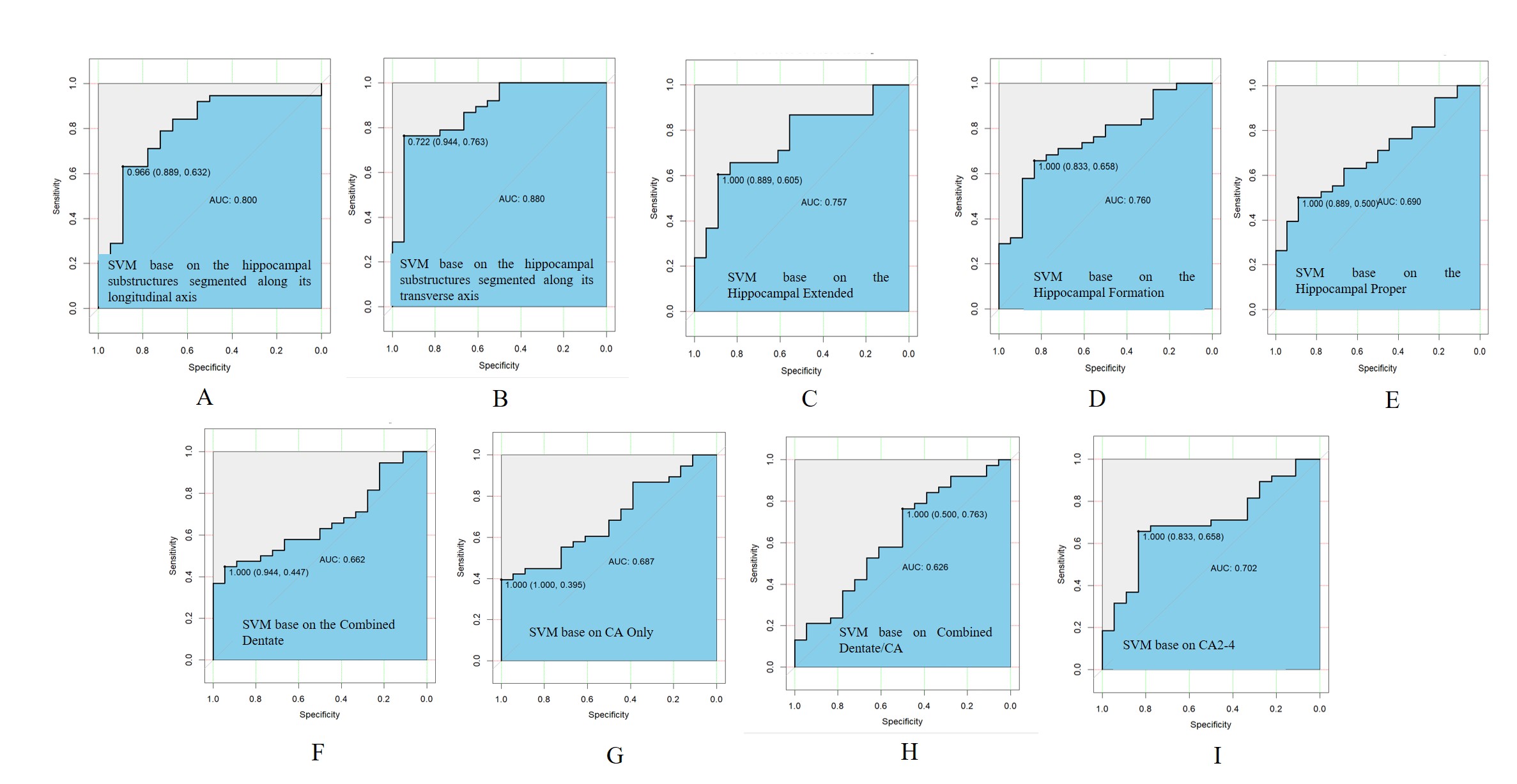
Results: We found hippocampal substructures segmented along its transverse axis exhibit best classification performance, and the most relevant region for treatments efficacy maybe located in the CA1 and ML.
Impact: We found hippocampal substructures segmented along its transverse axis exhibit best classification performance, and the most relevant region for treatments efficacy maybe located in the CA1 and ML.
Introduction
Major depressive disorder (MDD) is one of the most frequent and disabling psychiatric disorders. There are many treatment strategies available and the treatment response can vary wildly between individuals. Recent reviews reported that the pre-treatment hippocampal GMV can be a biomarker of response to antidepressant effects[1], and the volume of the hippocampus is one of the most replicated structural indicators for predicting treatment efficacy[2]. Few studies investigated if subfields or subregions of the hippocampus could better predict treatment response. Moreover, the results were heterogeneous. One of the reason could be that the hippocampus can be subdivided into different subfields or substructures according to different protocols, but there were no study compared and find which protocol could find a better biomarker to predict treatment response in depression. The most popular protocol segmented the hippocampus into several subfields along the transverse axis based on histological features, and another protocol segmented the hippocampus into the head, body, and tail along with its longitudinal axis based on its functional segregation[3]. In addition, few studies focused on the hippocampal composites, which created by summing certain component substructures. Thus, we aimed to explore which hippocampal substructure or composites volumes might predict an early response to antidepressants in medication-free adult patients with MDD.Methods
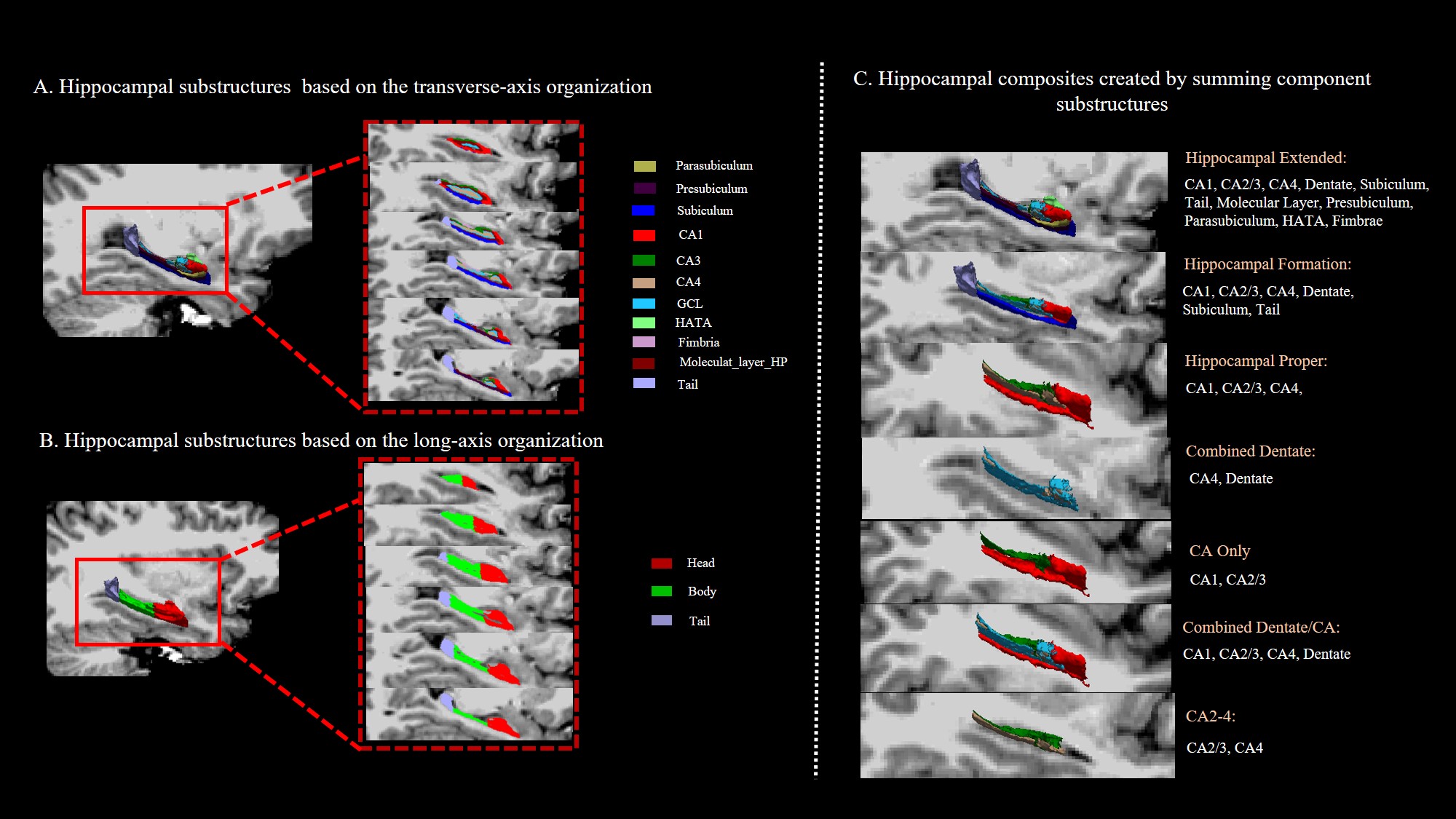
A total of fifty-six medication-free adult patients with MDD and seventy age- and gender-matched healthy controls (HCs) were recruited, the demographic and clinical characteristics of all participants are summarized in Table 1. The substructure segmentation was performed using FreeSurfer software automatically. The hippocampus were further divided into three subregions (head, body and tail) along its longitudinal axis() or eleven subfields (CA1,CA3,CA4, molecular layer, GC.ML.DG, subiculum, presubiculum, parasubiculum, fimbria, HATA, tail) according to its transverse axis. The hippocampal composites are created by summing component substructures derived form CA1,CA2,etc.(Figure 1). The response to antidepressants was evaluated by the reduction rate of the Hamilton Rating Scale for Depression score (RRS) after a 6-week routine clinical antidepressant treatment. Volume differences among early responding patients (ERP), nonresponding patients (NRP) and HCs were examined by general linear model (GLM) approaches. The relationship between hippocampal substructures volumes and RRS was explored using partial correlation analysis. Support vector machines (SVMs) was used to perform classification between the NRP and ERP group based on the different hippocampal organization.Results
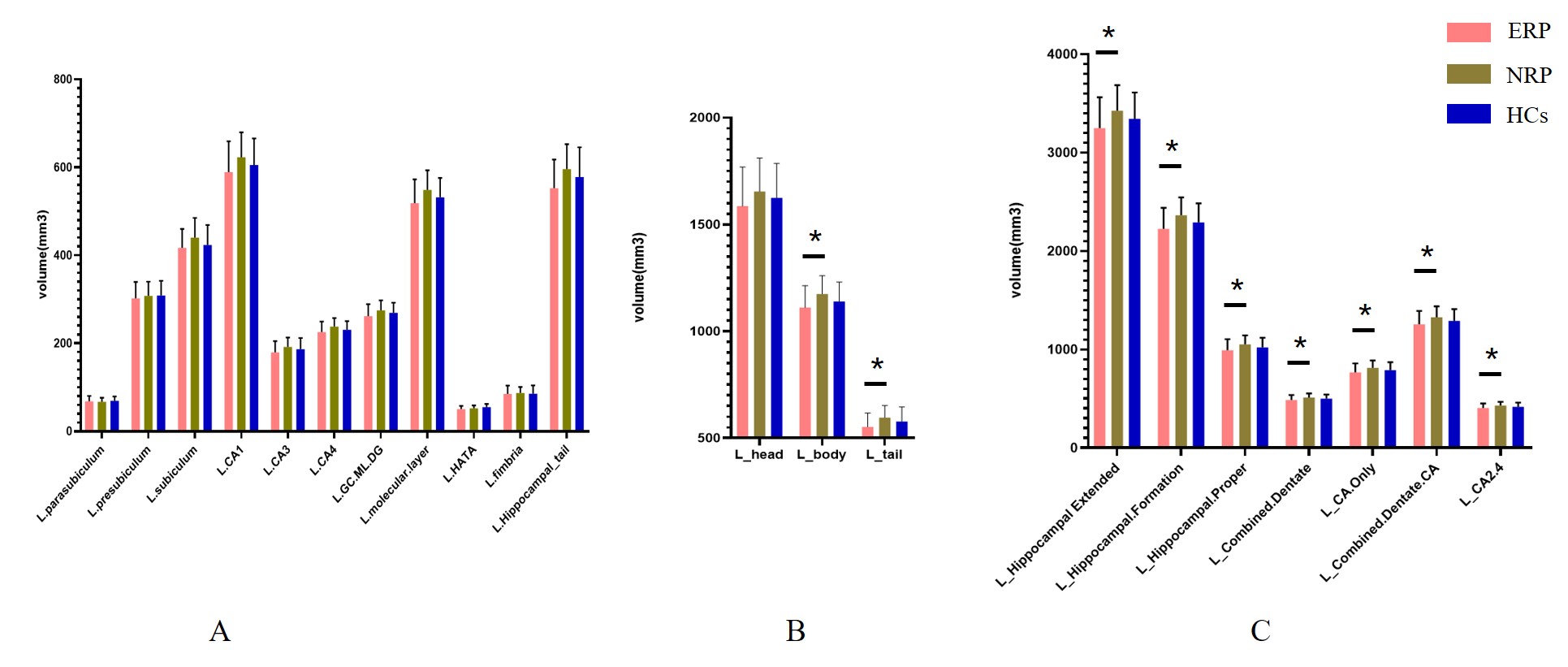
NRP had significantly larger volumes than ERP in the specific hippocampal substructures and composites in the left hemisphere, mainly including the hippocampal substructures (body and tail) and all composites except for the left Combined.Dentate.CA(Figure 2). Significant negative correlations of the RRS were found between the RRS and volumes of specific left volume parameters, mainly including the hippocampal substructures (the head, body and tail, CA1 and molecular.layer ) and all composites. SVMs model based on the substructures along with its transverse axis(CA1,CA2, etc.) gets the best performance classification. The area under the Receiver operating characteristic curve was 0.880, with a sensitivity of 76.3% and a specificity of 94.4%(Figure 3).Discussion and Conclusion
As far as we know, this is the first study to specifically investigate which hippocampal substructure or composites volumes might predict an early response to antidepressants in medication-free adult patients with MDD. We found hippocampal substructures obtained through segmentation along its transverse axis exhibit best classification performance, primarily located in the CA1 and ML. These findings contribute not only to the selection of a suitable hippocampal segmentation modalities in assessing the treatment efficacy for MDD patients, but also to deepening our comprehensive understanding of the relationship between the hippocampal substructure and antidepressant treatment efficacy.Acknowledgements
This study was supported by the Natural Science Foundation of Sichuan Province (Grant No. 2022NSFSC0052), the National Key R&D Program of China (Grant No. 2022YFF1202400) and Young Elite Scientists Sponsorship Program by CAST (2022QNRC001).References
1. Enneking V, Leehr EJ, Dannlowski U, Redlich R: Brain structural effects of treatments for depression and biomarkers of response: a systematic review of neuroimaging studies. Psychological medicine 2020, 50(2):187-209.
2. Gerlach AR, Karim HT, Peciña M, Ajilore O, Taylor WD, Butters MA, Andreescu C: MRI predictors of pharmacotherapy response in major depressive disorder. NeuroImage Clinical 2022, 36:103157.
3. Genon S, Bernhardt BC, La Joie R, Amunts K, Eickhoff SB: The many dimensions of human hippocampal organization and (dys)function. Trends in neurosciences 2021, 44(12):977-989.
Figures
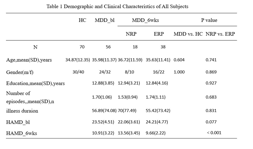
Note: MDD, major depressive disorder; bl, baseline; HCs, healthy control subjects; NRP, nonresponding patients; ERP, early responding patients; HAMD, Hamilton Rating Scale for Depression;