1721
Volumetric abnormalities in thalamic subnuclei in drug-naive patients with major depressive disorder1Department of Radiology and Huaxi MR Research Center (HMRRC), Functional and Molecular lmaging Key Laboratory of Sichuan Province, West China Hospital, Sichuan University, Chengdu, China, 2Institute of psychoradiology, West China Xiamen Hospital of Sichuan University, Xiamen, China
Synopsis
Keywords: Psychiatric Disorders, Psychiatric Disorders
Motivation: To investigate the specific changes of volumetric differences in Major Depressive Disorder (MDD) and healthy controls (HC) including their relationships with clinically related behavioral phenomena.
Goal(s): To investigate volumetric differences in the thalamic subnuclei of untreated MDD patients and HC groups.
Approach: We collected high-resolution 3D T1-weighted images from untreated MDD patients and age- and sex-matched healthy controls.
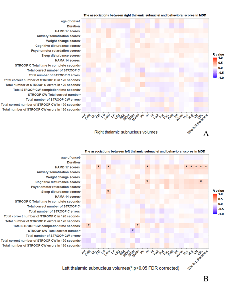
Results: No significant volume differences of the bilateral thalamus as a subnuclei between MDD patients and healthy controls. Found age-by-diagnosis interactions for specific thalamic subnuclei. Partial correlation analyses show several left thalamic subnuclei showed positive correlations with HAMD-17 scores and cognitive disturbance scores.
Impact: The study offers valuable insights into thalamic subnuclei changes and their impact on MDD. These findings underscore the significance of examining specific thalamic subnuclei and their potential impact on MDD's development.
Introduction
Major depressive disorder (MDD) is a common psychiatric illness, with clinical manifestations of persistently depressed mood, loss of interest, and cognitive dysfunction[1]. However, the regional distribution of anatomic alterations in this structure and their association with the symptoms of MDD remains to be established[2]. The thalamus engages in various functions including sensory processing, attention, decision making, and memory[3].The thalamus can be divided into five subnuclei groups[4], and each subnuclei of the thalamus has a unique function and are differentially associated with the pathological mechanisms of MDD[5]. However, few researches exist to describe the structural changes of each thalamic subnuclei in MDD. Hence, in this study, we aimed to investigate the specific and common changes of volumetric differences in MDD and HC and explore their relationships with clinically related behavioral phenomenon.Methods
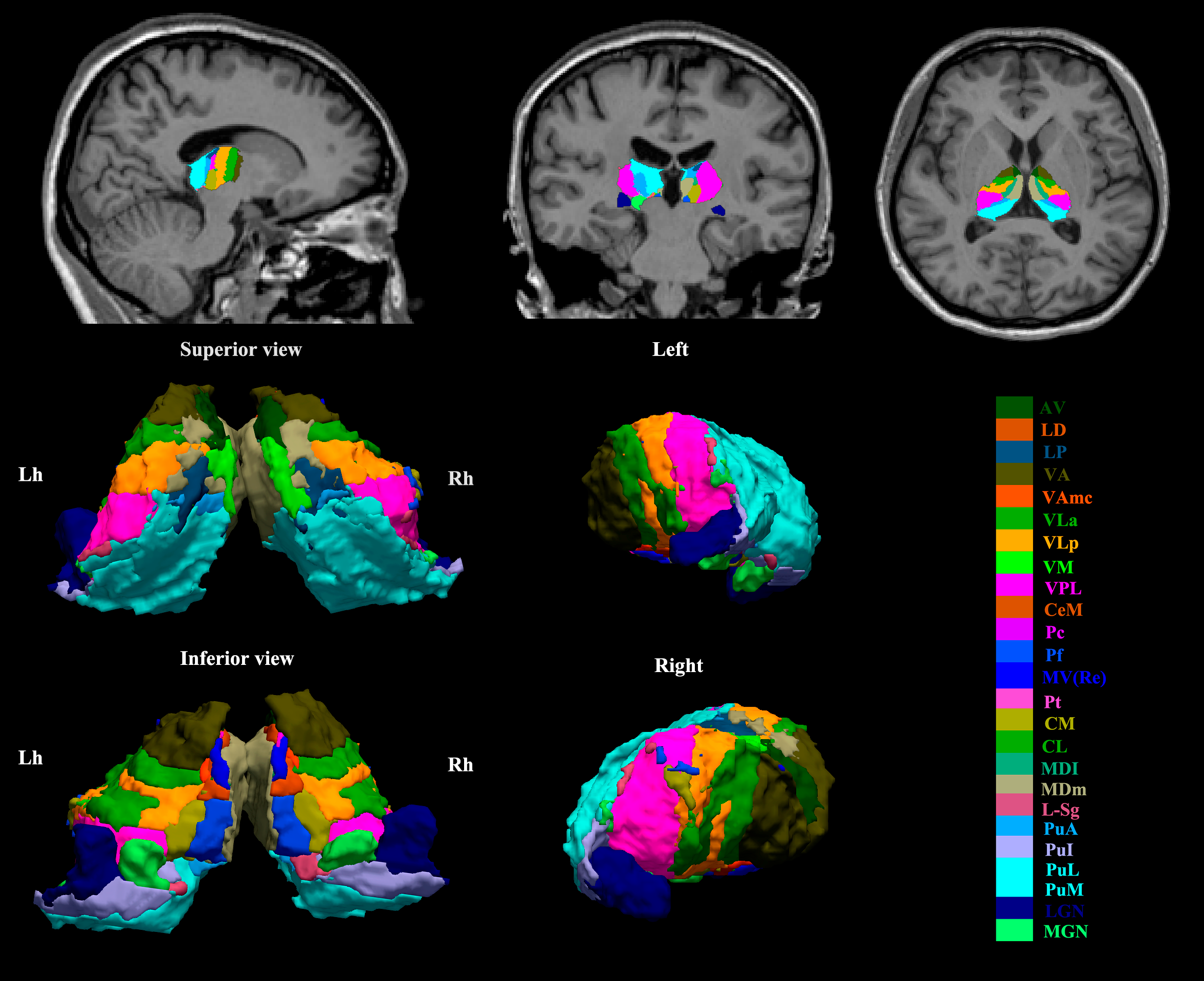
We collected high-resolution 3D T1-weighted images from 135 untreated patients with MDD and no lifetime history of comorbid psychotic, affective or anxiety disorders, and from 134 age- and sex-matched healthy controls (Table 1). We extracted the volume of 50 bilateral subnucleus of the thalamus and compared them across groups using FreeSurfer v7.3.2 (Fig 1). In exploratory analyses, we evaluated the association between HAMD-17, HAMA-14 scores, Stroop Color and Word Interference test (Stroop) frequently used to evaluate the severity of depression and thalamic nuclei volumes in MDD patients. We also tested diagnosis ×age and diagnosis × sex interactions with this model. We conducted multivariate analysis of covariance (MANCOVA) to test for groups differences in the thalamic subnucleus volumes, with age, sex and intracranial volume (ICV) as covariates. We additionally investigated the interaction of age-by-diagnosis and sex-by-diagnosis. A general linear model (GLM) was used to test for main effects of diagnosis and sex-by-diagnosis interactions on 25 thalamic subnuclei volumes, with diagnosis and sex as fixed factors, and age and ICV as covariates. Partial correlation analyses were performed to identify clinical associations of thalamic measures that showed significant group differences with age of onset illness duration, HAMD-17, HAMA-14 scores and Stroop scores in the patients with MDD, controlling for age, sex and ICV. A false discovery rate (FDR) correction was applied to correct for multiple hypothesis testing issues in between-group comparison and correlation analyses.Results
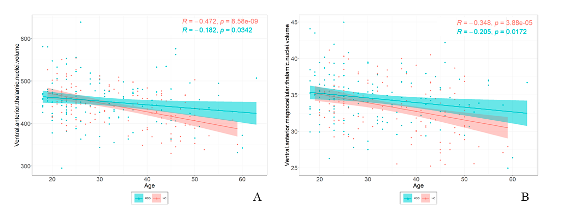
The demographic characteristics of subjects are shown in Table 1. There were no significant volume differences in the bilateral thalamus subnuclei between MDD patients and HCs. Influence of age and sex on MDD-related differences in thalamic nuclei volume. We found an age-by-diagnosis for the left Ventral anterior (p=0.000 FDR corrected, η2=0.435) and Ventral anterior magnocellular (p=0.026 FDR corrected, η2=0.390) (Figure 2). Group interaction with sex×diagnosis was not significant. Partial correlation analyses (Figure 3). Volume increase in the left Pt ,VPL,CL, LGN, Pf, Vla,VLp,VM,VPL and the entire left thalamus were related to higher HAMD-17 scores (when using age, sex and intracranial volume as covariates).Volume increase in the left ventral area were related to higher HAMD-17 scores and cognitive disturbance scores. However, the right thalamic nuclei did not exhibit significant correlations with average reaction times and Stroop effects.Discussion and Conclusion
Our findings provide a comprehensive profile of morphometric abnormalities of the amygdala in MDD and novel insights into how these abnormalities may contribute to MDD symptoms that are not confounded by current medication treatments or major psychiatric comorbidities. Subnuclei analysis revealed volume no differences in bilateral thalamus, consistent with our hypothesis. However, age-related differences were observed in the left anterior ventral nucleus and magnocellular nucleus. These findings highlight potential associations between specific thalamic subnuclei and clinical indicators in patients with depression, revealing the complex role of the thalamus in this disorder. Further research is necessary to comprehensively understand the structural changes in each thalamic subnucleus and their impact on patients with depression. Determining whether differences in the thalamus are primarily driven by specific nuclei may accurately indicate which specific circuits are more severely affected than others in the early stages of the disease. Changes volume in the left thalamic subnuclei may be particularly important for the early clinical dimensions of depression. There is a relationship between language processing ability and the size of specific brain areas.Acknowledgements
This study is supported by the Young Elite Scientists Sponsorship Program by CAST(2022QNRC001),The Research Project of Shanghai Science and Technology Commission (20dz2260300), and The Fundamental Research Funds for the Central Universities.References
[1] MARX W, PENNINX B, SOLMI M, et al. Major depressive disorder [J]. Nat Rev Dis Primers, 2023, 9(1): 44.
[2] NUGENT A, DAVIS R, ZARATE C, et al. Reduced thalamic volumes in major depressive disorder [J]. Psychiatry research, 2013, 213(3): 179-85.
[3] ROY D S, ZHANG Y, HALASSA M M, et al. Thalamic subnetworks as units of function [J]. Nat Neurosci, 2022, 25(2): 140-53.
[4] WEELAND C J, KASPRZAK S, DE JOODE N T, et al. The thalamus and its subnuclei-a gateway to obsessive-compulsive disorder [J]. Transl Psychiatry, 2022, 12(1): 70.
[5] CHILD N D, BENARROCH E E. Anterior nucleus of the thalamus: functional organization and clinical implications [J]. Neurology, 2013, 81(21): 1869-76.
Figures