1719
Hippocampal subfield and amygdala nuclei gray matter volume reduction in early-onset schizophrenia1Department of NMR, All India Institute of Medical Sciences, New Delhi, India, 2Department of psychiatry, All India Institute of Medical Sciences, New Delhi, India
Synopsis
Keywords: Psychiatric Disorders, Psychiatric Disorders
Motivation: Assessing the Gray matter volume changes in the hippocampal subfield and amygdala nuclei will help to understand the underlying mechanisms of the disease.
Goal(s): To assess hippocampal subfield and amygdala nuclei volume changes in patients with early-onset schizophrenia (EOS).
Approach: High-resolution 3D T1 images were acquired in both the control (34) and the EOS (31). Hippocampal subfield and amygdala nuclei segmentation was performed using FreeSurfer.
Results: Volume reduction in the bilateral basal, para-laminar, and right lateral amygdala nuclei, as well as molecular layer, subiculum, para-subiculum, CA4 hippocampal subfield.
Impact: The gray matter volume reduction of the hippocampus subfield and amygdala nuclei maybe associated with poor clinical outcomes in EOS.
Introduction
Early onset Schizophrenia (EOS) is a severe chronic mental health disorder in children and adolescents. Reduced gray matter (GM) volume in middle, superior temporal gyri with a negative correlation with PANSS score has been reported on neuroimaging1. Reduced volume of bilateral anterior cerebellar and total vermis in adult-onset schizophrenia (AOS) 2, morphological alterations in limbic system subregions (hippocampus, parahippocampus, anterior cingulate) of individuals at high risk for schizophrenia 3 has also been reported.The aim of the present study is to assess the reduction in gray matter volume of the hippocampal subfield and amygdala nuclei in patients with early-onset schizophrenia.
Material and Method
Thirty-one early-onset schizophrenia (EOS) patients (N=31; mean age 16.75±1.86 years; 11 females/20 males) who met ICD-10 criteria (code F20) with both positive symptoms (hallucinations, delusions, altered thoughts) and negative symptoms (social withdrawal, loss of speech) were recruited from psychiatric clinics. Thirty-four healthy subjects (N=34; mean age 17.78±1.71 years, 10 females/24 males) with no history of neurological or psychiatric disorders were recruited from the local community.High-resolution 3D T1 weighted data was obtained using a 3 T MR Scanner (Ingenia 3.0 T, M/s Philips Health Care, Netherlands) using 32 channel Head coil, with Turbo Field Echo (TFE) multi-shot spin echo sequence, TR = 8.2 ms; TE = 3.8 ms; matrix size = 240× 220×350; FOV = 240×240×175 mm; slice thickness = 1mm; number of slices = 350, Voxel Size=1×1×1.
Data Processing
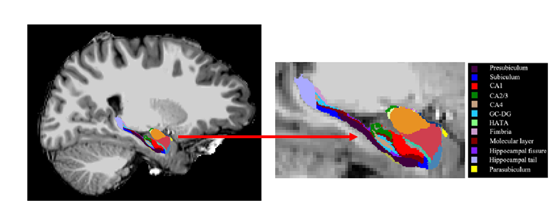
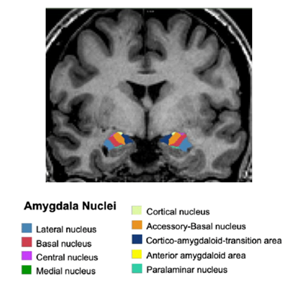
The measurement of hippocampal subfield and amygdala nuclei volumes for each participant was estimated using the Freesurfer software (v 7.2.0). We employed the automated volumetric approach, which was executed in accordance with the previously published protocol 4,5. Initially, brain segmentation was performed using the Freesurfer 'recon-all' function4. Subsequently, the automated subfield segmentation script 5 was utilized to delineate the hippocampal subfields and amygdala nuclei. The hippocampus is divided into 13 subfields in each hemisphere. These subfields include CA1, CA2/3, CA4, molecular layer, alveus, GC-ML-DG, HATA, subiculum, presubiculum, parasubiculum, fimbria, hippocampal tail and fissure. The subfields of the hippocampus were automatically segmented, and their volumes calculated. To ensure comprehensive data analysis, a thorough examination of all processed data by an investigator was conducted, revealing no errors in the automatic labeling of subjects. This confirms that the data generated by the Freesurfer analysis was entirely automated and unaffected from manual intervention. The statistical analysis consisted of multivariate analysis of covariance (MANCOVA), with age, gender, and estimated intracranial volume (eTIV) as covariates using in SPSS (version 20, IBM Corp, USA).Result
Significant volume reductions were observed in the bilateral basal, para-laminar, and right lateral amygdala nuclei, as well as significant reductions in hippocampal subfield nuclei, including the molecular layer, subiculum, para-subiculum, and CA4, in individuals with early-onset schizophrenia compared to healthy controls.Discussion
The lateral nucleus plays a pivotal role in fear-related responses and reported a lower mean total neuron count in schizophrenia6. Basal nucleus as an integral component of the threat processing circuitry7. The study has been reported reduced hippocampal subfield volumes associated with vulnerability to psychosis and memory function8,9. A correlation was reported in schizophrenia between decreased CA4 volume and visuospatial associative memory performance10. The reduced volume of amygdala and hippocampal subfield associated with poor clinical output related to altered memory, emotion and violence behaviour11. The reduction in gray matter volume in the hippocampal subfield and amygdala nuclei may be associated with adverse clinical outcomes in early-onset schizophrenia patients.Conclusion
Early identification of patients with poor clinical outcomes related to volume reduction of the hippocampus and amygdala nuclei will be helpful in the clinical management of EOS patients.Acknowledgements
We acknowledge that this research was supported by the National DBT-RA Program Biotechnology and Life Sciences (DBT/2020/January/72), Ministry science and technology, Government of IndiaReferences
1. Tang, J. et al. Decrease in temporal gyrus gray matter volume in first-episode, early onset schizophrenia: an MRI study. PloS One 7, e40247 (2012).
2. Moberget, T. et al. Cerebellar volume and cerebellocerebral structural covariance in schizophrenia: a multisite mega-analysis of 983 patients and 1349 healthy controls. Mol. Psychiatry 23, 1512–1520 (2018).
3. Hartberg, C. B. et al. Subcortical brain volumes relate to neurocognition in schizophrenia and bipolar disorder and healthy controls. Prog. Neuropsychopharmacol. Biol. Psychiatry 35, 1122–1130 (2011).
4. Fischl, B. et al. Whole Brain Segmentation: Automated Labeling of Neuroanatomical Structures in the Human Brain. Neuron 33, 341–355 (2002).
5. Iglesias, J. E. et al. A computational atlas of the hippocampal formation using ex vivo, ultra-high resolution MRI: Application to adaptive segmentation of in vivo MRI. NeuroImage 115, 117–137 (2015).
6. Johansen, J. P., Cain, C. K., Ostroff, L. E. & LeDoux, J. E. MOLECULAR MECHANISMS OF FEAR LEARNING AND MEMORY. Cell 147, 509 (2011).
7. Barth, C. et al. In Vivo Amygdala Nuclei Volumes in Schizophrenia and Bipolar Disorders. Schizophr. Bull. 47, 1431–1441 (2021).
8. Sasabayashi, D. et al. Reduced Hippocampal Subfield Volume in Schizophrenia and Clinical High-Risk State for Psychosis. Front. Psychiatry 12, 642048 (2021).
9. Yasuda, K. et al. Hippocampal Subfield Volumes and Cognitive Function in Schizophrenia and Mood Disorders. Neuropsychobiology 81, 204–214 (2022).
10. Wannan, C. M. J. et al. Hippocampal subfields and visuospatial associative memory across stages of schizophrenia-spectrum disorder. Psychol. Med. 49, 2452–2462 (2019).
11. Tesli, N. et al. Hippocampal subfield and amygdala nuclei volumes in schizophrenia patients with a history of violence. Eur. Arch. Psychiatry Clin. Neurosci. 270, 771–782 (2020).
Figures
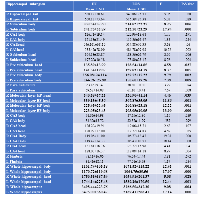
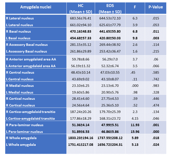
Figure 2. Amygdala nuclei volume changes in early onset Schizophrenia (EOS) patients as compared to healthy controls (HC) (R: right; L: left)