1718
Regional glutamine levels and cognition in patients with schizophrenia and healthy control subjects1Radiology, JHU SOM, Baltimore, MD, United States, 2Obstetrics & Gynecology, Weill Cornell Medicine, New York, NY, United States, 3JHU SOM, Baltimore, MD, United States
Synopsis
Keywords: Psychiatric Disorders, Metabolism
Motivation: To better understand the neurochemical correlates of cognitive impairment in subjects with schizophrenia and healthy control subjects.
Goal(s): Are regional brain metabolite levels and peripheral markers correlated with cognition in subjects with schizophrenia?
Approach: Regional brain metabolism using 7T MRS, detailed neuropsychological testing, and peripheral markers from venipuncture were obtained in 12 subjects with schizophrenia and 9 healthy control subjects.
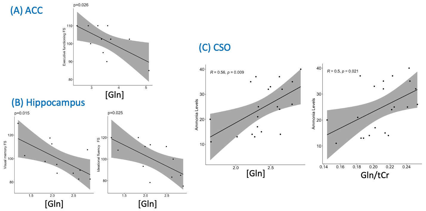
Results: In all subjects, significant positive correlations were found between regional glutamine (Gln) levels and blood ammonia levels, and, in subjects with schizophrenia, regional brain Gln levels were negatively correlated with measures of cognition.
Impact: These preliminary data support the hypothesized role of aberrant Gln metabolism as one of the factors associated with CI in schizophrenia, possibly by perturbation of the glutamate/GABA-glutamine neurotransmitter cycle.
Introduction
Some of the most disabling aspects of schizophrenia (SZ) are persistent negative symptoms and cognitive deficits, which do not respond well to antipsychotic treatments and are often among the key factors that preclude participation in steady employment or other activities of daily living. Cognitive impairment (CI) in patients with SZ occurs across multiple neuropsychological domains, including attention, working memory, verbal fluency, verbal learning, memory, and executive functioning (1).Although multiple risk factors for CI in patients with SZ have been identified, relatively little is understood about the underlying mechanisms. It has been hypothesized that CI in SZ may be the result of abnormal neurotransmission, including excitatory and inhibitory imbalance (2). Glutamate (Glu) and γ-aminobutyric acid (GABA) are major excitatory and inhibitory neurotransmitters in adult brain, and both can be measured non-invasively by proton magnetic resonance spectroscopy (MRS). MRS at 7T can measure brain glutamine (Gln) levels, giving information on the glutamate/GABA-glutamine cycle critical for neurotransmission.
In this study, the metabolic correlates of CI were investigated in both patients with SZ and healthy control (HC) subjects using 7T MRS, and compared with scores on a comprehensive battery of neuropsychological tests (3).
Methods
This study was approved by a Johns Hopkins Institutional Review Board. Each participant provided written informed consent. Participants (12 SZ, 9 HC) underwent 7T MRS and neuropsychological testing (4). Subjects with SZ (DSM-5 diagnosis) were required to be clinically stable for at least 4 weeks, and restricted to treatment with 2nd generation antipsychotic medications. Exclusion criteria for all subjects were recent substance dependence or use of valproate, regular benzodiazepine use, history of diabetes, liver disease, and obesity (BMI ≥ 35). Each participant also completed a blood draw for blood ammonia (NH3) level and complete metabolic panel, as well as urine toxicology.All participants were scanned using a 7T scanner (Philips ‘Achieva’, Best, Netherlands) equipped with a 32-channel receive head coil. 3D T1-weighted high resolution anatomical images were acquired using an MPRAGE sequence. Spectra were recorded from five regions: anterior cingulate cortex (ACC; 30×20×20 mm3), left dorsolateral prefrontal cortex (DLPFC; 25×20×20 mm3) left centrum semiovale (CSO; 40×20×15 mm3), bilateral thalamus (Thal, 20×30×15 mm3), and left hippocampus (35×15×15 mm3), using a STEAM sequence (TR/TE/TM = 3000/14/33 ms, 64 NEX, 4m 24s per VOI). VAPOR water suppression and 2nd order shim-correction used. In addition, a scan without water suppression was also acquired from each voxel (NEX = 2).
Water-scaling and eddy-current correction were applied using the unsuppressed water signal. Spectra were analyzed in the ‘LCModel’ software package (5) using a basis set containing 20 metabolites as described previously (3). Cramér-Rao lower bounds (CRLBs) and visual inspection of spectral artifacts were used to clean data prior to statistical analyses (performed with ‘R’ (version 3.5.3)). Two-sided Pearson correlation analysis was conducted to examine potential correlations between measures of CI, brain metabolite levels, and peripheral markers, with the level of significance set at p < 0.05.
Results
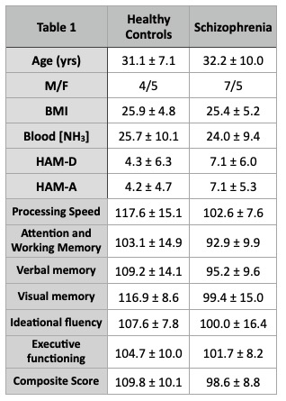
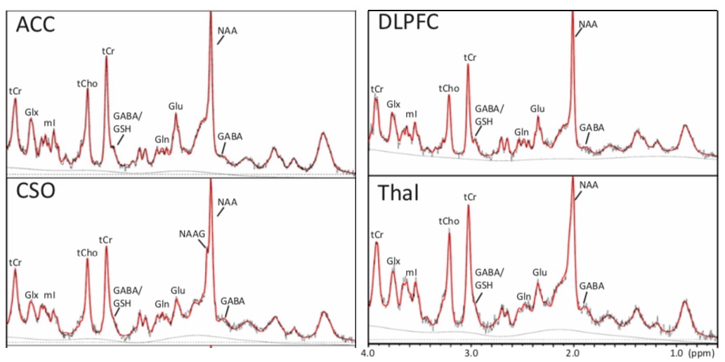
Table 1 gives the demographic information of the 2 groups and neuropsychological test scores. As expected, SZ patients performed significantly worse across neuropsychological tests, particularly in processing speed and overall composite score. Figure 1 shows representative LCModel analyses from 4 selected brain regions (ACC, DLPFC, CSO, Thal) in one subject with SZ.Figures 2A and 2B show significant negative correlations between brain Gln levels (ACC and hippocampus) and various measures of cognition (executive function, verbal memory, ideational fluency factor scores, p < 0.03) in patients with SZ. Across all participants (SZ and HC), CSO Gln levels correlated positively with blood [NH3] levels (p < 0.03).
Discussion
Altered Gln levels indicate a perturbation of the Glu/GABA-Gln cycle that is central to the energetics of glutamatergic neurotransmission, and which may be altered both in SZ and SZ-associated CI. Since Gln is synthesized from Glu and NH3, we propose a working model wherein CI in SZ associates with peripheral NH3 levels and brain Gln, as measured here using 7T MRS. Even with the relatively small sample size, these data support the hypothesized inverse relationship between Gln and CI in subjects with SZ. Furthermore, CSO Gln correlated with blood NH3 across all subjects, although no direct correlations were found between blood NH3 and cognitive performance. Future studies in larger samples and with other psychiatric comparison groups will be required to firmly establish the link between co-morbidities, peripheral NH3, regional Glu-Gln metabolism, and CI in SZ.Acknowledgements
Supported by NIH R21MH127285References
1. Schretlen DJ, Pena J, Aretouli E, Orue I, Cascella NG, Pearlson GD, Ojeda N. Confirmatory factor analysis reveals a latent cognitive structure common to bipolar disorder, schizophrenia, and normal controls. Bipolar Disord 2013;15(4):422-433.
2. McCutcheon RA, Keefe RSE, McGuire PK. Cognitive impairment in schizophrenia: aetiology, pathophysiology, and treatment. Mol Psychiatry 2023.
3. Wang AM, Pradhan S, Coughlin JM, Trivedi A, DuBois SL, Crawford JL, Sedlak TW, Nucifora FC, Jr., Nestadt G, Nucifora LG, Schretlen DJ, Sawa A, Barker PB. Assessing Brain Metabolism With 7-T Proton Magnetic Resonance Spectroscopy in Patients With First-Episode Psychosis. JAMA Psychiatry 2019;76(3):314-323.
4. Schretlen D, Testa SM, Pearlson G, Staff P. Calibrated neuropsychological normative system software portfolio (CNNS-SP). Psychological Assessment Resources Lutz, Florida; 2010.
5. Provencher SW. Estimation of metabolite concentrations from localized in vivo proton NMR spectra. Magn Reson Med 1993;30(6):672-679.
Figures