1714
NODDI revealed white matter microstructural changes are associated with global function and cognition decline in Schizophrenia Patients1Department of Radiology, West China Hospital, Sichuan University, Chengdu, China, 2GE HealthCare MR Research, Beijing, China
Synopsis
Keywords: Psychiatric Disorders, Diffusion/other diffusion imaging techniques, schizophrenia
Motivation: Schizophrenia is a complex psychiatric disorder with cognitive and behavioral disruptions. Previous studies have reported the abnormalities of white matter, but elucidating the biological mechanisms remains a challenge.
Goal(s): To investigate the changes of white matter in schizophrenia patients and correlate with changes of neuropsychological function.
Approach: NODDI was adopted to quantify the changes of neurite orientation and extracellular free water in white matter among schizophrenia patients. The correlation analyses between image indices and neuropsychological scale scores were performed.
Results: Significant changes of brain microstructure, particularly in the fornix with schizophrenia, was observed. Those changes were correlated with global function and cognition decline.
Impact: This study demonstrated abnormalities of white matter microstructure, especially in the fornix, among individuals with schizophrenia. NODDI could become a potential technique for finding biomarker in revealing the pathophysiological process of the disorder.
Introduction
Schizophrenia is a complex and severe psychiatric disorder characterized by a range of cognitive and behavioral disturbances[1]. Previous MRI studies have reported widespread cerebral white matter abnormalities in schizophrenia using T1-weighted imaging and diffusion tensor imaging[2]. However, whether these abnormalities are associated with biological changes still remains controversial. Neurite orientation dispersion and density imaging (NODDI) is designed to provide a biophysical characterization of white matter microstructure. The NODDI biophysical model adopts richer diffusion imaging data to quantify properties of microstructural environments, including neurite orientation dispersion and extracellular free water, which are potentially associated with morphological features of neural tissue[3]. Based on that, we deployed NODDI to investigate the changes of white matter microstructure in schizophrenia and to explore the association between microstructure and neuropsychological performance.Methods
A total of 37 participants, including 18 patients diagnosed with schizophrenia and 19 age- and sex-matched healthy controls, were enrolled for this study. All participants underwent a diffusion scan (NODDI, 120 directions, b = 1000 /1800 /2500 s/mm2, 2mm isotropic voxel size) and a 3D-T1-weighted scan (MPRAGE, 1 mm isotropic voxel size) using a GE Premier 3.0 Tesla MR imaging system and a 48-channel phased array head coil to assess cerebral microstructure. The Global Assessment of Functioning (GAF) and the Brief Assessment of Cognition in Schizophrenia (BACS) were also conducted as neuropsychological tests.NODDI data preprocessing was performed with MRtrix3 (https://www.mrtrix.org/) and FSL (https://fsl.fmrib.ox.ac.uk/fsl/). The preprocessed NODDI images were fed to Dmipy[4] to calculate quantitative maps based on the NODDI-Watson model. Using the same warp-fields, the FSL JHU-ICBM atlas was registered to NODDI and T1 space separately to obtain NODDI quantitative parameters, including the isotropic diffusion compartment (FISO) and the orientation dispersion index (ODI), and volume in each label. Group comparisons were performed for each modality to characterize structural changes. The correlation analyses were also conducted to investigate the association between diffusion parameters and neuropsychological scale ratings.
Results
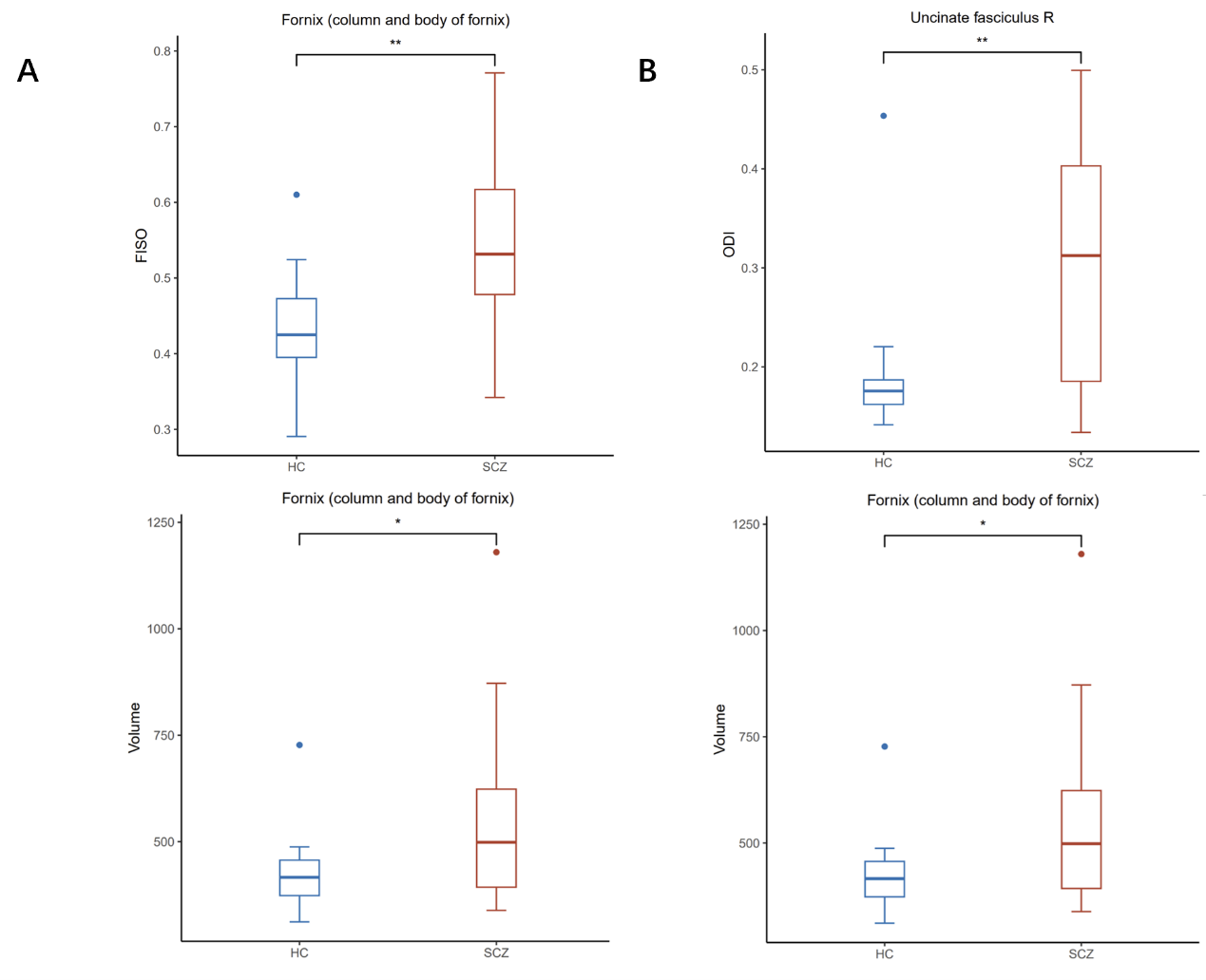
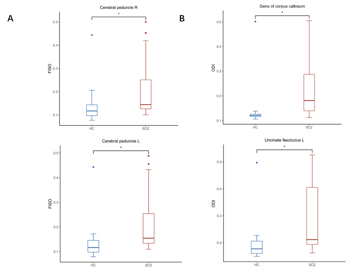
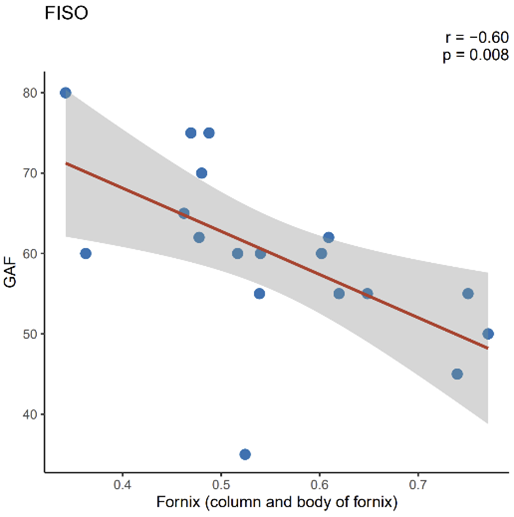
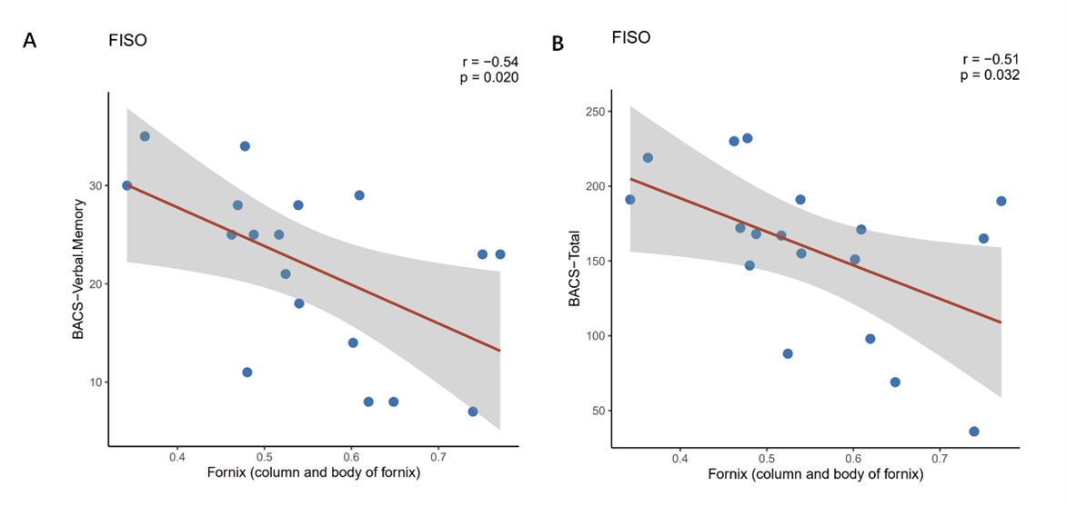
The schizophrenia group exhibited significantly increased FISO in the fornix (column and body of fornix) and increased ODI in the right uncinate fasciculus compared to healthy controls (p < 0.05), which is consistent with the volumetric changes measured using T1 imaging (Figure 1). In addition, increased FISO in the bilateral cerebral peduncle and increased ODI in the genu of the corpus callosum and the left uncinate fasciculus were also detected in schizophrenia patients (Figure 2). Furthermore, increased FISO in the fornix (column and body of the fornix) was associated with lower GAF (r=-0.60, p =0.008) (Figure 3), lower BACS verbal memory (r=-0.54, p=0.020), and BACS total scores in patients (r=-0.51, p =0.032) (Figure 4).Discussion
The study revealed significant differences of brain microstructure between individuals with schizophrenia and healthy controls using NODDI. Specifically, individuals with schizophrenia exhibited increased FISO in the fornix and bilateral cerebral peduncle, as well as increased ODI in the genu of the corpus callosum and bilateral uncinate fasciculus in comparison to the control group. Increased FISO values reflect an increase in free water, potentially leading to the early-stage edema in schizophrenia[5]. Increased ODI values reflect a more diverse and irregular distribution of neural fibers within the white matter region[6]. This pattern of findings indicates that schizophrenia is associated with microstructural anomalies contributing to the complexity of clinical manifestations.Patients with Schizophrenia exhibit increased FISO and volume in the fornix. Increased FISO in the fornix region are correlated with lower GAF score, which might serve as evidence of poorer clinical performance and indicative of a more severe psychopathological condition. The fornix is part of the limbic system and constitutes the main afferent and transmitting white matter fibers of the hippocampus, connecting the hippocampus with other brain areas such as the mammillary bodies, septal area, prefrontal cortex, etc. These brain areas are involved in processes of pathophysiological mechanisms in schizophrenia. Moreover, these structural connections mainly involve memory networks, so the fornix plays a critical role in memory tasks[7]. Our findings demonstrated that increased FISO in the fornix is associated with poorer verbal memory, which is consistent with previous studies[8].
Conclusion
This study revealed and quantified the changes of brain microstructure in patients with schizophrenia. The fornix emerges as a pivotal brain region linked to cognitive and overall functional changes, supporting its role in the pathophysiology of schizophrenia. As a novel imaging technique, NODDI presents promising clinical significance in monitoring the changes of white matter microstructure among schizophrenia patients. However, further investigation is warranted to provide more robust evidence on the clinical application of NODDI among schizophrenia patients.Acknowledgements
This work was supported by the National Natural Science Foundation of China (Project No. 82202110, 82120108014 and 82071908).References
1. Owen, M.J., A. Sawa, and P.B. Mortensen, Schizophrenia. Lancet, 2016. 388(10039): p. 86-97.
2. Kelly, S., et al., Widespread white matter microstructural differences in schizophrenia across 4322 individuals: results from the ENIGMA Schizophrenia DTI Working Group. Mol Psychiatry, 2018. 23(5): p. 1261-1269.
3. Kraguljac, N.V., et al., A longitudinal neurite and free water imaging study in patients with a schizophrenia spectrum disorder. Neuropsychopharmacology, 2019. 44(11): p. 1932-1939.
4. Fick, R.H.J., D. Wassermann, and R. Deriche, The Dmipy Toolbox: Diffusion MRI Multi-Compartment Modeling and Microstructure Recovery Made Easy. Front Neuroinform, 2019. 13: p. 64.
5. Wong, H.J., et al., Illness remission status and commissural and associative brain white matter fiber changes in schizophrenia. Psych J, 2020. 9(6): p. 894-902.
6. Gong, N.J., et al., Imaging microstructure with diffusion and susceptibility MR: neuronal density correlation in Disrupted-in-Schizophrenia-1 mutant mice. NMR Biomed, 2020. 33(10): p. e4365.
7. Douet, V. and L. Chang, Fornix as an imaging marker for episodic memory deficits in healthy aging and in various neurological disorders. Front Aging Neurosci, 2014. 6: p. 343.
8. Hovington, C.L., et al., Investigation of white matter abnormalities in first episode psychosis patients with persistent negative symptoms. Psychiatry Res, 2015. 233(3): p. 402-8.
Figures