1713
Disorganized Thalamic Subregional Functional Connectivity in Bipolar Disorder1Department of Radiology, The Second Affiliated Hospital, Zhejiang University School of Medicine, Hangzhou, China, 2Department of Psychiatry, the Fourth People’s Hospital of Chengdu, Chengdu, China, 3Department of Nuclear Medicine, West China Hospital of Sichuan University, Chengdu, China, 4Department of Radiology, West China Hospital of Sichuan University, Chengdu, China, 5University of Edinburgh, Edinburgh, United Kingdom, 6Department of Radiology, West China Xiamen Hospital of Sichuan University, Xiamen, Fujian, China, Chengdu, China
Synopsis
Keywords: Psychiatric Disorders, Psychiatric Disorders
Motivation: Compared to cortical regions, the involvement of functional connectivity of subcortical regions in bipolar disorder (BD) are less well known.
Goal(s): The potential involvement of subcortical and subregions of thalamus are explored in BD.
Approach: Seed-based subcortical and thalamic subregional functional connectivity was compared between the BD patients and HCs, as well as between patients with BD type I (BD-I) subgroup and HCs as a sub-group analysis.
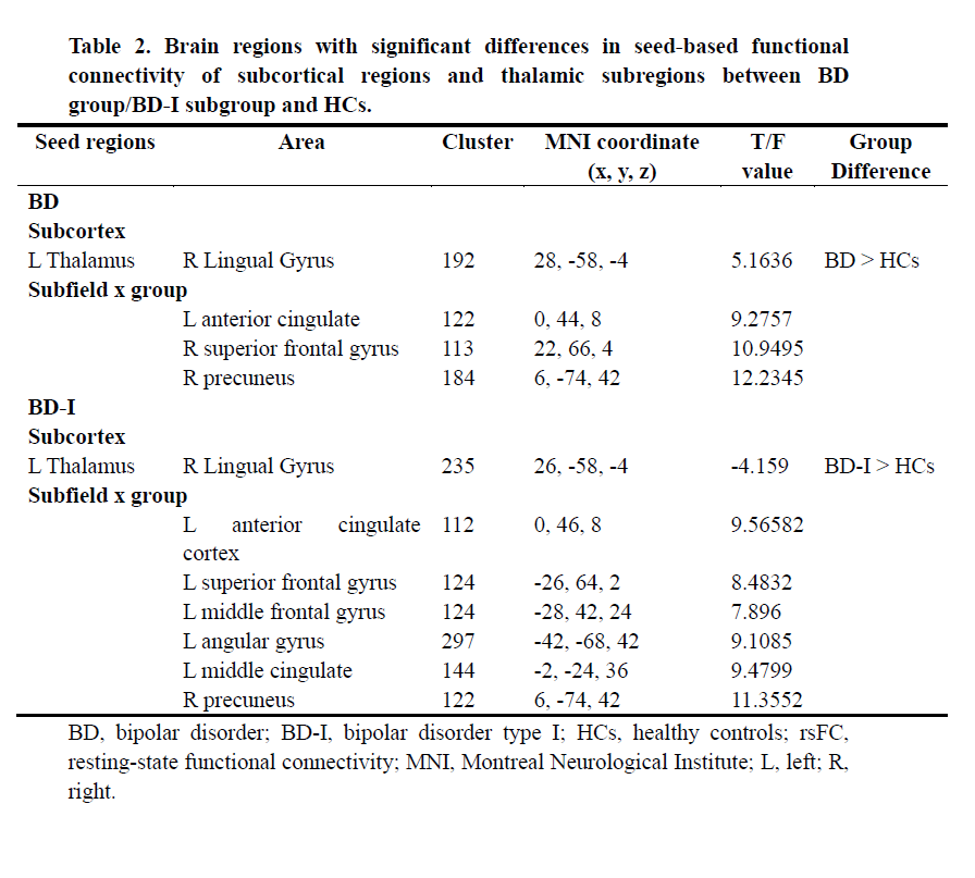
Results: BD patients showed increased functional connectivity between left thalamus and right lingual gyrus, between right anterior thalamus and superior frontal gyrus, and between right ventral anterior thalamus and precuneus.
Impact: These findings provide evidence of disorganized thalamo-cortical functional connectivity in BD, suggesting that the thalamus and its subregions may play important and specific roles in the neural circuitry of BD.
Introduction
Although alteration of brain connectivity in BD has been widely reported 1, 2, whether subcortical regions, which show extensive connections with cortical regions 3, are involved is less well studied 4, 5 and is an especial focus of the present study. Moreover, there have been very few investigations of how specific nuclei regions within subcortical structures may contribute to alterations in brain connectivity in BD. In terms of thalamic subregions, studies using manually delineated or prior ROIs or independent component analysis showed altered thalamic subregional functional connectivity with cortical regions. 6-8. However, these studies did not explore thalamic subregions from a topological perspective, therefore, the potential involvement of functional connectivity in thalamic subregions in BD still needs study. The main objective of the present study was to apply a new seed-based method to investigate the potential role of subcortical regions and subregions of the thalamus in each cerebral hemisphere in alterations in brain functional connectivity in patients with BD compared to HCs.Methods
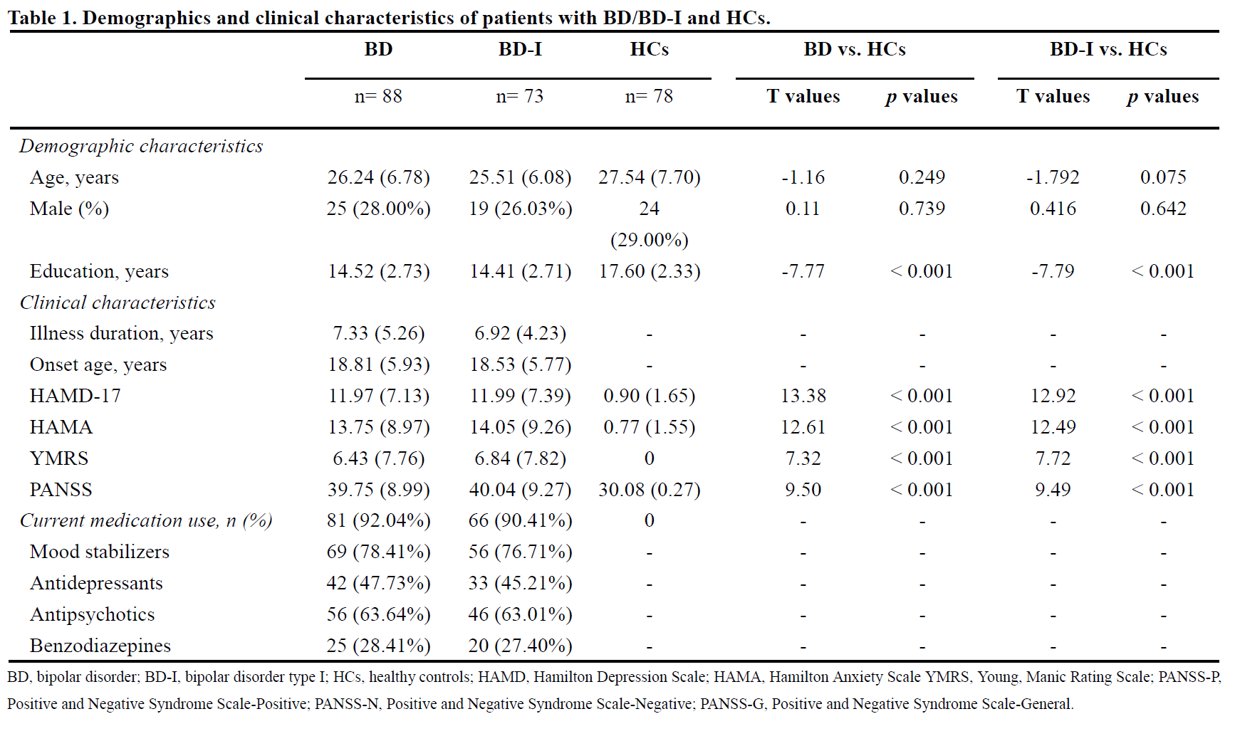
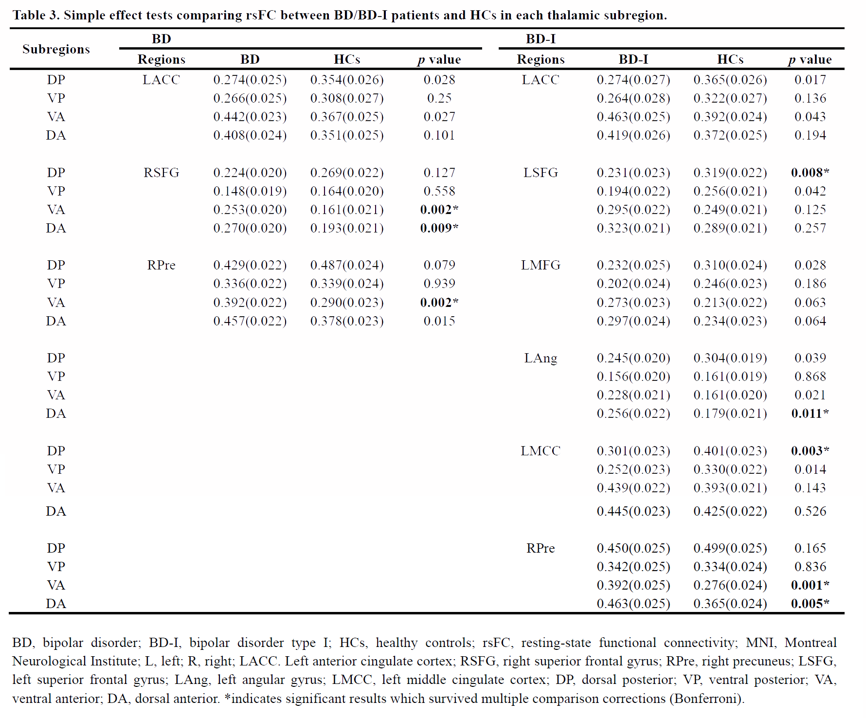
Eighty-eight BD patients and 78 HCs were recruited. Diagnosis of BD was made according to the Structured Clinical Interview for DSM-V diagnostic criteria (SCID). Magnetic Resonance Imaging (MRI) was performed on a 3.0 T system (Trio, Siemens Healthineers, Erlangen, Germany) equipped with a 32-channel head coil. T1 weighted MR images (3D MPRAGE) resting state-fMRI data (gradient-echo EPI sequence) were collected. The minimal pipeline recommended by the Human Connectome Project for (HCP)-style data was used for data pre-processing9. Seed-to-voxel functional connectivity analyses were performed using the Harvard-Oxford Atlas-based subcortical seed regions after removing subjects with excessive head motion. Thalamic subregions (dorsal anterior (DA), ventral anterior (VA), dorsal posterior (DP) and ventral posterior (VP)) were defined using an atlas based on functional gradientography10. Voxel-based z-value maps of the subcortical regions were compared between patient groups and HCs, with age, sex, and education years as covariates, by using a random-effect two-sample t-test for subcortical regions and a 2 by 2 by 4 full factorial analysis of variance, with group (BD vs. HCs) as a between-group factor and subregions (DP vs. VP vs. VA vs. DA) and hemispheres (left vs. right) as within group factors for thalamic subregions. For brain regions showing significant interaction effects, simple post-hoc tests were employed with Bonferroni correction for multiple comparisons to obtain details about which thalamus subregions contribute to the between group and within group effects. The GM volume of the thalamus and its subregions were calculated using voxel-based morphometry. Subgroup analysis was performed on patients with BD-I. Finally, a partial correlation analysis was performed to investigate the association between clinical information and rsFC data.Results
Comparison of subcortical functional connectivity in patients with BD and HCs has shown that (1) functional connectivity is significantly increased between left thalamus and right lingual gyrus in patients with BD and BD-I compared to HCs, and when subregions of the thalamus are considered (2) with regard to between group effects there is significantly increased functional connectivity between anterior thalamic subregions bilaterally and right superior frontal gyrus and precuneus in BD patients compared to HC and (3) with regard to within group effects, there is stronger functional connectivity in anterior subregions of the thalamus than in other subregions in BD patients and in dorsal posterior subregions of the thalamus than in other subregions in HCs. All the results remained significant after including lithium dose and medication load as covariates. No significant partial correlation was found.Discussion
The observation of increased functional connectivity between the left thalamus and right lingual gyrus in patients with BD and BD-I may be associated with characteristic symptoms of disturbances in relaying and integrating sensory information in patients with BD 11. The different functional connectivity pattern in BD and HC suggests a disrupted thalamic subregional functional connectivity pattern in patients with BD, especially in the anterior-posterior axes. This disorganized functional pattern may be related to the abnormal integration of visual, sensorimotor and emotional processing in BD. Alteration in the function of the thalamus in BD may not be accompanied by structural changes or perhaps it is the case that functional alterations precede structural changes.Conclusion
In conclusion, using a new seed-based approach, significant differences have been observed in the functional connectivity of subregions of the thalamus and prefrontal cortex and the default mode network in patients with BD patients and BD-I, relative to HCs. These findings added to the accumulating evidence that the thalamus and especially anterior-posterior subregions, is involved in the neuropathology of BD.Acknowledgements
We want to thank MS. Yuxin Cui and Mr. Hongkun Zhai from Nankai University for assisting in the statistical analysis.References
1. Wang, X., et al. Brain grey-matter volume alteration in adult patients with bipolar disorder under different conditions: a voxel-based meta-analysis. J Psychiatry Neurosci. 2019; 44(2): p. 89-101.
2. Wang, Y., et al. Large-scale network dysfunction in the acute state compared to the remitted state of bipolar disorder: A meta-analysis of resting-state functional connectivity. EBioMedicine. 2020; 54: p. 102742.
3. Li, J., et al. Mapping the subcortical connectivity of the human default mode network. Neuroimage. 2021; 245: p. 118758.
4. Angelescu, I., et al. The magnitude and variability of brain structural alterations in bipolar disorder: A double meta-analysis of 5534 patients and 6651 healthy controls. J Affect Disord. 2021; 291: p. 171-176.
5. Ching, C.R.K., et al. What we learn about bipolar disorder from large-scale neuroimaging: Findings and future directions from the ENIGMA Bipolar Disorder Working Group. Hum Brain Mapp. 2022; 43(1): p. 56-82.
6. Anticevic, A., et al. Mediodorsal and visual thalamic connectivity differ in schizophrenia and bipolar disorder with and without psychosis history. Schizophr Bull. 2014; 40(6): p. 1227-43.
7. Anand, A., et al. Resting state corticolimbic connectivity abnormalities in unmedicated bipolar disorder and unipolar depression. Psychiatry Res. 2009; 171(3): p. 189-98.
8. Skåtun, K.C., et al. Thalamo-cortical functional connectivity in schizophrenia and bipolar disorder. Brain Imaging Behav. 2018; 12(3): p. 640-652.
9. Glasser, M.F., et al., The minimal preprocessing pipelines for the Human Connectome Project. Neuroimage, 2013. 80: p. 105-24.
10. Tian, Y., et al. Topographic organization of the human subcortex unveiled with functional connectivity gradients. Nat Neurosci. 2020; 23(11): p. 1421-1432.
11. Anticevic, A., et al. Characterizing thalamo-cortical disturbances in schizophrenia and bipolar illness. Cereb Cortex. 2014; 24(12): p. 3116-30.
Figures
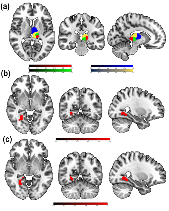
Atlas of thalamic subregions and results of group comparison of subcortical rsFC. (a) Functional gradient parcellation of the thalamus in the current study. (b) BD patients showed increased rsFC between the left thalamus and right lingual gyrus compared to HCs. (c) BD-I patients showed increased rsFC between the left thalamus and right lingual gyrus compared to HCs. BD, bipolar disorder; BD-I, bipolar disorder type I; HC, healthy controls; rsFC, resting-state functional connectivity.
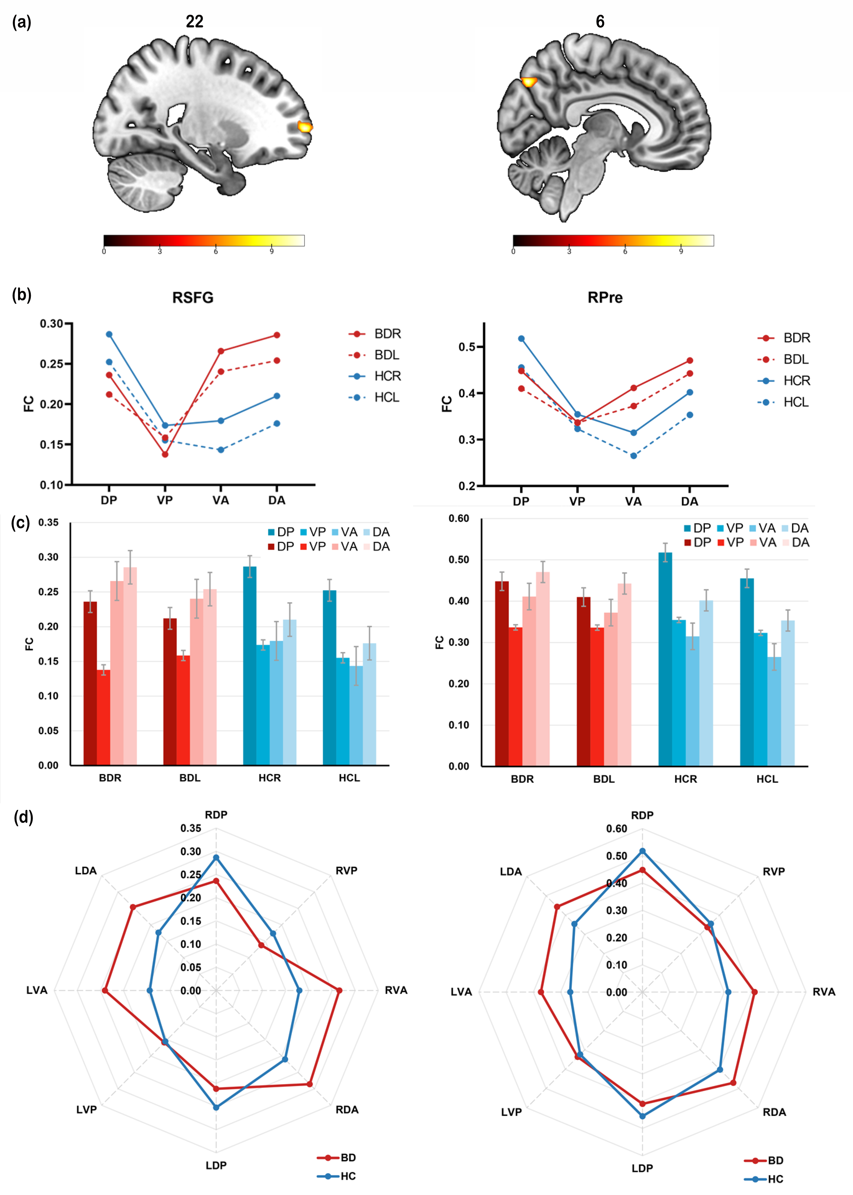
Thalamic subregional rsFC in BD patients and HCs. (a) Full factorial analysis revealed significant interactions between group (BD vs. HC) and thalamic subregions in the RSFG and RPre. (b) Significant interactions between group and thalamic subregions. (c) Simple effect tests comparing rsFC between-group and within-group. (d) Polar plots illustrating thalamic subregional rsFC showing between-group and within-group differences in BD patients and HCs. RSFG, right superior frontal gyrus; RPre, right precuneus.