1712
Emotional regulation mediates the protective role of resting-state brain activity against depression symptoms in late adolescence1Department of Radiology and Huaxi MR Research Center (HMRRC), West China Hospital, Sichuan University, Chengdu, China, 2Department of Radiology, West China Xiamen Hospital of Sichuan University, Xiamen, China
Synopsis
Keywords: Psychiatric Disorders, Adolescents, depression, emotional regulation, PFC
Motivation: The neural correlation between emotional regulation and adolescent depressive symptoms has not yet been elucidated.
Goal(s): Our goal was to identify the neural substrates of emotional regulation ability (ERA) and its association with depressive symptoms in adolescents.
Approach: We conducted whole-brain correlation, prediction and mediate analyses with resting-state fMRI and behavioral measurements data.
Results: The results indicated that ERA was associated with brain activity in right ventromedial prefrontal cortex (VMPFC) and cuneus. Additionally, ERA mediated the link between spontaneous activity of VMPFC and depressive symptoms.
Impact: Emotional regulation ability may indirectly modulate depressive symptoms in adolescents through affecting neural activity in the VMPFC, which may provide some neurobiological implications for early intervention of adolescent depression.
Introduction
Depression is a highly prevalent negative mental state characterized by low mood and aversion to daily activities 1. Adolescence is a rapid developmental period characterized by enhanced emotional reactivity, neurobiological changes and increased rates of anxiety and depression 2. During the transition to adolescence, suicidal ideation increases dramatically 3. Emotional regulation (ER) is crucial in adolescence, and the inability to effectively regulate emotions is associated with most forms of psychopathology 4,5. Given that adolescence is a critical period of neuroplasticity, it is important to understand the neurobiological basis of emotion regulation and its link to psychopathology, making it an ideal period for the delivery of emotion-focused interventions. Therefore, this study aims to explore the neural basis of ER ability (ERA) and its role in depressive symptoms in adolescence.Methods
We recruited 234 12-th grade general high school students from local public high schools with no history of psychiatric and neurological diseases. Three participants were excluded due to having brain cysts. Finally, 231 participants (121 women; mean age = 18.48 ± 0.54 years) were included. We employed Regulation of Emotion Scale (ROES, a subscale of the Wong and Law Emotional Quotient Scale) to evaluate ERA 6, and Beck Depression Inventory to access participants' depressive symptoms 7. In addition, we investigated the Socioeconomic Status Scale to eliminate the potential effect of family socioeconomic status (SES) on other variables 8.We obtained rs-fMRI data using a 3.0T Siemens Trio Erlangen MRI scanner equipped with a 12-channel head coil at West China Hospital, Sichuan University, and then calculated the fractional amplitude of low-frequency fluctuations (fALFF) values for each participant. To identify the brain regions significantly correlated with ERA, we performed whole-brain correlation analyses, with age, gender, head motion and family SES as covariates. Moreover, we performed mediation analysis to explore the potential role of ERA in the association between intrinsic brain activity and depressive symptoms.
Results
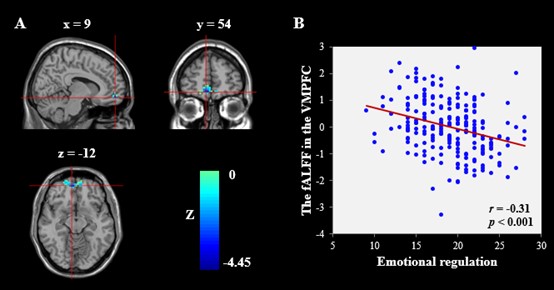
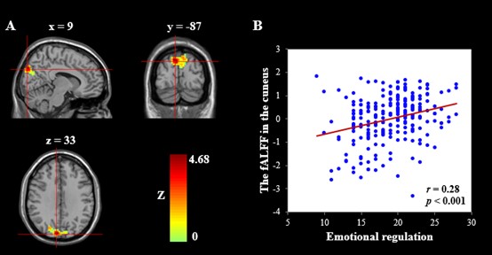
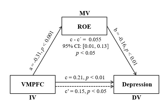
After controlling for covariates and adjusting for multiple comparisons, we found that ROES scores was negatively correlated with fALFF in the right ventromedial prefrontal cortex (VMPFC) (r = -0.28, p < 0.001, Figure 1), and positively correlated with fALFF in the right cuneus (r = 0.31, p < 0.001, Figure 2). Depressive symptoms were inversely related to ERA, and only showed a positive link to fALFF in the right VMPFC. Moreover, we found that ERA had a significant indirect effect on the association between spontaneous activities of VMPFC and depressive symptoms (indirect effect = 0.06, 95%CI: 0.012, p < 0.05). This indirect effect remained significant after adjusting for age, sex, head motion and family SES (Figure 3).Discussion
The current study investigated the correlation between ERA and brain activity at rest, and determined that the spontaneous activity of two brain regions, the right VMPFC and cuneus, is associated with individuals ERA. This result is consistent with previous studies of functional neuroimaging in patients with major depressive disorder 9,10. Interestingly, only the brain activity of VMPFC was positively associated with depressive symptoms. The prefrontal cortex is a key hub of DMN and involved in regulating attention, cognitive control, motivation, and emotion 11. ERA also exerts a significant mediating effect on the interaction of brain activity in VMPFC with adolescent depressive symptoms. These results imply that the intrinsic activity of VMPFC may plays a vital role in the neural substrate of trait ER, individuals with higher ERA may perform better to protect against depressive symptoms.Conclusion
This study indicates ERA and right VMPFC activity as neuropsychological contributors for the depressive symptoms. It deepens our understanding of the neural basis of ER, and may have clinical potential to develop targeted brain interventions. It is necessary to strengthen psychological guidance and training for vulnerable individuals to improve their ability of emotional regulation and keep adolescents away from depression.Acknowledgements
We would like to express our deep appreciation to all the participants in this study.References
1. Malhi GS, Mann JJ. Depression. Lancet (London, England) 2018;392(10161):2299-312. doi: 10.1016/s0140-6736(18)31948-2
2. Crone EA, Konijn EA. Media use and brain development during adolescence. Nature communications 2018;9(1):588. doi: 10.1038/s41467-018-03126-x
3. Nock MK, Green JG, Hwang I, et al. Prevalence, correlates, and treatment of lifetime suicidal behavior among adolescents: results from the National Comorbidity Survey Replication Adolescent Supplement. JAMA psychiatry 2013;70(3):300-10. doi: 10.1001/2013.jamapsychiatry.55
4. Aldao A, Nolen-Hoeksema S, Schweizer S. Emotion-regulation strategies across psychopathology: A meta-analytic review. Clinical psychology review 2010;30(2):217-37. doi: 10.1016/j.cpr.2009.11.004
5. Ellehauge E, Thoustrup C, Nielsen MN, et al. Frequency and types of emotional dysregulation in referrals to child and adolescent mental health services. Acta psychiatrica Scandinavica 2023;148(2):165-78. doi: 10.1111/acps.13544
6. Kong F. The validity of the Wong and Law Emotional Intelligence Scale in a Chinese sample: Tests of measurement invariance and latent mean differences across gender and age. Personality and Individual Differences 2017;116:29-31. doi: https://doi.org/10.1016/j.paid.2017.04.025
7. Richter P, Werner J, Heerlein A, et al. On the validity of the Beck Depression Inventory. A review. Psychopathology 1998;31(3):160-8. doi: 10.1159/000066239
8. Wang S, Zhao Y, Li J, et al. Neurostructural correlates of hope: dispositional hope mediates the impact of the SMA gray matter volume on subjective well-being in late adolescence. Social cognitive and affective neuroscience 2020;15(4):395-404. doi: 10.1093/scan/nsaa046
9. Gong J, Wang J, Qiu S, et al. Common and distinct patterns of intrinsic brain activity alterations in major depression and bipolar disorder: voxel-based meta-analysis. Translational psychiatry 2020;10(1):353. doi: 10.1038/s41398-020-01036-5
10. Kaiser RH, Andrews-Hanna JR, Wager TD, et al. Large-Scale Network Dysfunction in Major Depressive Disorder: A Meta-analysis of Resting-State Functional Connectivity. JAMA psychiatry 2015;72(6):603-11. doi: 10.1001/jamapsychiatry.2015.0071
11. Smallwood J, Bernhardt BC, Leech R, et al. The default mode network in cognition: a topographical perspective. Nature reviews Neuroscience 2021;22(8):503-13. doi: 10.1038/s41583-021-00474-4
Figures