1711
Quantitative measurements of masseter fat infiltration in head and neck cancer using Dixon conjugated with machine learning auto-segmentation1Department of Medical Imaging and Radiological Sciences, Chung Shan Medical University, Taichung, Taiwan, Taichung, Taiwan, 2Department of Medical Imaging, Chung Shan Medical University Hospital, Taichung, Taiwan, Taichung, Taiwan, 3Division of Medical Oncology, Department of Internal Medicine, Chung Shan Medical University Hospital, Taichung, Taiwan, Taichung, Taiwan, 4Department of Medical Imaging and Radiological Sciences, Chung Shan Medical University, Taichung, Taiwan, Department of Radiation Oncology, Chung Shan Medical University Hospital, Taichung, Taiwan, Taichung, Taiwan, 5Department of Medical Imaging, Chung Shan Medical University Hospital, Taichung, Taiwan, Department of Medical Imaging and Radiological Sciences, Chung Shan Medical University, Taichung, Taiwan, Taichung, Taiwan, 6Department of Medical Imaging and Radiological Sciences, Chung Shan Medical University, Taichung, Taiwan, Department of Medical Imaging, Chung Shan Medical University Hospital, Taichung, Taiwan, Taichung, Taiwan
Synopsis
Keywords: Muscle, Aging, Dixon, fat fraction, texture analysis
Motivation: Pathological changes in the masseter muscle have been associated with head and neck cancer (HNC). Nevertheless, investigations on the quantification of fatty infiltration in the masseter muscle and its correlation with HNC is limited.
Goal(s): We aim to assess fatty infiltration, morphological characteristics, and texture features of the masseter muscle in HNC.
Approach: This study sought to employ the Dixon method for fat fraction estimation conjugated with a machine learning-based auto-segmentation of the masseter muscle.
Results: Our analysis revealed an elevated level of fatty infiltration in the masseter muscle among patients with head and neck cancer.
Impact: Dixon method conjugated with machine learning-based auto-segmentation should facilitate in reliably assessing masseter fat alteration in head and neck cancer (HNC), this may be beneficial in response prediction in HNC treatment.
Introduction
Several studies emphasize the pivotal role of the masseter muscle in influencing the treatment and prognosis of individuals with head and neck cancer1. It has been observed that the progression and spread of cancer involve the involvement of adipose cells2. As individuals age, fat accumulates progressively within their muscles, leading to a decline in muscle function and muscle wasting. Recently, fat-suppression techniques, such as Dixon method, allows for the visualization of distinct water and fat images, facilitating the quantification of fat content via fat fraction images, which have proven effective in assessing fat infiltration in muscles3. However, the measurement of fat fraction in individual muscles typically necessitates laborious manual marking or segmentation, which can be time-consuming and subjective. While taking advantage of machine learning (ML) technology, image segmentation and processing have become more accessible, reducing the demand for extensive human intervention, these methods have seen limited application in the study of the masseter muscle and its relevance to head and neck cancer patients. Therefore, this study employs the UNet++ neural network, trained through ML, to automate the segmentation of masseter muscles for further investigation on the connection between the altered masseter fat fraction and head and neck cancer by using the Dixon method and texture analysis4.Material and methods
The study was conducted using a 3T MR scanner (MAGNETON Skyra, Siemens Healthcare), equipped with a 20-channel head and neck coil for signal acquisition. Data were collected from a total of 70 participants, comprising 17 normal individuals and 53 patients diagnosed with head and neck cancer. The magnetic resonance imaging (MRI) protocols employed in this research encompassed 32 axial T1-weighted and T2-weighted Dixon images of the head and neck. For the Dixon sequence, parameters were as follows: TR= 4000 ms, TE= 86 ms, matrix size= 320×224, bandwidth= 400 Hz/pixel, slice thickness= 5 mm, one average, and flip angle= 1760. After the acquisition, the fat fraction maps were derived from the fat and water images using a provided formula. Then the masseter muscle region was manually delineated for the subsequent machine learning model training. The neural network, based on UNet++, was constructed and trained using Matlab, employing the Adam optimizer, 20 maximum epochs, shuffling at each epoch, a mini-batch size of 5, a learning rate drop period of 10, a learning rate drop factor of 0.01, validation frequency of 50, gradient decay factor of 0.7, and squared gradient decay factor of 0.8. A comparison was made between the manual segmentation and machine segmentation using the Dice score. Additionally, quantitative parameters on the segmented regions, including contrast, correlation, energy, and homogeneity, were computed to provide more objective data for heterogeneity analysis.Results
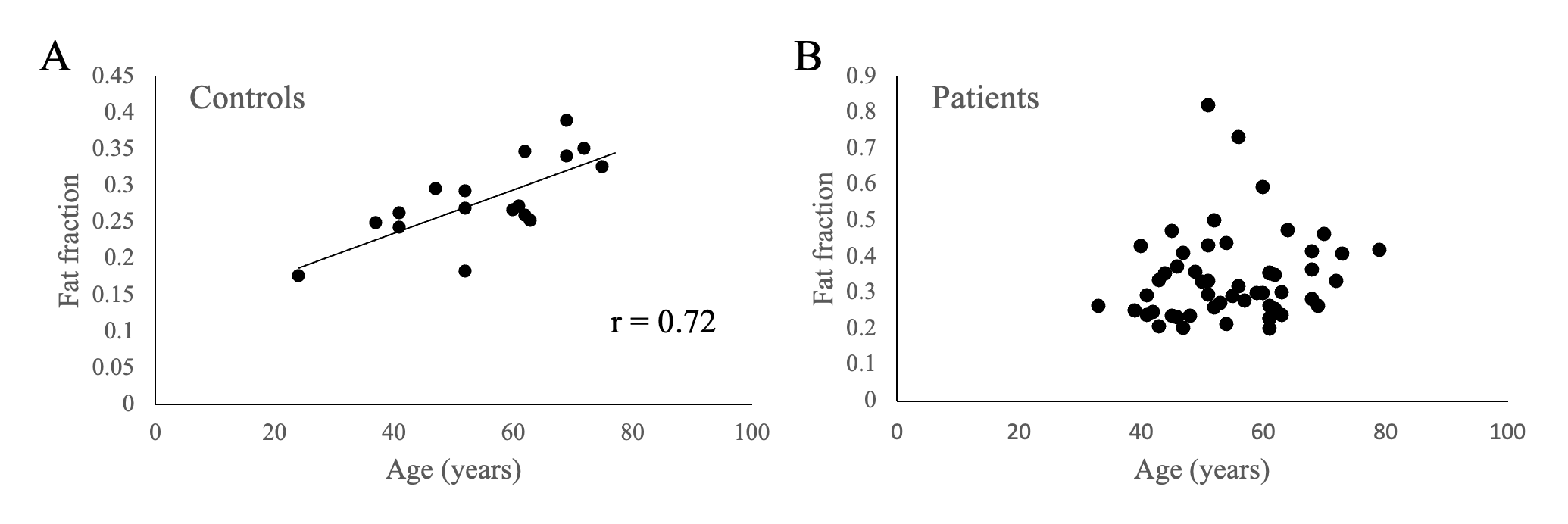
Figure 1 demonstrated the neural network architecture of the enrolled UNet++ scheme and an illustration of the auto-segmentation of the masseter muscles. The fat fraction maps, covering the bilateral masseter muscles of 2 normal controls (24- and 41-year old) and a HNC patient (40-year old) were illustrated in Figure 2A-C. Comparisons of the fat fraction, morphological and texture features in the segmented masseter muscles between the HNC patients and normal controls were shown in Table 1, respectively. While no significant differences of the morphological features in the masseter muscles were found between the two groups (p > 0.05), the fat fraction and most of the texture features in the muscles were significantly greater in HNC patients than that in controls (p < 0.05). Moreover, the derived fat fraction tends to increase with aging in normal controls (Figure 3, r=0.72).Discussions
This present study demonstrated that the feasibility of using ML-facilitated Dixon method to evaluate the altered masseter fat content in patient with HNC. While the cause of the cancer-related muscle dysfunction is complex, the HNC patients tend to exhibit higher levels of fat infiltration in the masseter muscles compared to the controls, supportive of the crucial relationships between the HNC tumor and its surrounding tissues5. In addition, in the control groups, the masseter fat fraction significant correlates with ages, which is in coincidence with the previous reports. To sum up, our preliminary findings may contribute to advancements in the treatment of head and neck cancer and enhancing the overall quality of life for those affected by this condition. The outcomes of this research are anticipated to offer clinical practitioners valuable insights into the interplay between fat cells and cancer cells, thereby assisting in the diagnosis and treatment decision-making process for individuals with head and neck cancer.Acknowledgements
This study is supported by the National Science and Technology Council, Taipei, Taiwan (NSTC 112-2221-E-040 -001 -MY2).References
- McGoldrick DM, Yassin Alsabbagh A, Shaikh M, Pettit L, Bhatia SK. Masseter muscle defined sarcopenia and survival in head and neck cancer patients. Br J Oral Maxillofac Surg. 2022;60(4):454-458.
- Mukherjee A, Bilecz AJ, Lengyel E. The adipocyte microenvironment and cancer. Cancer Metastasis Rev. 2022;41(3):575-587.
- Lee SK, Jung JY, Kang YR, Jung JH, Yang JJ. Fat quantification of multifidus muscle using T2-weighted Dixon: which measurement methods are best suited for revealing the relationship between fat infiltration and herniated nucleus pulposus. Skeletal Radiol. 2020;49(2):263-271.
- Zongwei Zhou, Md Mahfuzur Rahman Siddiquee, Nima Tajbakhsh, Jianming Liang. UNet++:A Nested U-Net Architecture for Medical Image Segmentation. arXiv:1807.10165, 2018.
- Wang S, Chen Y, She D, et al. Evaluation of lateral pterygoid muscle in patients with temporomandibular joint anterior disk displacement using T1-weighted Dixon sequence: a retrospective study. BMC Musculoskelet Disord. 2022;23(1):125. Published 2022 Feb 8.
Figures
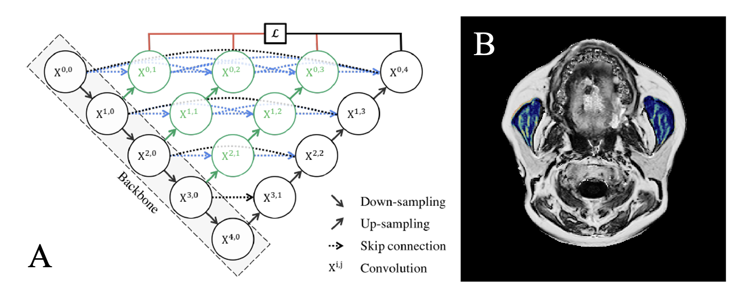
Figure. 1 Illustration of the UNet++ neural network architecture (A), and the fat fraction within the auto-segmented masseter muscles (B)
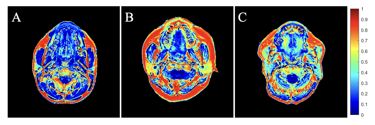
Fig 2. Fat fraction maps covering the bilateral masseter muscles in two controls (A: 24-year old, B: 41-year old) and a HNC patient (C)
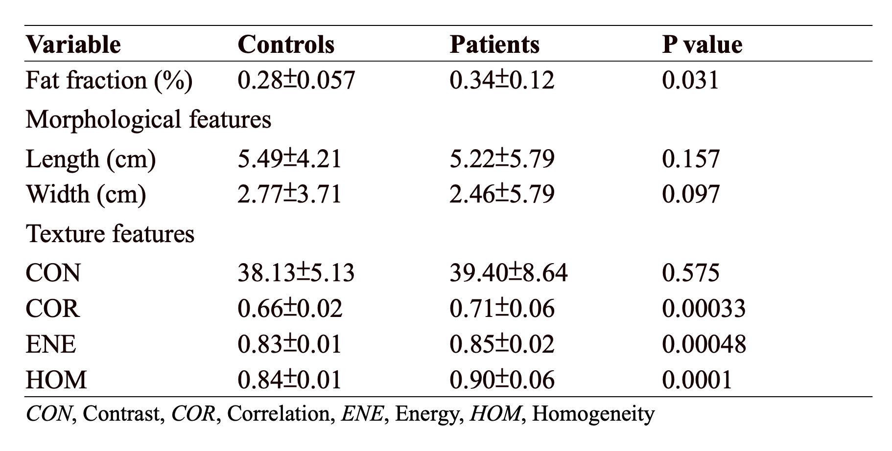
Table 1 Comparisons of the derived quantitative indices, including fat fraction, morphological and texture features of masseter muscles between control and HNC groups