1708
Relationship between degree of resorption of lumbar disc herniation and fat content of paraspinal muscles1Sichuan Province Orthopedic Hospital, Chengdu, China, 2GE HealthCare MR Research, Beijing, China
Synopsis
Keywords: Muscle, MSK
Motivation: Conservative treatment can lead to lumbar disc herniation (LDH) regression. However, the relationship between LDH regression and paraspinal muscle (PSM) fat content remains unclear.
Goal(s): To assess radiological changes in LDH with conservative treatment and investigate the connection between LDH regression and PSM fat content.
Approach: 80 LDH patients underwent MRI before and after conservative treatments. They were divided into resorption and non-resorption groups. PSM fat content and age were analyzed.
Results: 64% experienced LDH regression, mainly those with lower PSM fat content. Age-related muscle atrophy played a role. We highlights PSM fat content as a predictor of LDH regression during conservative treatment.
Impact: The fat content of paraspinal muscles serves as an important predictor of the conservative treatment outcomes for LDH patients. Patients with lower fat content in their paraspinal muscles are more likely to experience lumbar disc reabsorption.
Introduction
Lumbar disc herniation (LDH) can lead to pain, disability. It also imposes substantial economic burdens on individuals and healthcare systems. Recent evidence suggests that LDH can spontaneously undergo resorption over time without the need for surgical intervention [1-4]. Traditional Chinese medicine, as part of conservative treatment [5], has been found to provide relief to many LDH patients. The lumbar paravertebral muscles are crucial for maintaining upright posture and the dynamic stability of the lumbar spine. However, the relationship between the degree of LDH resorption and the fat content of paraspinal muscles remains controversial. This study aims to evaluate radiological changes in lumbar disc herniation using MRI following conservative treatment and investigate the correlation between the degree of disc resorption and the proton density fat fraction (PDFF) of paraspinal muscles using the IDEAL-IQ MR sequence.Methods
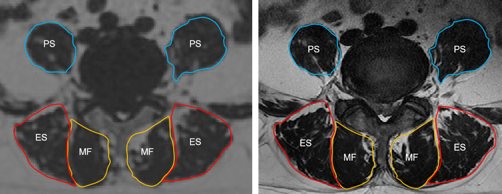
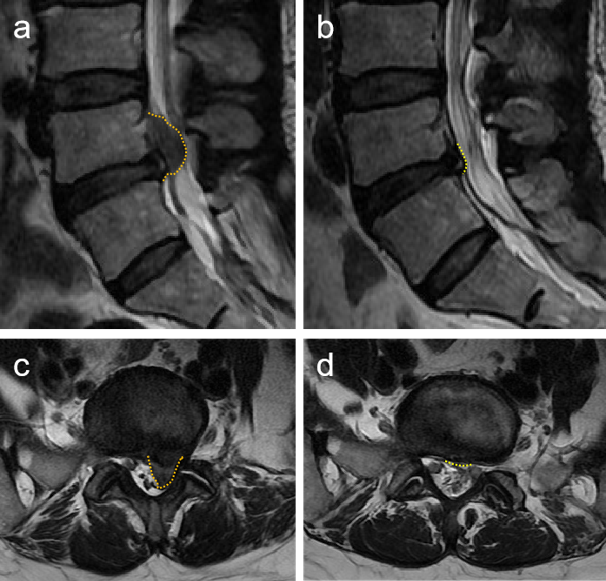
After IRB-approved written informed consent was obtained, 80 patients diagnosed with lumbar disc herniation (LDH) at the L4-L5 and L5-S1 regions were enrolled. All participants underwent MR examinations before and after conservative treatment, utilizing a 3T scanner (SIGNA Architect, GE Healthcare) equipped with a 32-channel torso coil. The scan protocol consisted of axial and sagittal T2-weighted imaging sequences, as well as the IDEAL-IQ sequence. Subjects were categorized into two groups: the resorption group, defined as those displaying a decreased grade level according to the Michigan State University (MSU) classification [6], and the non-resorption group. Additionally, to mitigate the potential influence of age on the PDFF, subjects were further categorized into different age groups. The average fatty infiltration of bilateral multifidus (MF), erector spinae (ES), and psoas major (PS) muscles was quantified at the L4-5 level using PDFF maps derived from IDEAL-IQ (Figure 1 and Figure 2). Statistical analyses were conducted with IBM SPSS 26.0. PDFF of paraspinal muscles was compared among different groups using one-way analysis of variance (ANOVA) or independent-samples t-test. P < 0.05 was considered statistically significant.Results
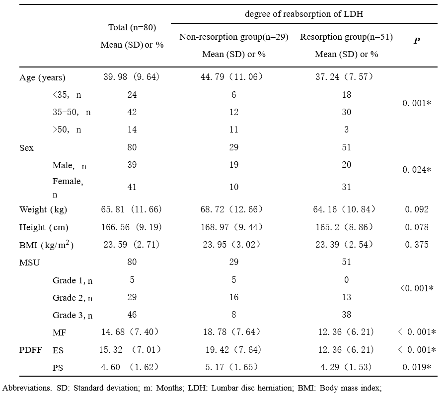
The demographic characteristics of the study participants are detailed in Table 1. Among the 80 patients included in the study, 51 patients (64%) exhibited partial or complete resorption of their lumbar disc herniation, while 29 patients (36%) did not respond to the treatment. The study found that height (p = 0.092), weight (p = 0.078), and BMI (p = 0.375) were not significantly correlated with the degree of lumbar disc herniation resorption.In terms of the PDFF of MF, ES, and PS muscles, the non-resorption group exhibited higher values compared to the resorption group (Table 1). The resorption of intervertebral disc herniation was significantly associated with the PDFF of paraspinal muscles (MF, ES, and PS) and was primarily observed in patients with lower fat content.
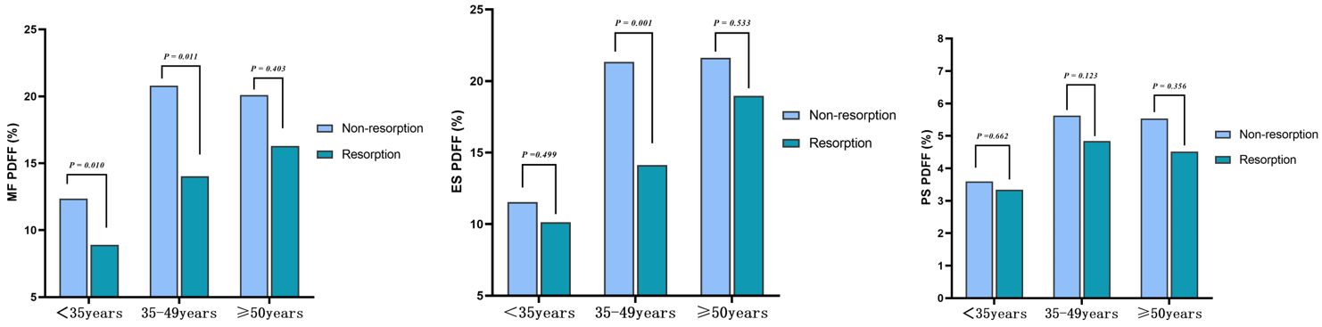
Regarding the reabsorption of lumbar disc herniation, women demonstrated comparable or better radiological outcomes than men (p = 0.024). There were differences in the degree of reabsorption of lumbar disc herniation among different age groups and sexes (p = 0.01). Changes in the reabsorption of lumbar disc herniation predominantly occurred in individuals under the age of 50 with lumbar disc herniation. The difference in fat fractions of MF/ES/PS between the non-resorption and resorption groups exhibited varying trends in different age groups (Figure 3).
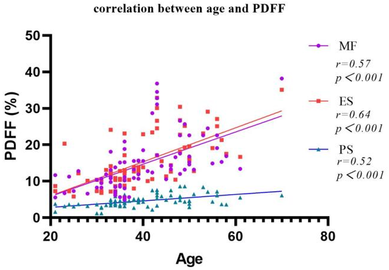
Patients in the resorption group displayed lower fat content than those in the non-resorption group. The fat fractions of the MF/ES/PS (p < 0.001) were positively correlated with age, indicating that these muscles became more atrophic with increasing age (Figure 4).
Discussion
While extensive research has explored the relationship between disc herniation reabsorption and various factors [1] such as age, duration of symptoms, sex, and the size of the herniated disc, limited attention has been given to the impact of the fat content of paraspinal muscles on the degree of disc herniation regression. Our study reveals that the extent of fat infiltration in paraspinal muscles is indeed associated with disc herniation reabsorption. Fat infiltration in paraspinal muscles has a positive predictive effect on the degree of disc herniation regression.We observed that changes in the reabsorption of lumbar disc herniation primarily occur in individuals under the age of 50, implying an association with age-related muscle degeneration. It's noteworthy that LDH patients with complete reabsorption tended to be younger and have a smaller baseline volume of herniation [3].
Conclusion
In conclusion, in addition to age and sex, the fat content of paraspinal muscles serves as an important predictor of the conservative treatment outcomes for LDH patients. Patients with lower fat content in their paraspinal muscles are more likely to experience lumbar disc reabsorption.Acknowledgements
This work was supported in part by the Research Program of Sichuan provincial administration of traditional Chinese medicine (2021MS053).
References
[1] C.-C. Chiu, T.-Y. Chuang, K.-H. Chang, C.-H. Wu, P.-W. Lin, W.-Y. Hsu, The probability of spontaneous regression of lumbar herniated disc: a systematic review, Clin Rehabil. 29 (2015) 184–195. https://doi.org/10.1177/0269215514540919.
[2] K. Kawaguchi, K. Harimaya, Y. Matsumoto, M. Hayashida, S. Okada, K. Iida, G. Kato, K. Tsuchiya, T. Doi, Y. Oda, Y. Iwamoto, Y. Nakashima, Effect of cartilaginous endplates on extruded disc resorption in lumbar disc herniation, PLoS ONE. 13 (2018) e0195946. https://doi.org/10.1371/journal.pone.0195946.
[3] B. Kesikburun, E. Eksioglu, A. Turan, E. Adiguzel, S. Kesikburun, A. Cakci, Spontaneous regression of extruded lumbar disc herniation: Correlation with clinical outcome: Spontaneous regression of extruded lumbar disc herniation, Pak J Med Sci. 35 (2019). https://doi.org/10.12669/pjms.35.4.346.
[4] A.R. Elkholy, A.M. Farid, E.A. Shamhoot, Spontaneous Resorption of Herniated Lumbar Disk: Observational Retrospective Study in 9 Patients, World Neurosurgery. 124 (2019) e453–e459. https://doi.org/10.1016/j.wneu.2018.12.115.
[5] Z. Fan, N. Xu, J. Qi, H. Su, H. Wang, Regression of a large prolapsed lumbar disc herniation achieved by conservative treatment: A case report and literature review, Heliyon. 9 (2023) e20041. https://doi.org/10.1016/j.heliyon.2023.e20041.
[6] Mysliwiec LW, Cholewicki J, Winkelpleck MD, et al. MSU Classification for herniated lumbar discs on MRI: toward developing objective criteria for surgical selection. Eur Spine J 2010;19(7):1087–1093; doi: 10.1007/s00586-009-1274-4.
Figures