1706
Diffusion Tensor MRI Analysis of Hamstring Muscle Architecture Following 9-Week Eccentric Training1Department of Bioengineering, Stanford University, Stanford, CA, United States, 2School of Human Movement and Nutrition Sciences, The University of Queensland, Brisbane, Australia, 3Department of Bioengineering, Clemson University, Clemson, NC, United States, 4School of Health and Rehabilitation Sciences, The University of Queensland, Brisbane, Australia, 5Deaprtment of Radiology, Stanford University, Stanford, CA, United States, 6Deaprtment of Radiology, New York University, New York, NY, United States, 7Department of Radiology, Stanford University, Stanford, CA, United States
Synopsis
Keywords: Muscle, Diffusion Tensor Imaging, Hamstring muscle, diffusion tensor imaging, axial diffusivity, mean diffusivity, radial diffusivity, nordic hamsrting exercise, eccentric training
Motivation: To unveil the mechanism of preventative action offered by eccentric exercise regimes towards hamstring strain injuries, understanding muscle adaptations at microstructural level is crucial.
Goal(s): To investigate microstructural adaptations in hamstring muscles post 9-weeks of eccentric NHE using diffusion tensor imaging (DTI) metrics like axial (AD), mean (MD), and radial (RD) diffusivities.
Approach: Ten participants underwent Dixon and DTI scans pre and post 9-weeks of supervised eccentric NHE training.
Results: Post intervention, significant increases in AD, MD, and RD were observed, suggesting muscle hypertrophy, exercise-induced microtrauma, structural remodelling and potential Type II muscle fiber adaptations.
Impact: This study explored the ability of DTI to provide novel insights into microstructural adaptations of hamstring muscle to eccentric training. The findings highlight hypertrophy, structural remodelling, and fiber type shifts, advancing injury prevention and rehabilitation strategies through a fiber-level perspective.
Introduction
Hamstring strains are the most common non-contact injuries1, accounting for 37% of all muscle injuries in sports2. Although, eccentric Nordic hamstring exercise (NHE) has shown potential in reducing hamstring injuries3–5, the adaptations in the muscles that contribute to the underlying preventative mechanism remains unknown. Many operator-dependent6 2D ultrasound-based studies have reported an increase in muscle fascicle length, but mostly in Biceps Femoris long head (BFlh)7–9. Moreover, these studies do not highlight microstructural fiber level changes in all the hamstring muscles in response to NHE. Diffusion tensor imaging (DTI) has demonstrated the ability characterize muscle micro trauma and exercise effects10–14, insights that are beyond macro-level information provided by ultrasound or anatomic scans of the muscle. To further explore microstructural hamstring muscle adaptations to long periods of NHE training, we aim to investigate the effect of 9-weeks of NHE intervention on DTI metrics such as axial diffusivity (AD), mean diffusivity (MD), and radial diffusivity (RD). We hypothesize that AD, MD, and RD would increase in response to the intervention.Methods
Subjects: Ten recreationally active participants (4 males / 6 females, age = 27.9 ± 3.7 years, mass = 70.9 ± 13.23 kg, no injury or NHE training in the past 18 months) underwent supervised eccentric Nordic hamstring exercise (NHE) training (~867 repetitions) for 9 weeks.Data Collection: All subjects underwent a Magnetic Resonance Imaging (MRI) scan pre and post training on a 3T MR scanner (GE Healthcare, Wi, USA) using a 21-channel blanket Air coil in prone, feet-first posture. The scan protocol consisted of DTI (3 scans at b = 0 s/mm2 and 15 directional diffusion-weighted scans at b = 400 s/mm2) and Dixon (IDEAL IQ) sequences with parameters as shown in Fig. 1.
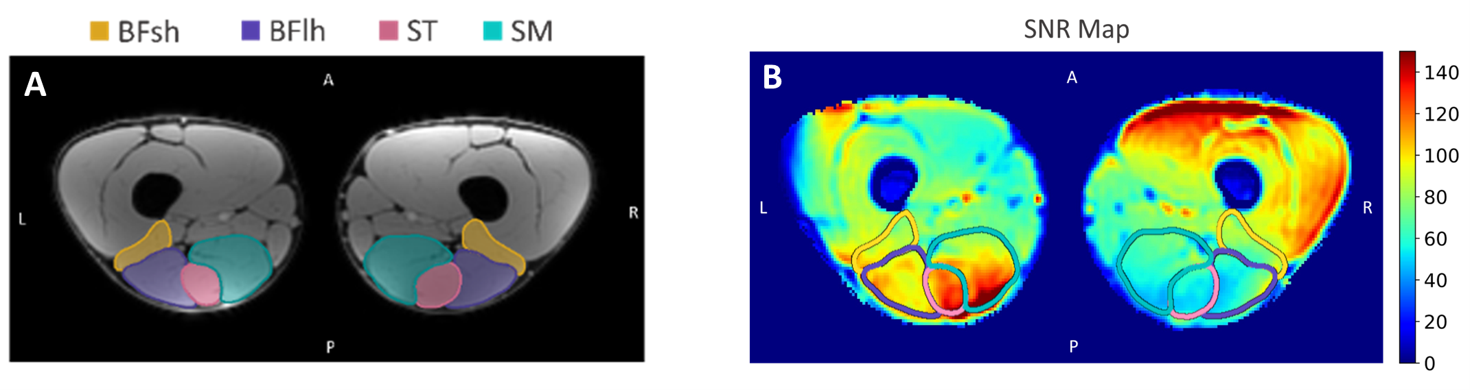
Image Processing: Four hamstring muscles, biceps femoris short head (BFsh), biceps femoris long head (BFlh), semitendinosus (ST), and semimembranosus (SM) were manually segmented on the Dixon scan using 3DSlicer15, blinded to pre/post intervention status (Fig. 2 A). DTI scans were denoised using non-local means16,17, corrected for eddy current deformation, and susceptibility-induced EPI distortion by registering to anatomic Dixon scans using DIPY18 and DOSMA19. Outcome metrics, AD, MD, and RD were computed and averaged across the volume of each muscle excluding values in pixels that had an SNR < 3020. Since many pixels on the posterior-right side had SNR< 30, only the hamstrings on the left leg were considered for analysis (Fig.2 B).
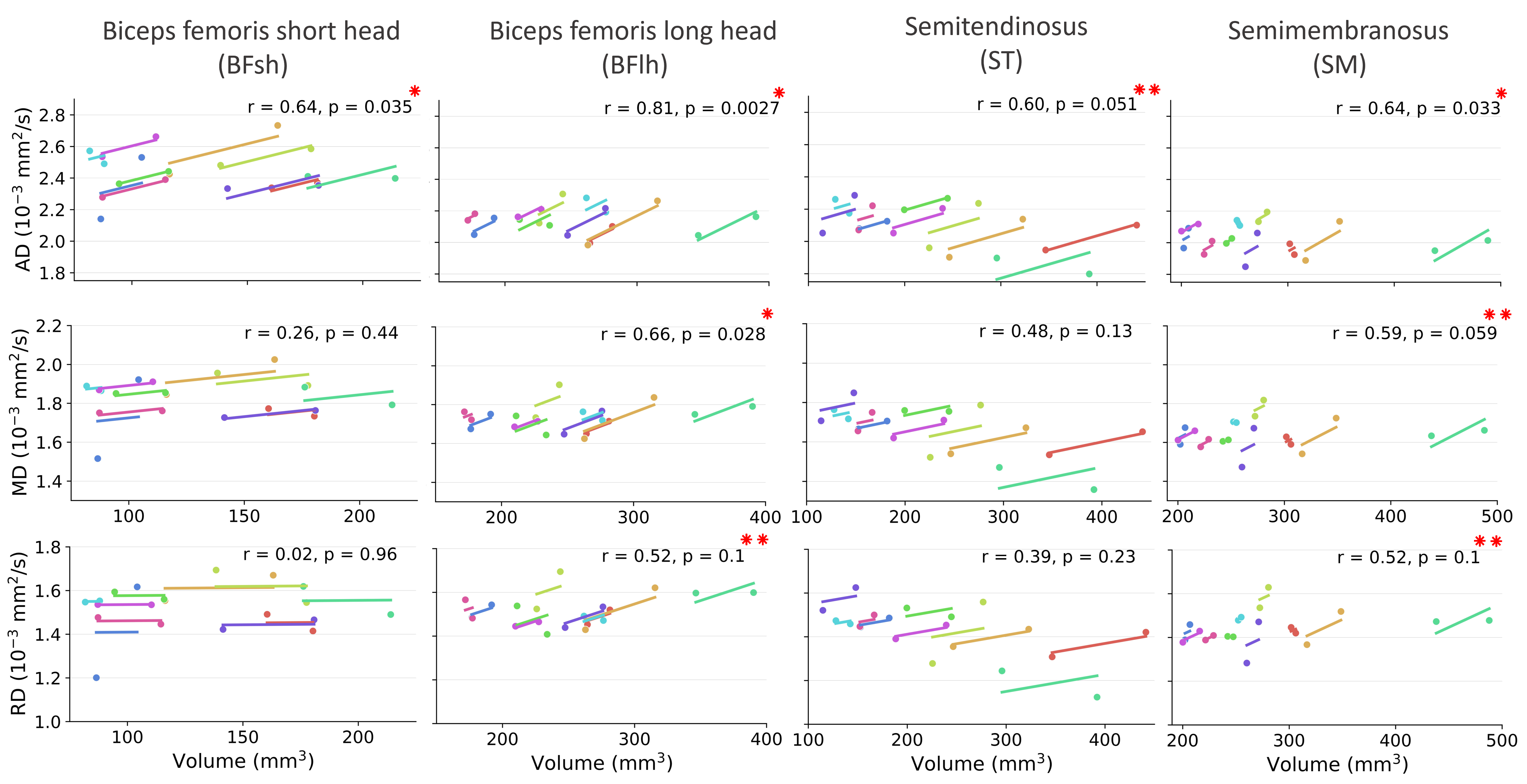
Statistical Analysis: Linear mixed effects model was used to analyse the outcome measures with “Timepoints” and “Muscles” as fixed effects, and “Subjects” as a random effect. Effect sizes are reported as partial eta squared (ηp2, where 0.2, 0.5, and 0.8 are as small, medium, and large, respectively). A repeated measures correlation model was used to correlate AD, MD, RD with muscle volume. Values with p<0.05 and p<0.1 were considered significant and approaching significance, respectively.
Results
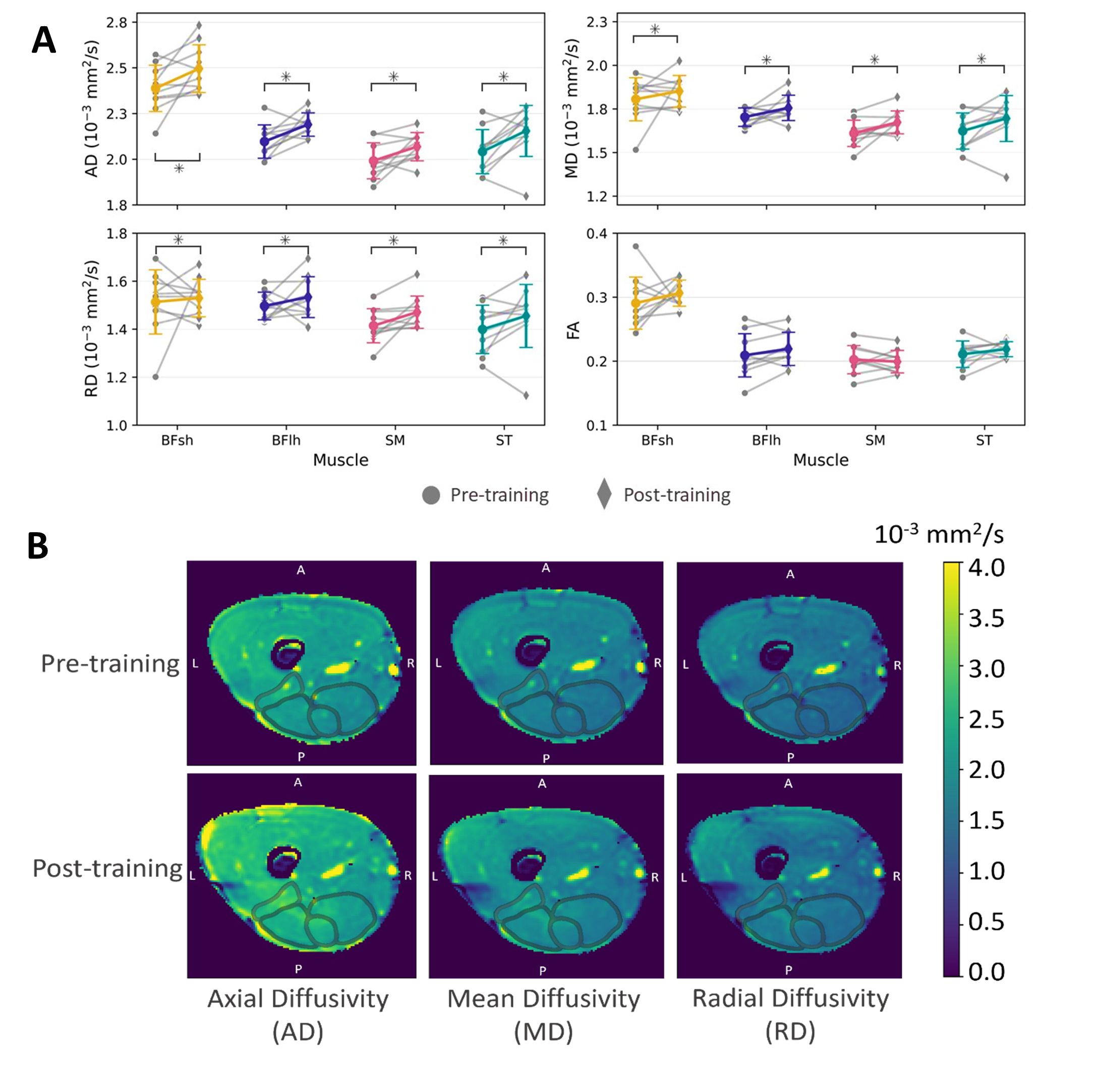
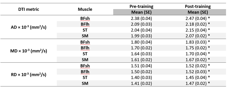
AD (p<0.001, ηp2=0.23), MD (p=0.003, ηp2=0.12), and RD (p=0.029, ηp2=0.07) were significantly higher post-training compared to pre-training for all the muscles (Figs. 3 and 4). No significant interactions were found between muscles and timepoints. Furthermore, AD significantly and positively correlated with muscle volume (Fig. 5).Discussion
An increase in AD (diffusion along the long axis of the muscle fiber) and its positive correlation with volume suggests that AD is attributed to hamstring muscle hypertrophy; a hallmark of muscle adaptation to NHE training14,21. An increase in AD could also suggest structural remodelling of the muscle fibers post NHE. An increase in MD (overall diffusion in the muscle) could imply exercise-induced microtrauma22 or local edema (increased intra- and extra-cellular water content) associated with altered muscle fiber microstructure (water diffusion pathways)23 as a consequence of NHE. The observed increase in RD, indicative of the diffusion along the short-axis of the muscle, suggests an increase in muscle fiber radius (or cross-sectional area) or adaptation towards a type II muscle fiber phenotype post-intervention. This is in line with existing literature; higher value of RD corresponds to larger proportion of Type II fibers in skeletal muscles owing its larger diameter and lower mitochondrial density24. Studies have also shown that eccentric training not only enhances the cross-sectional area of type II muscle fibers but also induces a structural transition towards fast-twitch muscle phenotype25. Small sample size, DTI data with low SNR leading to large variability in computed metrics are a few limitations of this study.Conclusion
In this study, we explored the potential of DTI metrics to characterise hamstring muscle micro adaptations to long-term eccentric training. Understanding these muscle adaptions is crucial to improve hamstring injury prevention and rehabilitation strategies.Acknowledgements
We received research support from GE Healthcare, NIH grants R01 AR077604, R01 EB002524, R01 AR079431, P41 EB027060, Stanford Graduate Fellowship, Wu Tsai Human Performance Alliance at Stanford University and the Joe and Clara Tsai Foundation, Australian Research Council Discovery Project (DP200101476).References
1. Malliaropoulos, N. G. Non contact Hamstring injuries in sports. Muscles Ligaments Tendons J 2, 309–311 (2013).
2. Ernlund, L. & Vieira, L. de A. Hamstring injuries: update article. Rev Bras Ortop 52, 373–382 (2017). 3. van der Horst, N., Smits, D.-W., Petersen, J., Goedhart, E. A. & Backx, F. J. G. The preventive effect of the nordic hamstring exercise on hamstring injuries in amateur soccer players: a randomized controlled trial. Am J Sports Med 43, 1316–1323 (2015).
4. Cuthbert, M. et al. The Effect of Nordic Hamstring Exercise Intervention Volume on Eccentric Strength and Muscle Architecture Adaptations: A Systematic Review and Meta-analyses. Sports Med 50, 83–99 (2020).
5. Rudisill, S. S., Varady, N. H., Kucharik, M. P., Eberlin, C. T. & Martin, S. D. Evidence-Based Hamstring Injury Prevention and Risk Factor Management: A Systematic Review and Meta-analysis of Randomized Controlled Trials. Am J Sports Med 51, 1927–1942 (2023).
6. Klimstra, M., Dowling, J., Durkin, J. L. & MacDonald, M. The effect of ultrasound probe orientation on muscle architecture measurement. Journal of Electromyography and Kinesiology 17, 504–514 (2007).
7. Pincheira, P. A., Boswell, M. A., Franchi, M. V., Delp, S. L. & Lichtwark, G. A. Biceps femoris long head sarcomere and fascicle length adaptations after 3 weeks of eccentric exercise training. Journal of Sport and Health Science 11, 43–49 (2022).
8. Alonso-Fernandez, D., Docampo-Blanco, P. & Martinez-Fernandez, J. Changes in muscle architecture of biceps femoris induced by eccentric strength training with nordic hamstring exercise. Scandinavian Journal of Medicine & Science in Sports 28, 88–94 (2018).
9. Bourne, M. N. et al. Impact of the Nordic hamstring and hip extension exercises on hamstring architecture and morphology: implications for injury prevention. Br J Sports Med 51, 469–477 (2017).
10. Froeling, M. et al. Muscle Changes Detected with Diffusion-Tensor Imaging after Long-Distance Running. Radiology 274, 548–562 (2015).
11. Mazzoli, V. et al. Assessment of passive muscle elongation using Diffusion Tensor MRI: Correlation between fiber length and diffusion coefficients. NMR Biomed 29, 1813–1824 (2016).
12. Monte, J. R. et al. Diffusion tensor imaging and quantitative T2 mapping to monitor muscle recovery following hamstring injury. NMR in Biomedicine 36, e4902 (2023).
13. Okamoto, Y. et al. Changes in diffusion tensor imaging (DTI) eigenvalues of skeletal muscle due to hybrid exercise training. Magnetic Resonance Imaging 32, 1297–1300 (2014).
14. Suskens, J. J. M. et al. Effect of two eccentric hamstring exercises on muscle architectural characteristics assessed with diffusion tensor MRI. Scandinavian Journal of Medicine & Science in Sports n/a,.
15. Fedorov, A. et al. 3D Slicer as an image computing platform for the Quantitative Imaging Network. Magn Reson Imaging 30, 1323–1341 (2012).
16. Coupe, P. et al. An Optimized Blockwise Nonlocal Means Denoising Filter for 3-D Magnetic Resonance Images. IEEE Trans. Med. Imaging 27, 425–441 (2008).
17. Coupé, P., Manjon, J., Robles, M. & Collins, L. Adaptive multiresolution non-local means filter for three-dimensional magnetic resonance image denoising. Image Processing, IET 6, 558–568 (2012).
18. Garyfallidis, E. et al. Dipy, a library for the analysis of diffusion MRI data. Front. Neuroinform. 8, (2014).
19. Guan, B. et al. Deep learning approach to predict pain progression in knee osteoarthritis. Skeletal Radiol 51, 363–373 (2022).
20. Froeling, M., Nederveen, A. J., Nicolay, K. & Strijkers, G. J. DTI of human skeletal muscle: the effects of diffusion encoding parameters, signal-to-noise ratio and T2 on tensor indices and fiber tracts. NMR Biomed 26, 1339–1352 (2013).
21. Maeo, S. et al. Greater Hamstrings Muscle Hypertrophy but Similar Damage Protection after Training at Long versus Short Muscle Lengths. Medicine & Science in Sports & Exercise 53, 825 (2021).
22. Heemskerk, A. M., Strijkers, G. J., Drost, M. R., van Bochove, G. S. & Nicolay, K. Skeletal muscle degeneration and regeneration after femoral artery ligation in mice: monitoring with diffusion MR imaging. Radiology 243, 413–421 (2007).
23. Zaraiskaya, T., Kumbhare, D. & Noseworthy, M. D. Diffusion tensor imaging in evaluation of human skeletal muscle injury. Journal of Magnetic Resonance Imaging 24, 402–408 (2006).
24. Scheel, M. et al. Fiber type characterization in skeletal muscle by diffusion tensor imaging. NMR Biomed 26, 1220–1224 (2013).
25. Vogt, M. & Hoppeler, H. H. Eccentric exercise: mechanisms and effects when used as training regime or training adjunct. Journal of Applied Physiology 116, 1446–1454 (2014).
Figures