1705
Using deep learning to predict muscle function based on anatomical MRI scans from the OAI and SOMMA datasets1Athinoula A. Martinos Center for Biomedical Imaging, Radiology, Massachusetts General Hospital, Boston, MA, United States, 2Radiology, Harvard Medical School, Boston, MA, United States, 3Medicine, Boston University, Boston, MA, United States, 4Computer Science and Faculty of Computing & Data Sciences, Boston University, Boston, MA, United States, 5Section of Rheumatology, Yale School of Medicine, New Haven, CT, United States, 6Section of Rheumatology, VA Connecticut Healthcare System, West Haven, CT, United States
Synopsis
Keywords: Muscle, Muscle, Data Analysis, MSK
Motivation: Estimating physical performance including muscle function is conventionally done by having a subject complete physical tasks. MRI-based estimates leveraging deep learning could complement such measures.
Goal(s): To investigate the feasibility of predicting measures physical performance including muscle strength from MRI scans of the leg using deep learning.
Approach: We used large MRI databases (OAI and SOMMA) to train a neural network for classification into high or low physical performance. We also tried the method on a small prospective cohort.
Results: We obtained over 70% accuracy for estimating high or low muscle function, indicating high predictive power.
Impact: We demonstrate the feasibility of predicting muscle function from anatomical MRI scans using deep learning, leveraging advances in deep learning and musculoskeletal MRI databases that include functional measures. Such MRI-based predictions could complement conventional methods for estimating muscle function.
Introduction
With an aging population, musculoskeletal disorders associated with aging are expected to increase in prevalence. This includes sarcopenia, the loss of muscle mass, strength, and performance. Muscle strength and performance are often measured by having the subject complete tasks such as walking or pressing a device. However, such reliance on patient cooperation can introduce noise in the measurements. Here, we developed machine learning algorithms to predict physical function from MRI. Neural networks were trained to make such predictions based on large databases containing MR images and various physical function measures. After training the network on acquired images, we tested the network on scan data acquired at our own institution.Methods
As training data, we used images and physical function measurements from the Osteoarthritis Initiative (OAI) and the Study of Muscle, Mobility, and Aging (SOMMA)1,2. We obtained 15 bilateral axial MRI slices in the mid-thigh from each of 3,515 OAI participants and 835 SOMMA participants. Each cohort also collected measures of leg strength (isometric strength chair for OAI, Keiser leg press for SOMMA), walking speed over short and long distances (20 m and 400 m for OAI, 4 m and 400 m for SOMMA), and the chair stand test. For our network architecture, we used a ResNet18 neural network, tuning it to our data set by training the weights of the last network layer. OAI and SOMMA data were used separately to train two separate networks with this same methodology, splitting each dataset into 90% for training and 10% for validation, with the validation accuracy used as the performance metric. For the predicted outcome, we trained the network to predict whether the subject had a high or low physical function for each functional measure, defined as being above or below the median value of the cohort. For further investigation, the OAI network was also trained separately on males and females from the database, still using the median of the combined cohort as the classification threshold. Upon the completion of the training, we used the best performing network to predict the physical function of 8 healthy volunteers scanned at our own institution. For these subjects, self-reported exercise (“yes”/”no”) was used as the physical performance measure to be predicted and the imaging was designed to replicate the OAI imaging settings for the thigh. Sample inputs are shown in Figure 1, with image parameters in Table 1. The network was implemented in PyTorch. Training was performed over 50 epochs using an Nvidia GeForce GTX 1080 GPU, with training taking about 7 hours for the OAI dataset and about 2 hours for the SOMMA dataset. Other training parameters included using the SGD optimizer with a learning rate of 0.0001 and a momentum of 0.9, using the Cross Entropy Loss function, and a batch size of 8. The network structure is shown in Figure 2.Results
Of the four physical performance methods, the leg strength measure was found to yield the best predictive accuracy for both the OAI and SOMMA networks, with over 70% validation accuracy for both datasets (OAI results shown in Figure 3). For the whole cohort as well as for the male- and female-specific networks, the predictive success rate exceeded the baseline of the relative size of the larger class within each cohort (e.g., for the female cohort, 60% were in the “low strength” class, a baseline exceeded by the network). Less predictive accuracy was obtained for other functional measures such as walking speed and chair stands. Saliency maps did not show an obvious pattern of heavily weighted regions, but may indicate an emphasis on the tissue periphery (Figure 4). For the prospective scans at our own institution, the OAI network successfully classified 5 out of the 8 subjects.Discussion
The results demonstrate that a neural network based on the ResNet18 architecture can classify subjects into categories of high or low leg strength based on thigh MRIs with an accuracy of over 70%. While not conclusive, the observed pattern in the saliency maps of weighting the tissue periphery may point to the network using thigh size as a determinant. This is further supported by the reduced accuracy above baseline when stratifying by gender. Further investigation to control for body size, for instance by stratifying by BMI, is ongoing.Conclusion
Axial thigh images can be used to classify subjects into having high or low physical function with over 70% accuracy using deep neural networks. This approach could complement standardized methodologies for assessing function.Acknowledgements
This work was supported by 5P30AR072571. Its contents are solely the responsibility of the authors and do not necessarily represent the official views of the NIAMS. This work was also funded by R00AG066815. Imaging was performed at the Athinoula A. Martinos Center for Biomedical Imaging at the Massachusetts General Hospital using resources provided by the Center for Functional Neuroimaging Technologies (P41EB015896) and the Center for Mesoscale Mapping (P41EB030006), Biotechnology Resource Grants supported by the National Institute of Biomedical Imaging and Bioengineering, National Institutes of Health (NIH).References
1. Eckstein F., Wirth W. & Nevitt M. Recent advances in osteoarthritis imaging—the Osteoarthritis Initiative. Nat Rev Rheumatol. 2012;8:622-630.
2. Cummings SR, Newman AB, Coen PM, et al. The Study of Muscle, Mobility and Aging (SOMMA): A Unique Cohort Study About the Cellular Biology of Aging and Age-related Loss of Mobility. J Gerontol A Biol Sci Med Sci. 2023;78(11):2083-2093.
Figures
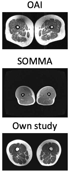
Figure 1: Sample slices from sample subjects from the OAI, the SOMMA, and own data, all used as inputs to the network.
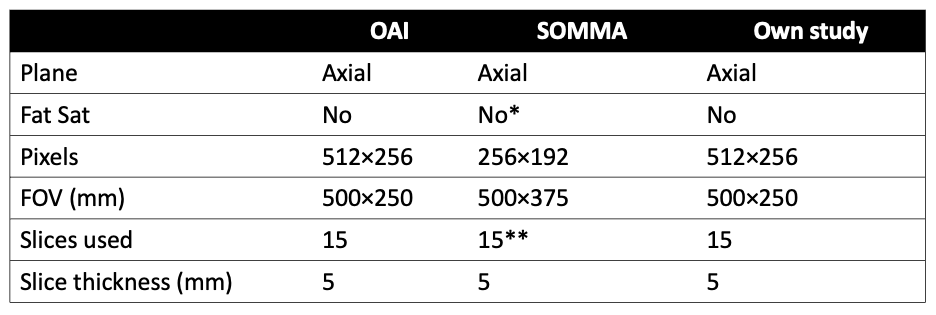
Table 1: Imaging parameters for the three types of input. (*) In SOMMA, a two-point Dixon acquisition was performed. We used the in-phase image containing both water and fat. (**) SOMMA acquired a whole-body scan. We selected 15 slices from the thigh region, based on distance from the shoulders relative to the whole body.
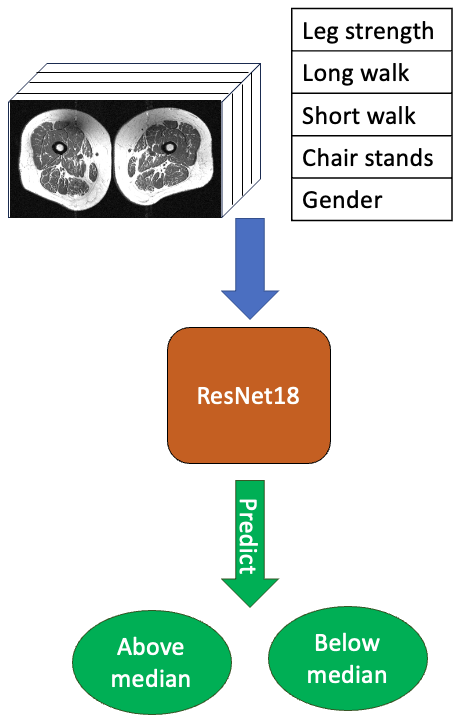
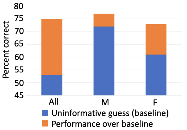
Figure 3: Bar graphs showing the prediction accuracy rate and what was considered the prediction baseline, defined as the relative size of the larger class within each cohort.
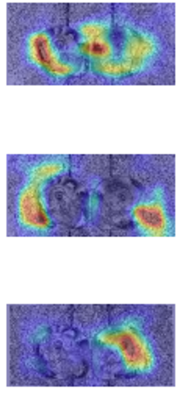
Figure 4: Saliency maps from sample slices in a sample OAI subject. While not conclusive, the network often seemed to give high weighting to the tissue periphery.