1698
Free-breathing Magnetic Resonance Fingerprinting for fat fraction and water T1 quantification of upper body muscles1NMR Laboratory, Institute of Myology, Neuromuscular Investigation Center, Paris, France
Synopsis
Keywords: Muscle, Motion Correction
Motivation: Respiratory muscles are often altered in subjects with neuromuscular diseases. Characterizing their structure using quantitative MRI is then crucial but challenging due to respiratory motion.
Goal(s): We developed a 3D free-breathing Magnetic Resonance Fingerprinting sequence for quantifying fat fraction (FF) and water T1 (T1H2O) of upper body muscles at 3T.
Approach: We estimated the free-form respiration motion deformation on a 3D pre-scan using VoxelMorph and subsequently applied it in an iterative reconstruction framework to retrieve the MRF image series.
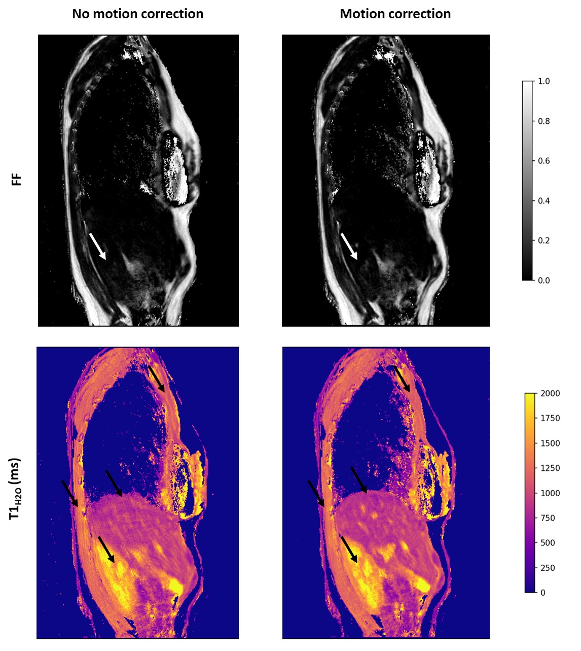
Results: This method allows a significant reduction of motion artefacts on parametric FF and T1H2O maps.
Impact: Free-breathing MRF T1-FF on 3T scanners paves the way for high resolution quantification of FF and T1H2O in the upper body muscles for monitoring their structural alterations in subjects with neuromuscular diseases with high precision.
Introduction
In the field of neuromuscular disorders, radial MR Fingerprinting with water and fat separation (MRF T1-FF) has been proposed to simultaneously quantify fat fraction (FF) and water T1 (T1H2O), which can be used as biomarkers of muscle tissue alterations 1,2 . However, upper body muscles such as abdominal, diaphragm, pectoral and intercostal muscles are prone to breathing artefacts and cannot be evaluated using this approach. Free-form deformation estimation and iterative reconstruction have been introduced for free-breathing cardiac and liver spiral MRF at 1.5T 3. In their proposal, authors estimated a motion field on MRF singular volumes. However larger B1 variations through the field of view at 3T, combined with greater undersampling artefacts due the radial acquisition scheme of the MRF T1-FF sequence complicates this task. For qualitative imaging, 4D joint motion-compensated high-dimensional total variation (MoCo-HDTV) has also been proposed 4 to iteratively refine deformation fields estimation and volumes reconstruction. In this work, we propose to estimate the motion field iteratively on a pre-scan and apply it to reconstruct the MRF T1-FF singular volumes before pattern matching.Methods
Acquisitions were performed on a healthy volunteer on a 3T Siemens PrismaFit scanner (Erlangen, Germany).The pre-scan consisted in a stack of star radial 3D-FLASH (TE/TR = 3.45/5.67 ms, FA = 10º) and the 3D radial MRF T1-FF sequence was presented in previous studies 5. For both acquisitions, 1-dimensional navigators were inserted every 28 spokes to track the liver-lung interface 6, FOV was 400x400x320mm3 and spatial resolution 1x1x5mm3.
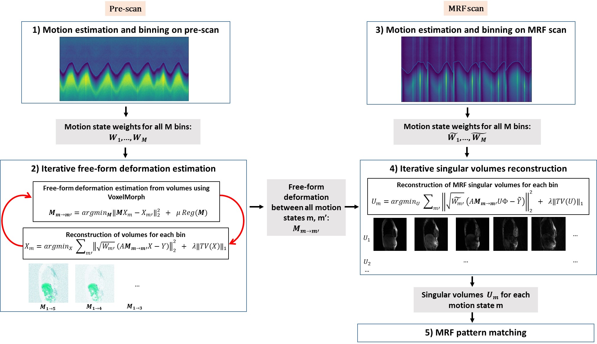
The post-processing steps are summarized in Figure 1 and consisted in: 1) Motion detection and binning on the pre-scan; 2) Iterative free-form deformation estimation between all motion-resolved pre-scan volumes ; 3) Motion detection and binning on the MRF T1-FF; 4) Iterative reconstruction of MRF singular volumes using the deformation fields estimated on the pre-scan; 5) MRF T1-FF bi-component matching for parametric mapping 7.
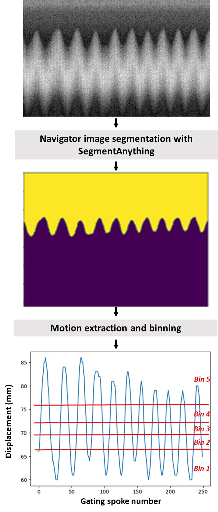
Motion detection (steps 1 and 3) used the neural network SegmentAnything 8 to segment the navigator images in two regions and estimate the liver-lung interface displacement (Figure 2). The data were split into M=5 respiratory bins with equivalent number of spokes.
The free-form deformation field (step 2) was estimated iteratively by refining the volumes reconstruction for each bin m through registration from all the other bins using the VoxelMorph neural network 9 and spatial total variation (TV) regularization in the 3 directions:
$$X_m=argmin_X ∑_{motion\ state\ m'}‖\sqrt{W_{m'}} (A\boldsymbol{M_{m→m'}}X-Y)‖_2^2 + λ‖TV(X)‖_1$$
Where $$$X_m$$$ is the rebuilt volume for bin m, $$$Y$$$ is the acquired k-space data, $$$M_{m→m'}$$$ is the motion field for registering bin m on bin m’, $$$W_{m'}$$$ are the weights associated with motion state m’, A is the MRI acquisition encoding operator combining radial k-space sampling, Fourier transform and coil sensitivities, and $$$\lambda$$$ is the regularization penalty.
$$$M_{m→m'}$$$ estimated on the pre-scan were used for iterative reconstruction of the MRF singular volumes for each bin m (step 4):
$$U_m=argmin_U ∑_{motion\ state\ m'}‖\sqrt{\tilde{W_{m'}}} (A\boldsymbol{M_{m→m'}}U\Phi-\tilde{Y})‖_2^2 + λ‖TV(U)‖_1$$
Where $$$\tilde{Y}$$$ is the acquired k-space data,$$$\tilde{W_{m'}}$$$ are the weights associated with motion state m and $$$\Phi$$$ is the temporal basis extracted from the MRF dictionary.
Results
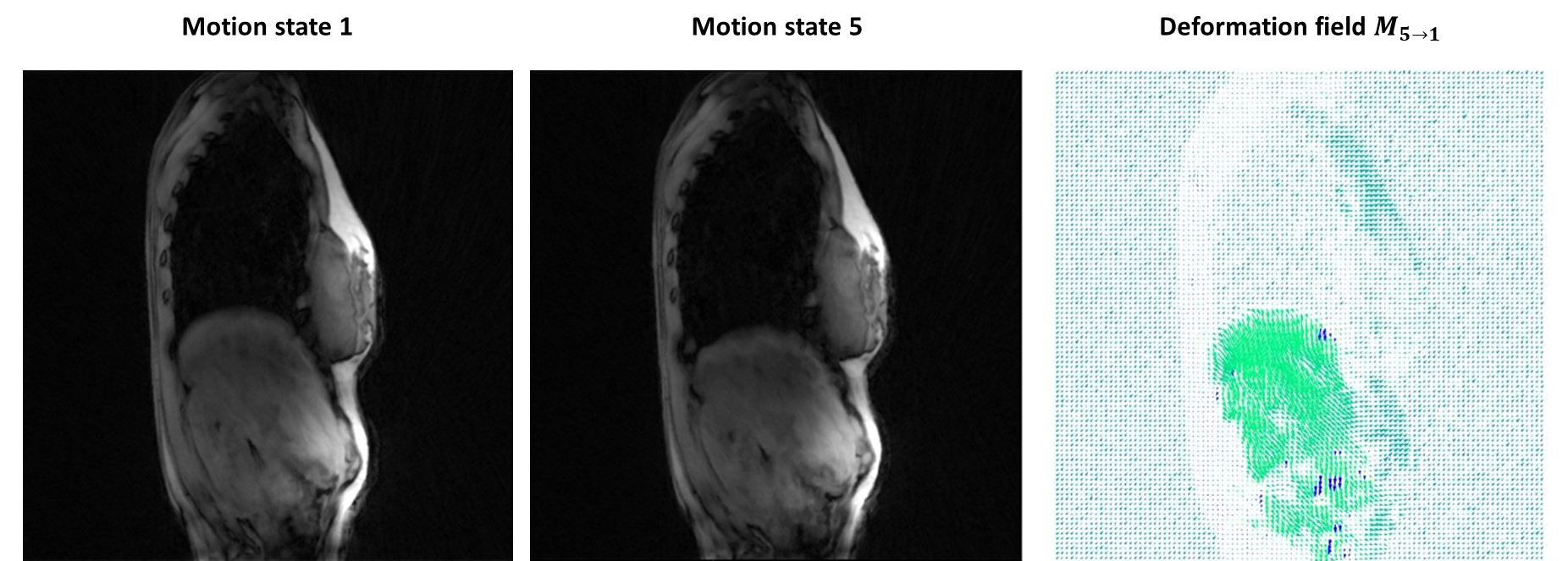
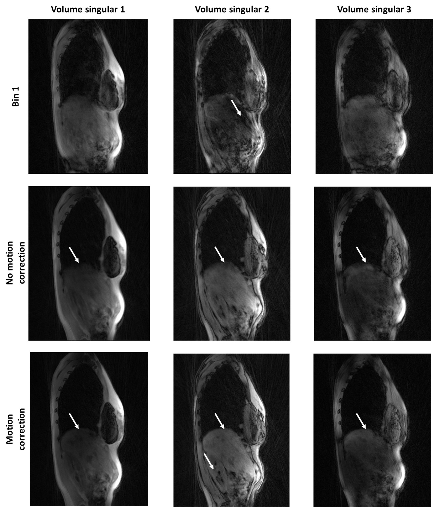
In figure 3, we show the two extreme motion-resolved volumes reconstructed from the pre-scan, together with the deformation field bridging them. The most significant displacements manifest in the abdominal region (liver and kidney), as well as in the anterior thoracic region. The singular volumes rebuilt for the first respiratory bin without any post-processing are heavily artefacted and not suitable for motion field estimation (Figure 4). The MRF T1-FF singular volumes reconstructed for the first respiratory bin with the proposed method are less affected by motion artefacts than the reference singular volumes reconstructed with all spokes without motion correction.This obviously translates into more anatomical details visible on the quantitative maps, particularly evident in the case of T1H2O (Figure 5). Streaking artifacts are significantly reduced in the pectoral and intercostal muscles. The lung-liver interface where the diaphragm lies is clearly visible on the maps with the proposed method.Discussion & Conclusion
We proposed an acquisition and post-processing framework to efficiently detect and correct respiratory motion on 3D radial MRF acquisitions. From an iterative estimate of free-form deformation fields using VoxelMorph on a pre-scan, we could reconstruct the MRF singular volumes, and subsequent parametric FF and T1H2O maps without motion artefacts. This paves the way toward quantitative imaging of the abdominal and thoracic muscles at high spatial resolution.Acknowledgements
This study was funded by ANR-20-CE19-0004.References
1. Marty B, Reyngoudt H, Boisserie JM, et al. Water-fat separation in MR fingerprinting for quantitative monitoring of the skeletal muscle in neuromuscular disorders. Radiology. 2021;300(3):652-660. doi:10.1148/radiol.2021204028
2. Marty B, Carlier PG. MR fingerprinting for water T1 and fat fraction quantification in fat infiltrated skeletal muscles. Magn Reson Med. 2020;83(2):621-634. doi:10.1002/mrm.27960
3. Cruz G, Qi H, Jaubert O, et al. Generalized low‐rank nonrigid motion‐corrected reconstruction for MR fingerprinting. Magnetic Resonance in Med. 2022;87(2):746-763. doi:10.1002/mrm.29027
4. Rank CM, Heußer T, Buzan MTA, et al. 4D respiratory motion‐compensated image reconstruction of free‐breathing radial MR data with very high undersampling. Magnetic Resonance in Med. 2017;77(3):1170-1183. doi:10.1002/mrm.26206
5. Marty B. 3D MR fingerprinting with water and fat separation. In: Proc. 28th ISMRM. Virtual Congress; 2020:1140.
6. Ehman RL, Felmlee JP. Adaptive technique for high-definition MR imaging of moving structures. Radiology. 1989;173(1):255-263. doi:10.1148/radiology.173.1.2781017
7. Slioussarenko C, Baudin P, Reyngoudt H, Marty B. Bi‐component dictionary matching for MR fingerprinting for efficient quantification of fat fraction and water T 1 in skeletal muscle. Magnetic Resonance in Med. October 2023:mrm.29901. doi:10.1002/mrm.29901
8. Kirillov A, Mintun E, Ravi N, et al. Segment Anything. 2023. doi:10.48550/ARXIV.2304.02643
9. Balakrishnan G, Zhao A, Sabuncu MR, Guttag J, Dalca AV. VoxelMorph: A Learning Framework for Deformable Medical Image Registration. IEEE Trans Med Imaging. 2019;38(8):1788-1800. doi:10.1109/TMI.2019.2897538
Figures