1697
Improved Quantitative Spatial Analysis of Spontaneous Muscular Activities using Label Uncertainty and Feature Analysis1Section on Experimental Radiology, University Hospital of Tübingen, Tübingen, Germany, 2Institute of Signal Processing and System Theory, University of Stuttgart, Stuttgart, Germany
Synopsis
Keywords: Muscle, Muscle
Motivation: Understanding the visual representation of spontaneous activities in DWI.
Goal(s): Automatically identifying visual differences in patterns of spontaneous muscular activities.
Approach: Deep-learning based detection and segmentation with subsequent feature analysis.
Results: Feasibility of feature-based clustering in individual subjects was shown.
Impact: Investigation of a pipeline for automated image processing for exploring differences in spontaneous muscular activities visible in DWI.
Introduction
Spontaneous muscular activity (SMAMs) can be detected and visualized in several different muscle groups of the human musculature system using time-series diffusion-weighted imaging (DWI).1 Spatial analysis of the activity patterns and distribution of spontaneous activities could provide important findings in healthy subjects and patients suffering from amyotrophic lateral sclerosis (ALS)2,3. Analysis of SMAMs visible in DWI might be challenging due to the very time-consuming task of manual annotation, the reduced signal-to-noise ratio and large partial volume effects in low-resolution DWI.In this work, an improved neural network-based detection and segmentation approach as well as an introduction of a cluster-based investigation of these individual spontaneous muscular activities is presented.
MR Imaging
For training of the neural network, DWI sequences with spin-echo (SE) and stimulated-echo (STE) diffusion sensitizing were applied on the lower leg of 25 subjects (age: 33±12) on 3T systems (MAGNETOM Trio/Skyra/Prismafit/Vida, Siemens Healthcare, Erlangen, Germany) with following parameters: TE: 26-31 ms (SE: 37-53 ms), TR: 500/1000 ms, Matrix: 64/80, FoV: 180-210 mm², b-value: 100s/mm².Spontaneous Activity Detection and Segmentation
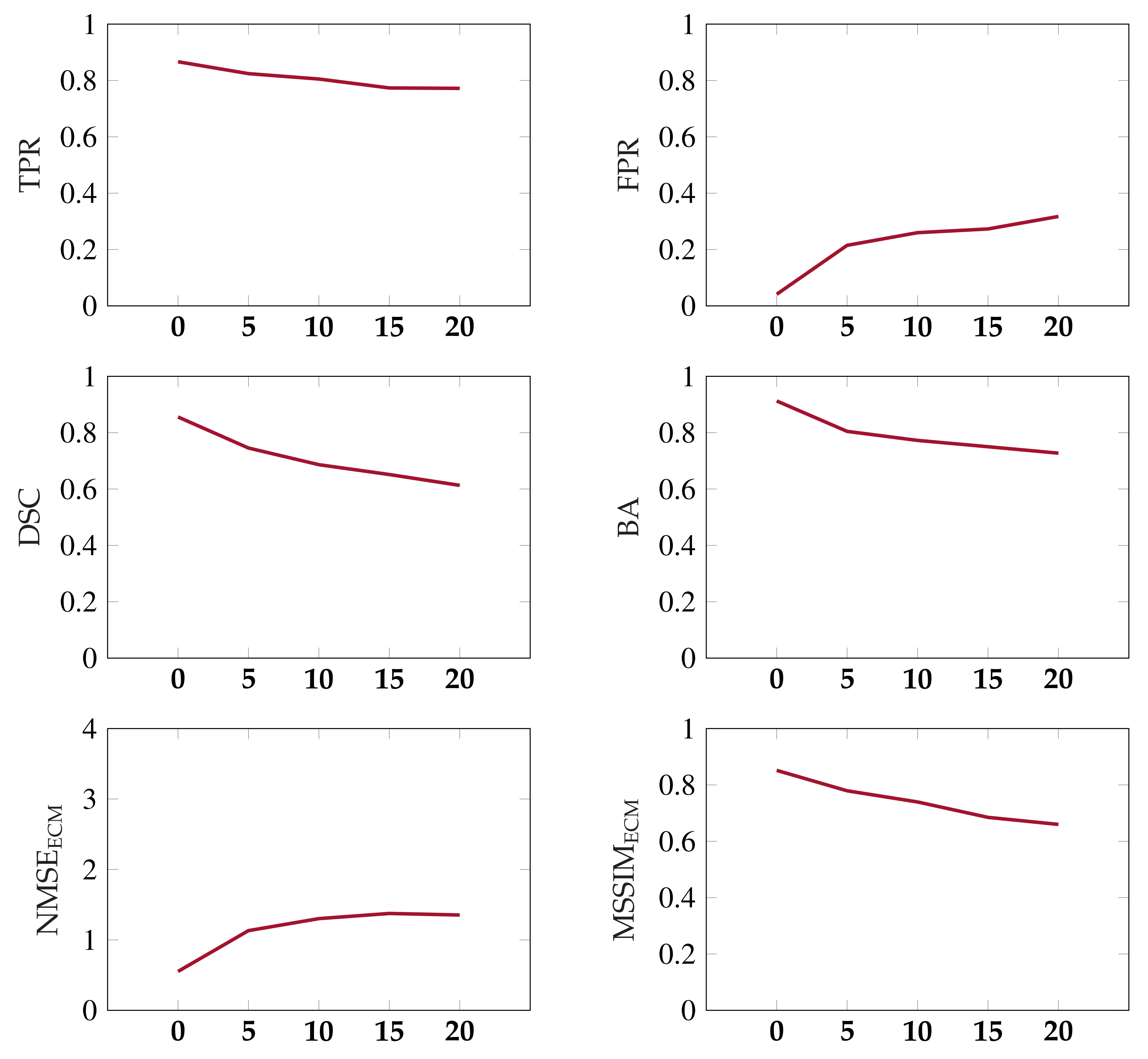
Manual annotation of SMAMs with irregular patterns in DWI and a high number of image repetitions is a time-consuming and error-prone task. Furthermore, a large amount of partial volume effects due to the rather low resolution of DWI might hamper this process. For this, a neural network-based approach from a former work4 was applied. The neural network consists of following building blocks: encoder-decoder structure5,6, residual blocks7, attention-gates8, and prediction smoothing by convolutional long short-term memory9,10 blocks. This architecture has shown rather good detection capabilities, but a reduced Dice-Score (DSC).4 This might be related to the segmentation problem of partial volume effected regions.11 In case of inconsistencies in the annotation with a more random appearance at the boundary region of SMAMs, the neural network learns the underlying distribution by trying to fit the more non-random parts of the data sets. Label uncertainty is used in this work to overcome this effect by adjusting the last dense layer to output probabilities and model the aleatoric uncertainty12. For the initial coarse SMAM annotation, output segmentations of a graph-based approach13 are utilized. Furthermore, a random variation of the entire signal intensity of an imaging slice was induced to train the neural network only to detect SMAMs and not regional signal intensity changes typical in diffusion-tensor imaging. Therefore, each imaging slice is multiplied by a factor following a uniform distribution in the range of 0.9-1.1. The image processing pipeline is given in Fig. 1. Performance of the neural network is evaluated using standard metrics (TPR: true positive rate, FPR: false positive rate, BA: balanced accuracy) and NMSE (normalized mean squared error) and MSSIM14 (mean structural similarity) on event count maps (ECM), i.e., summation of activities over time.Feature-based Analysis
For feature extraction, the region around the segmented SMAM is reformatted to a fixed size to overcome the size-dependency of some features15,16. Feature extraction was performed using the open-source PyRadiomics Python package17 in accordance to the IBSI (Image Biomarker Standardisation Initiative) guidelines. 93 texture features were calculated from each segmented SMAM. These textural features were further processed by a clustering method (DBSCAN: Density-Based Spatial Clustering of Applications with Noise)18,19 using scikit-learn (v1.2.2).Results
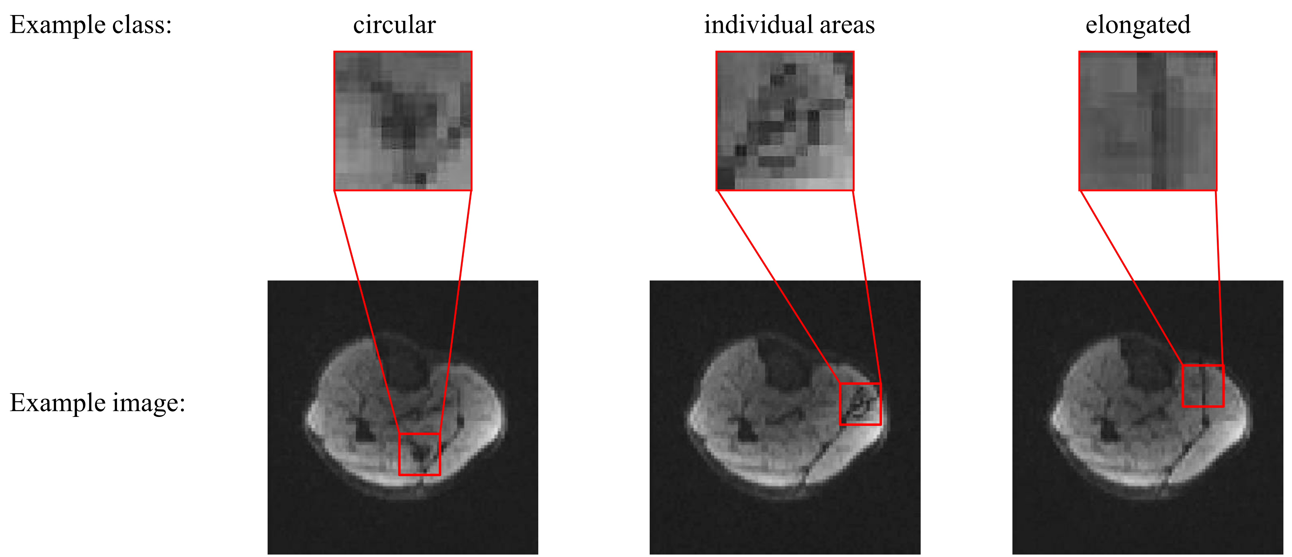
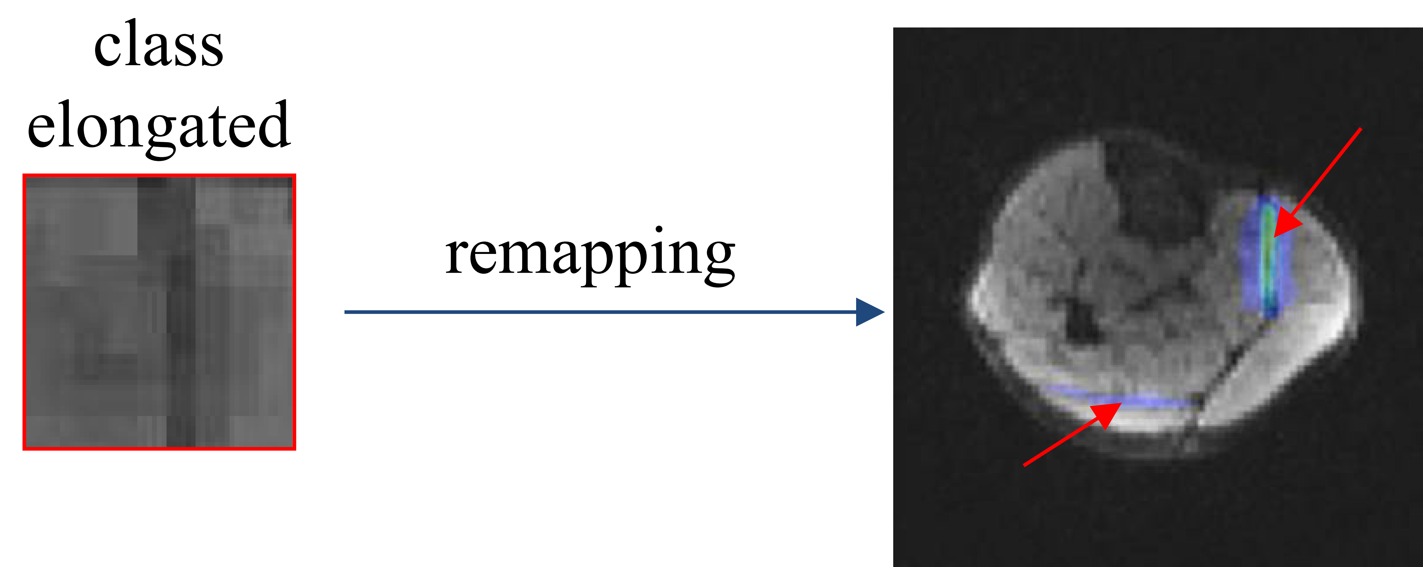
It can be seen in Fig. 2 that the neural network is able to detect and segment the visible SMAMs in DWI with a mean TPR of 89%, FPR of 3.9%, DSC of 87% and BA of 91% without additional noise. In this case, label uncertainty is able to increase the segmentation quality compared to a common training approach. Fig. 3 shows an example of three different patterns of spontaneous muscular activity in one subject after textural clustering. The differences in the shape of the recruitment pattern can be clearly seen. Remapping of the elongated class in Fig. 4 shows a clear relationship to the region between m. soleus and m. gastrocnemius medialis, i.e., at the fascia. In some cases DBSCAN overestimated the number of classes showing unstable behavior.Conclusion
Analysis of spontaneous muscular activities recorded by DWI with respect to spatial patterns revealed distinct individual differences. It has been shown that the automatic detection, segmentation and clustering of spontaneous muscular activities within a subject is feasible. However, more robust features or new methods, e.g., clustering methods based on deep learning, need to be investigated. The proposed segmentation and cluster analysis was limited to one imaging slice of the musculature of the lower leg. Further imaging regions, e.g., in the region of shoulder or back muscles2, might be of interest.Acknowledgements
This work was supported and funded by the German Research Foundation (DFG) under grant: SCHI 498/11-2, YA 28/16-2.References
1. G. Steidle and F. Schick. Addressing spontaneous signal voids in repetitive single-shot DWI of musculature: spatial and temporal patterns in the calves of healthy volunteers and consideration of unintended muscle activities as underlying mechanism. NMR in Biomedicine, 28:801–810, July 2015. ISSN 1099-1492. doi: 10.1002/nbm.3311.
2. M. Schwartz, P. Martirosian, G. Steidle, T. Küstner, B. Yang, A. Stemmer, T. Feiweier, L. Schöls, M. Synofzik, and F. Schick. Measuring Spontaneous Muscular Activities in Neuromuscular Disease: Preliminary Results. In Proceedings of the Annual Meeting International Society for Magnetic Resonance in Medicine (ISMRM), 2020.
3. R. G. Whittaker, P. Porcari, L. Braz, T. L. Williams, I. S. Schofield, and A. M. Blamire. Functional magnetic resonance imaging of human motor unit fasciculation in amyotrophic lateral sclerosis. Annals of Neurology, 85:455–459, Mar. 2019. ISSN 1531-8249. doi:10.1002/ana.25422.
4. Schwartz M, Küstner T, Martirosian P, Machann J, Steidle G, Yang B, Schick F. Robust Quantification of Spontaneous Muscular Activities by Simultaneous Interpretation of sEMG Data. Proceedings of the 36 th Annual Scientific Meeting ESMRMB, Rotterdam, Netherlands, 2019.
5. Ronneberger O, Fischer P, Brox T. U-Net: Convolutional Networks for Biomedical Image Segmentation. Medical Image Computing and Computer-Assisted Intervention (MICCAI), Munich, Germany, 2015.
6. Milletari F, Navab N, Ahmadi SA. V-Net: Fully Convolutional Neural Networks for Volumetric Medical Image Segmentation. arXiv:1606.04797, 2016.
7. He K, Zhang X, Ren S, Sun J. Deep Residual Learning for Image Recognition. IEEE Conference on Computer Vision and Pattern Recognition (CVPR), Las Vegas, USA, 2016.
8. Oktay O, Schlemper J, Le Folgoc L, Lee M, Heinrich M, Misawa K, Mori K, McDonagh S, Hammerla NY, Kainz B, Glocker B, Rueckert D. Attention U-Net: Learning Where to Look for the Pancreas. Conference on Medical Imaging with Deep Learning (MIDL), Amsterdam, Netherlands, 2018.
9. Hochreiter S, Schmidhuber J. Long Short-Term Memory. Neural Computation 9(8):1735-80, 1997.
10. Shi X, Chen Z, Wang H, Yeung DY, Wong Wk, Woo Wc. Convolutional LSTM Network: A Machine Learning Approach for Precipitation Nowcasting. arXiv:1506.04214, 2015.
11. Martin Schwartz. Recording and Processing of Magnetic Resonance Imaging and Electromyographic Data for Assessment of Spontaneous Neuromuscular Activities, ISBN 978-3-8439-5252-1
12. A. Kendall und Y. Gal. What Uncertainties Do We Need in Bayesian Deep Learning for Computer Vision? Part of Advances in Neural Information Processing Systems 30 (NIPS 2017)
13. M. Schwartz, G. Steidle, P. Martirosian, B. Yang, F. Schick. Graph-based segmentation of signal voids in time series of diffusion-weighted images of musculature in the human lower leg. Proceedings of the Annual Meeting ISMRM 2016, May 2016
14. Z. Wang, A. C. Bovik, H. R. Sheikh, and E. P. Simoncelli. Image quality assessment: from error visibility to structural similarity. IEEE Transactions on Image Processing, 13(4):600–612, 2004.
15. Roy S, Whitehead TD, Quirk JD, Salter A, Ademuyiwa FO, Li S, An H, Shoghi KI. Optimal co-clinical radiomics: Sensitivity of radiomic features to tumour volume, image noise and resolution in co-clinical T1-weighted and T2-weighted magnetic resonance imaging. EBioMedicine. 2020 Sep;59:102963. doi: 10.1016/j.ebiom.2020.102963. Epub 2020 Sep 2. PMID: 32891051; PMCID: PMC7479492.
16. Jensen LJ, Kim D, Elgeti T, Steffen IG, Hamm B, Nagel SN. Stability of Radiomic Features across Different Region of Interest Sizes-A CT and MR Phantom Study. Tomography. 2021 Jun 8;7(2):238-252. doi: 10.3390/tomography7020022. PMID: 34201012; PMCID: PMC8293351.
17. van Griethuysen, J. J. M., Fedorov, A., Parmar, C., Hosny, A., Aucoin, N., Narayan, V., Beets-Tan, R. G. H., Fillon-Robin, J. C., Pieper, S., Aerts, H. J. W. L. (2017). Computational Radiomics System to Decode the Radiographic Phenotype. Cancer Research, 77(21), e104–e107.
18. Ester, M., H. P. Kriegel, J. Sander, and X. Xu. A Density-Based Algorithm for Discovering Clusters in Large Spatial Databases with Noise. In: Proceedings of the 2nd International Conference on Knowledge Discovery and Data Mining, Portland, OR, AAAI Press, pp. 226-231. 1996
19. Schubert, E., Sander, J., Ester, M., Kriegel, H. P., & Xu, X. (2017). DBSCAN revisited, revisited: why and how you should (still) use DBSCAN. ACM Transactions on Database Systems (TODS), 42(3).
Figures
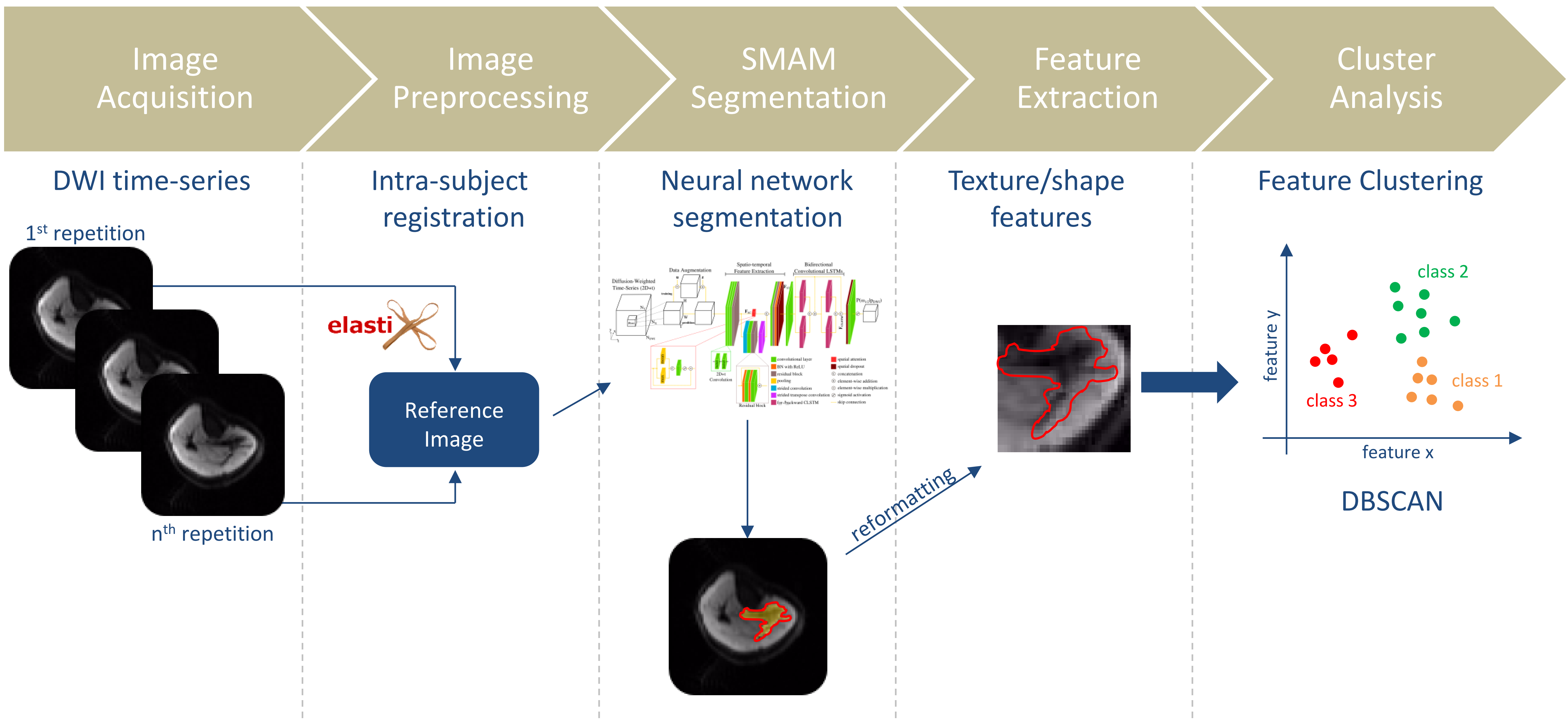
Figure 1: Concept of quantitative spatial analysis of spontaneous muscular activities.