1693
Prediction of Backfill Progression at the Sacroiliac Joint in Patients with Axial Spondyloarthritis using a radiomics method1Tianjin Frist Center Hospital, Tianjin, China, 2Philips Healthcare, Beijing, China
Synopsis
Keywords: Cartilage, MSK
Motivation: Identifying patients with a backfill progression is crucial for predicting clinical prognosis and adjusting treatment approaches in the disease process.
Goal(s): This study aimed to extract radiomics features for the sacroiliac joint on MRI images in patients with axSpA to predict backfill progression within one year.
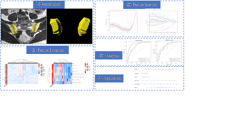
Approach: This retrospective study analyzed 257 patients diagnosed with axSpA. The radiomics and clinical models were combined to construct a nomogram model through multivariable logistic regression analysis.
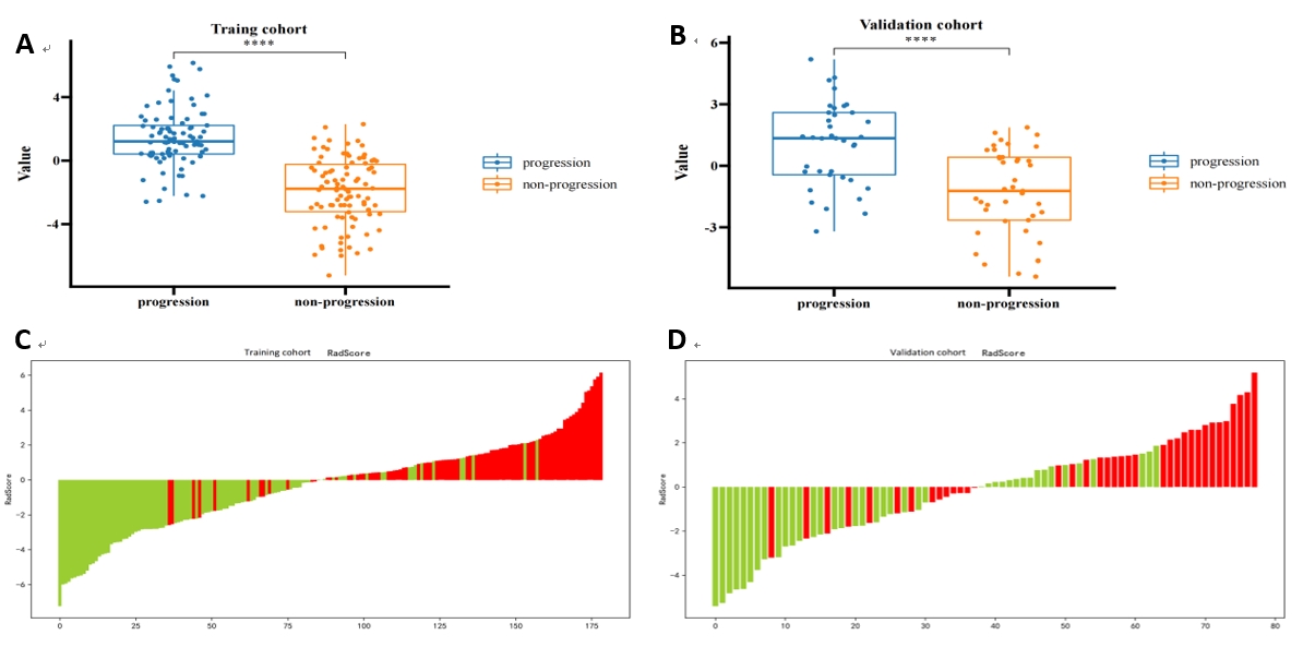
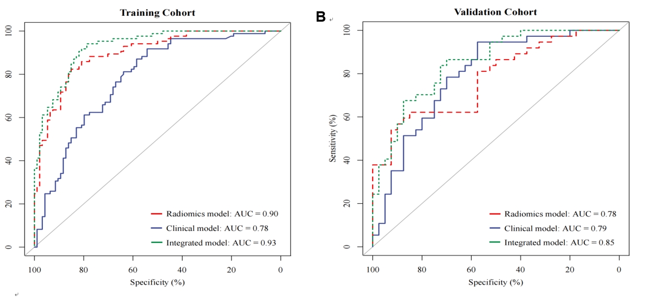
Results: Seven radiomics features were extracted to generate a Rad-score. The AUCs of the radiomics, clinical, and nomogram models in the training cohort were 0.90, 0.78 and 0.93, respectively.
Impact: The built radiomics-based nomogram has good predictive value for structural progression in patients with axial spondyloarthritis.
Aim
To evaluate the performance of a nomogram based on magnetic resonance imaging (MRI) for predicting backfill progression at the sacroiliac joint (SIJ) in patients with axial spondyloarthritis (axSpA).Materials and methods
This retrospective study analyzed 257 patients diagnosed with axSpA with baseline and one-year follow-up sacroiliac joint MRI images. The patients were randomized into the training (n=179) and validation (n=78) cohort at the ratio of 7:3. Totally 1691 radiomics features were extracted from the SIJ MRI images of each patient, and Pearson’s correlation, F test, and least absolute shrinkage and selection operator (LASSO) analyses were performed for feature selection in the training cohort to construct an optimal radiomics model. In parallel, 7 clinical risk factors were used to construct a clinical model. Then, the radiomics and clinical models were combined to construct a nomogram model through multivariable logistic regression analysis. The performances of the three models were evaluated by ROC curve, calibration curve, and decision curve (DCA) analyses.Results
Seven radiomics features were extracted to generate a Rad-score. The AUCs of the radiomics, clinical, and nomogram models in the training cohort were 0.90, 0.78 and 0.93, respectively; these values were 0.78, 0.79 and 0.85 in the validation cohort, respectively. Calibration curve analysis (P>0.05) and DCA proved that the nomogram model was useful for predicting backfill progression in axSpA.Conclusion
This study developed a radiomics model for the prediction of backfill progression at the SIJ in axSpA patients, and constructed a radiomics-based nomogram with elevated predictive efficacy.Acknowledgements
No acknowledgement found.References
No reference found.Figures