1692
Magnetic Resonance Imaging Texture Analysis for Evaluating Radiation Induced Femoral Head Changes in Rectal Cancer after Radiotherapy1Hangzhou Cancer Hospital, Hangzhou, China
Synopsis
Keywords: Bone, Bone, Radiation, MRI, Texture Analysis
Motivation: To promote a non-invasive method to evaluate the radiation induced femoral head changes.
Goal(s): To investigate the value of MRI texture analysis for evaluating radiation induced femoral head changes in rectal cancer patients after radiotherapy.
Approach: The texture parameters of T1WI, T2WI and DWI images were extracted in femoral head. And the differences of texture parameters before and after radiotherapy were compared.
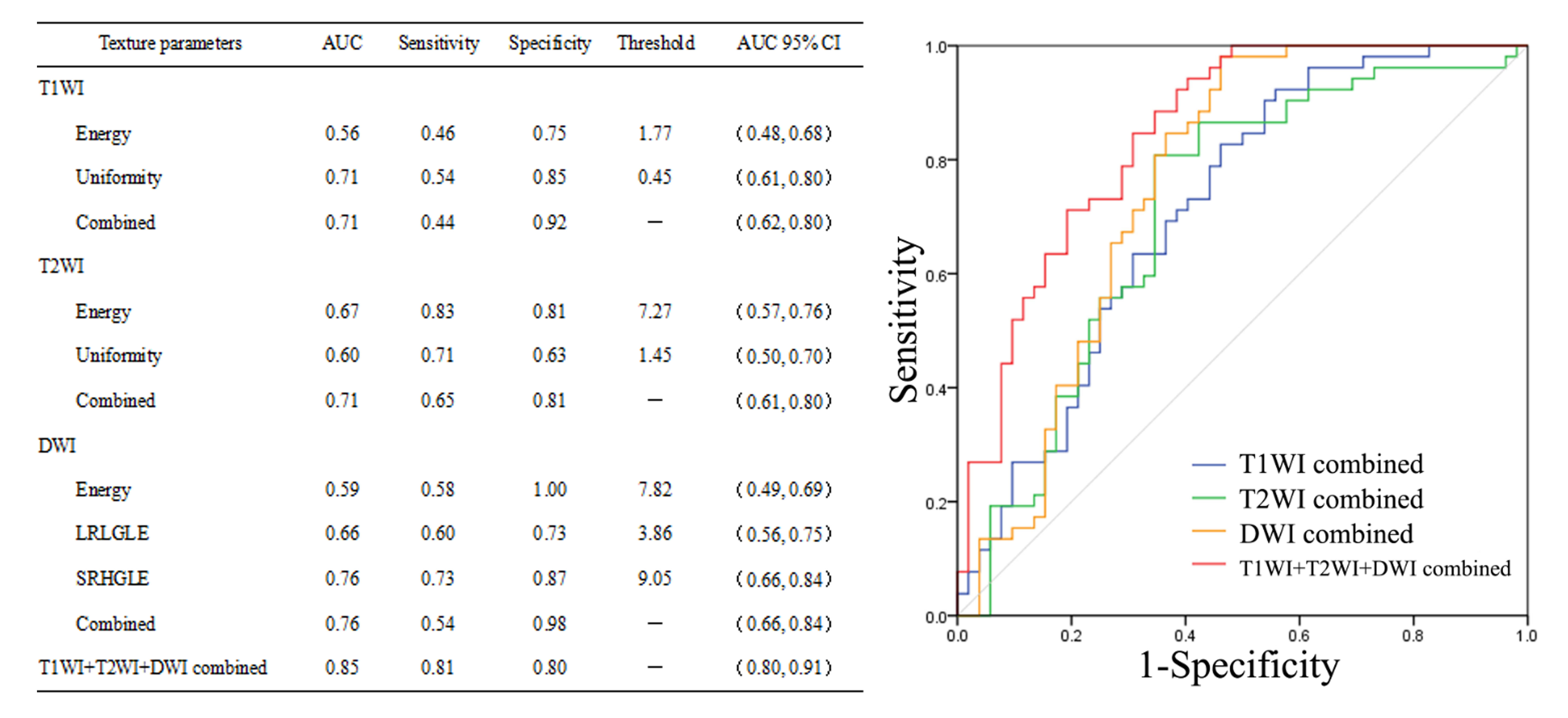
Results: We found that when combined the texture parameters of T1WI, T2WI and DWI images, the diagnostic efficiency of femoral head changes was optimal with AUC of 0.85 (sensitivity,0.81; specificity,0.80).
Impact: MRI texture analysis may provide a non-invasive method to evaluate the radiation induced femoral head changes, providing imaging clues for monitoring the potential radiation-induced femoral head injury.
Introduction
Radiotherapy is an important treatment strategy for rectal cancer, but the adverse events of the femoral head caused by radiation have also become a widely concerned issue[1]. Radiation-induced femoral head injury may occur several months to years after radiotherapy, and about 1.5% patients may experience insufficiency femoral head fractures within 2 years after radiotherapy[2]. Therefore, the early detection of radiation-induced femoral head changes is particularly important for clinicians. Texture analysis can provide quantitative imaging biomarkers for diagnosis and prognosis[3]. In this study, we aim to investigate the value of magnetic resonance imaging (MRI) texture analysis for evaluating radiation induced femoral head changes in rectal cancer patients after radiotherapy.Methods
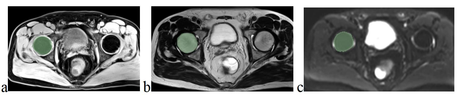
The 52 rectal cancer patients who have underwent pelvic radiotherapy were retrospectively collected in present study, then their MRI images including T1WI, T2WI and DWI within two weeks before radiotherapy and three months after radiotherapy were collected. The T1WI, T2WI and DWI images before and after radiotherapy were imported into 3D-slicer software to segment the region of interest (unilateral femoral head) and extract texture parameters. The regions of interest were shown in Fig.1. The texture parameters included maximum, minimum, mean, median, deviation, skewness, kurtosis, entropy, energy, uniformity, LongRunHighGrayLevelEmphasis (LRHGLE), LongRunLowGrayLevelEmphasis (LRLGLE), ShortRunHighGrayLevelEmphasis (SRHGLE), ShortRunLowGrayLevelEmphasis (SRLGLE). The differences of texture parameters before and after radiotherapy were compared, and the diagnostic efficiency was evaluated by receiver operating characteristic (ROC) analysis.Results
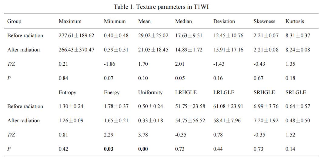
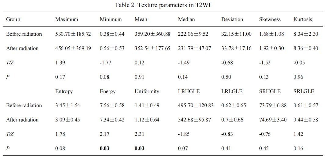
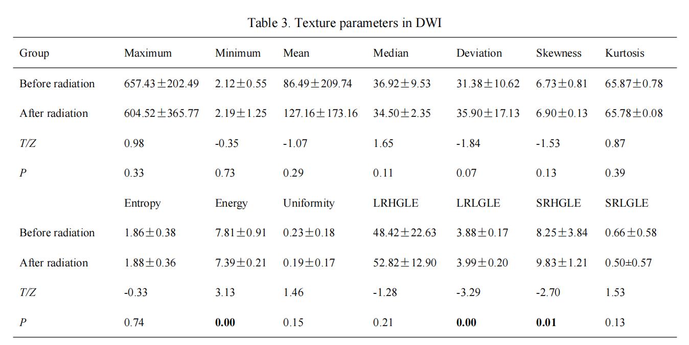
In T1WI and T2WI images, the energy and uniformity of the femoral head before radiotherapy were significantly higher than those after radiotherapy (Fig.2, Fig.3). In DWI image, the energy of the femoral head before radiotherapy was significantly higher than that after radiotherapy, and the LRLGLE and SRHGLE were significantly lower than those after radiotherapy (Fig.4). When combined the texture parameters of T1WI, T2WI and DWI images, the diagnostic efficiency was optimal with AUC of 0.85 (sensitivity,0.81; specificity,0.80). ROC analysis were shown in Fig.5.Discussion
The feasibility of MRI texture analysis to evaluate the radiation-induced femoral head changes has been proved in present study. We found the texture parameters including energy, uniformity, LRLGLE and SRHGLE were potential imaging biomarkers, and these texture parameters suggested that the heterogeneity of femoral head was increased after radiation. These results were considered reasonable because the biological changes (such as vascular fibrosis, bone fibroblast damage, and cell proliferation inhibition) may caused the increased heterogeneity in femoral head after radiation[4, 5].Conclusion
MRI texture analysis may provide a non-invasive method to evaluate the radiation induced femoral head changes, providing more clues for monitoring the potential radiation-induced femoral head injury.Acknowledgements
No acknowledgement.References
[1]Almendros-Abellán VM, Castro-García M, Canales-Vázquez J, et al. Alterations in femoral neck strength following pelvic irradiation. A finite element analysis of simulated eccentric forces using bone density data derived from CT[J]. Bone. 2021,145:115865.
[2]Sapienza LG, Salcedo MP, Ning MS, et al. Pelvic insufficiency fractures after external beam radiotherapy for gynecologic cancers: a meta-analysis and meta-regression of 3,929 patients[J]. Int J Radiat Oncol Biol Phys. 2020,106(03):475-484.
[3]Giganti Francesco,Antunes Sofia,Salerno Annalaura et al. Gastric cancer: texture analysis from multidetector computed tomography as a potential preoperative prognostic biomarker.[J] .Eur Radiol, 2017, 27: 1831-1839.
[4]Zhong Xi,Dong Tianfa,Tan Yu, et al. Pelvic insufficiency fracture or bone metastasis after radiotherapy for cervical cancer? The added value of DWI for characterization[J].Eur Radiol. 2020, 30(04): 1885-1895.
[5]Deloch Lisa,Derer Anja,Hueber Axel J, et al. Low-Dose Radiotherapy Ameliorates Advanced Arthritis in hTNF-α tg Mice by Particularly Positively Impacting on Bone Metabolism[J].Front Immunol. 2018, 18(09): 1834.
Figures