1687
Detective Ability of pH in Osteosarcoma Microenvironment Using Chemical Exchange Saturation Transfer(CEST) Imaging1Shanghai Ninth People's Hospital, Shanghai Jiao Tong University School of Medicine, Shanghai, China, 2Philips healthcare, Beijing, China, 3Philips Healthcare, Shanghai, China
Synopsis
Keywords: Other Musculoskeletal, Tumor, CEST
Motivation: The precise assessment of the efficacy of preoperative neoadjuvant chemotherapy in osteosarcoma is important for clinical decision making, which requires the consideration of complex factors.
Goal(s): In this study, we investigated the ability of ioversol chemical exchange saturation transfer (CEST) imaging in detecting pH in the microenvironment of rat osteosarcoma.
Approach: Assessing pH in the microenvironment of osteosarcoma using CEST imaging.
Results: It was found that the lower pH value, the lower ratiometric value acquired by two different saturation power of 1μT and 3μT.
Impact: In addition, CEST can detect the pH-dependent ratiometric value of tumor microenvironment non-invasively and sensitively. It is expected to become one of the methods for early evaluation of the effect of preoperative neoadjuvant chemotherapy in cancer patients.
Introduction
Osteosarcoma is the most common malignant tumor in the musculoskeletal system, and its five-year relative survival rates remain poor[1]. The most important reason for its poor five-year relative survival rate is tumor heterogeneity[2]. Because of the microenvironment of most malignant tumors is acidic[3] as its extracellular pH (pHe) is lower than normal tissue, the change of extracellular pHe can be used to assess the effect of preoperative neoadjuvant chemotherapy and to guide the choice of subsequent treatment and surgery[4]. Therefore, early detection of the change of tumor acidic microenvironment is crucial for clinical decision making. CEST is a non-invasive method which can be used to display the pHe of tumors[5, 6]. In order to enhance the effect of CEST imaging, exogenous contrast agents were introduced.Ioversol, a widely used CT agent, was selected for this study because of its high water solubility and low toxicity. Potentially due to the complexity of tumors in the musculoskeletal system, the characteristics of osteosarcoma microenvironment under CEST imaging has not been comprehensively studied. Therefore, this study aimed to evaluate the ability of ioversol CEST imaging in detecting pHe in the microenvironment of osteosarcoma.
Methods
This study was approved by ethical committee of Shanghai Ninth People’s Hospital. In vitro, we used 15 phantom tubes filled with PBS solution with different pH and different concentration of ioversol to verified the relation between pH and CEST ratiometric values. And in vivo, ten rats were used in this study to test whether ioversol could be used as contrast agent for pH imaging on osteosarcoma. Rat osteosarcoma cells (UMR-106) were injected subcutaneously into the hind limbs of Sprague-Dawley (SD) rats. 2 weeks after cell implantation, the rats were scanned by a 3T MR system (Ingenia CX, Philips Healthcare, Best, Netherland) with a 32-channel head coil. For 9 out of 10 rats, ioversol (4g I/kg body weight) was injected through tail vein (Figure 1). CEST images were acquired using a 2d turbo spin echo sequence with the saturation duration of 1.5s and saturation powers of 1μT and 3μT. The other parameters were as follows: TR = 11800ms, TE = 7.3ms, FOV = 180 × 180mm, slice thickness = 6mm, matrix = 100 × 98. All CEST images were postprocessed via MATLAB (The Mathworks, Inc., Natick, MA, USA). The pH weighted ratiometric values were calculated by the ratio of MTRasym (3.5 ppm) in 1 μT to MTRasym (3.5 ppm) in 3 μT using a custom Matlab code.Results
To test whether CEST imaging could distinguish the ioversol solutions at different pH level, 10 tubes of 30mM ioversol at pH 5.0, 5.8, 6.1, 6.4, 6.7, 7.0, 7.3, 7.6, 7.9, 8.2 were used. The result showed that the pH value and CEST effect were linearly correlated in the pH range of 5.8-7.0 (Figure 2B,C, R2=0.86, p<0.0001). To test the relationship between ioversol concentration and ratiometric value, 5 tubes of pH 7.2 with different ioversol concentrations (30, 50, 70, 90, 110 mM) were prepared, Figure 2D showed that the ratiometric values of ioversol solution were not concentration-dependent on in the concentration range tested. Therefore, low concentration (30mM) was used in the in vivo test. Two example images of osteosarcoma with and without necrosis were shown in Figure 3. The CEST effect was heterogeneous in the tumor ROI.Discussion
Our findings provide the first evidence that ioversol-enhanced CEST imaging can actually measure pH-related ratiometric value in osteosarcoma in a concentration-independent manner, with excellent spatial resolution. In phantom studies, the results of 10 tubes of 30mM ioversol showed that the log10 ratio of the CEST effect was linearly proportional to the pH value in the range of 5.8-7.0, which is wider than the physiological and pathological pH range in normal tissue and tumor. Therefore, it is feasible to use Ioversol-enhanced CEST imaging for pH mapping. Moreover, the results of 5 tubes of pH 7.2 ioversol solution with different ioversol concentrations confirm that ioversol is concentration-independent. In the in vivo studies, the calculated ratiometric value of the majority of the tumor region was relatively low, suggesting the acidic and solid part of the tumor, while there was also small region with high value, suggesting the necrotic and cystic portion of the tumor. This finding needs to be confirmed by histopathology.Conclusion
By using CEST imaging, the status of osteosarcoma can be correlated with the actual pH value. CEST imaging potentially can be a novel method for assessing the early effect of preoperative neoadjuvant chemotherapy in the future.Acknowledgements
No acknowledgement found.References
1. Cole, S., et al., Osteosarcoma: A Surveillance, Epidemiology, and End Results program-based analysis from 1975 to 2017. Cancer, 2022. 128(11): p. 2107-2118.
2. Boedtkjer, E. and S.F. Pedersen, The Acidic Tumor Microenvironment as a Driver of Cancer. Annu Rev Physiol, 2020. 82: p. 103-126.
3. Corbet, C. and O. Feron, Tumour acidosis: from the passenger to the driver's seat. Nat Rev Cancer, 2017. 17(10): p. 577-593.
4. Rosen, G., et al., Primary osteogenic sarcoma: the rationale for preoperative chemotherapy and delayed surgery. Cancer, 1979. 43(6): p. 2163-77.
5. Chen, M., et al., Extracellular pH is a biomarker enabling detection of breast cancer and liver cancer using CEST MRI. Oncotarget, 2017. 8(28): p. 45759-45767.
6. Tang, Y., et al., Noninvasive Detection of Extracellular pH in Human Benign and Malignant Liver Tumors Using CEST MRI. Front Oncol, 2020. 10: p. 578985.
Figures
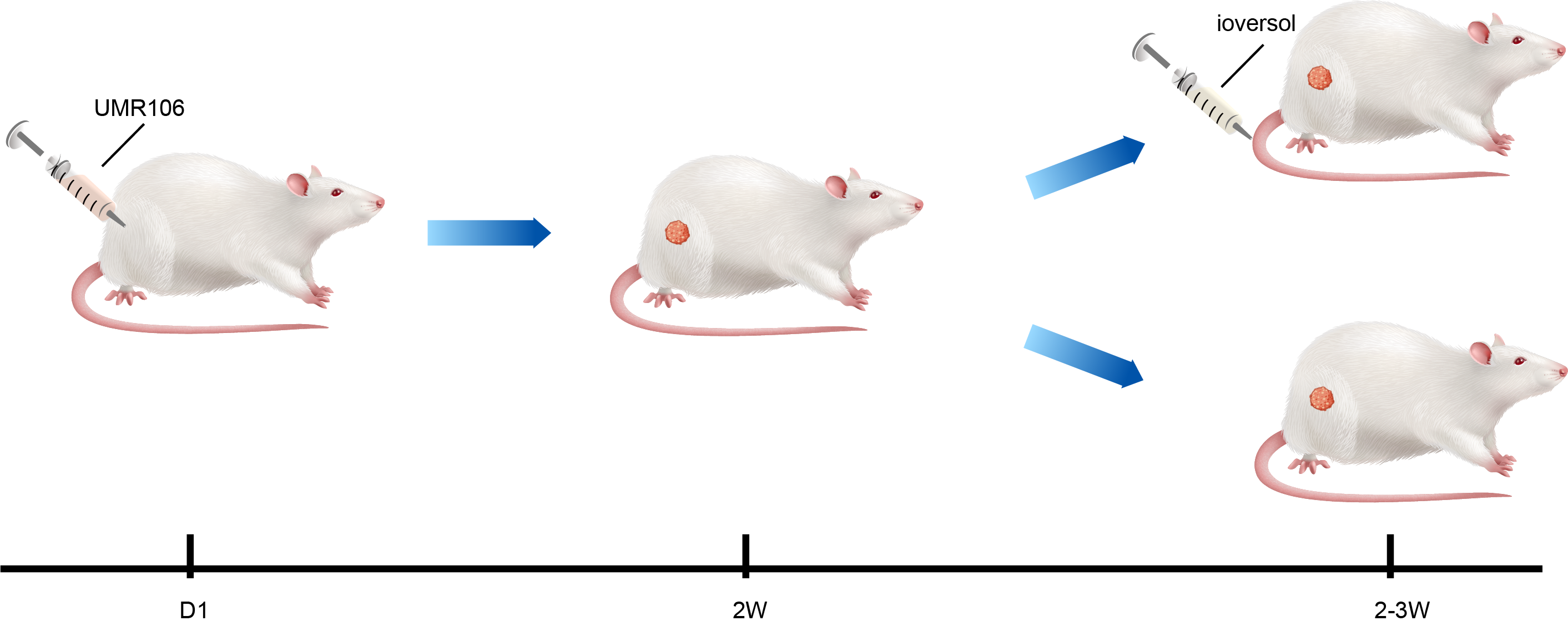
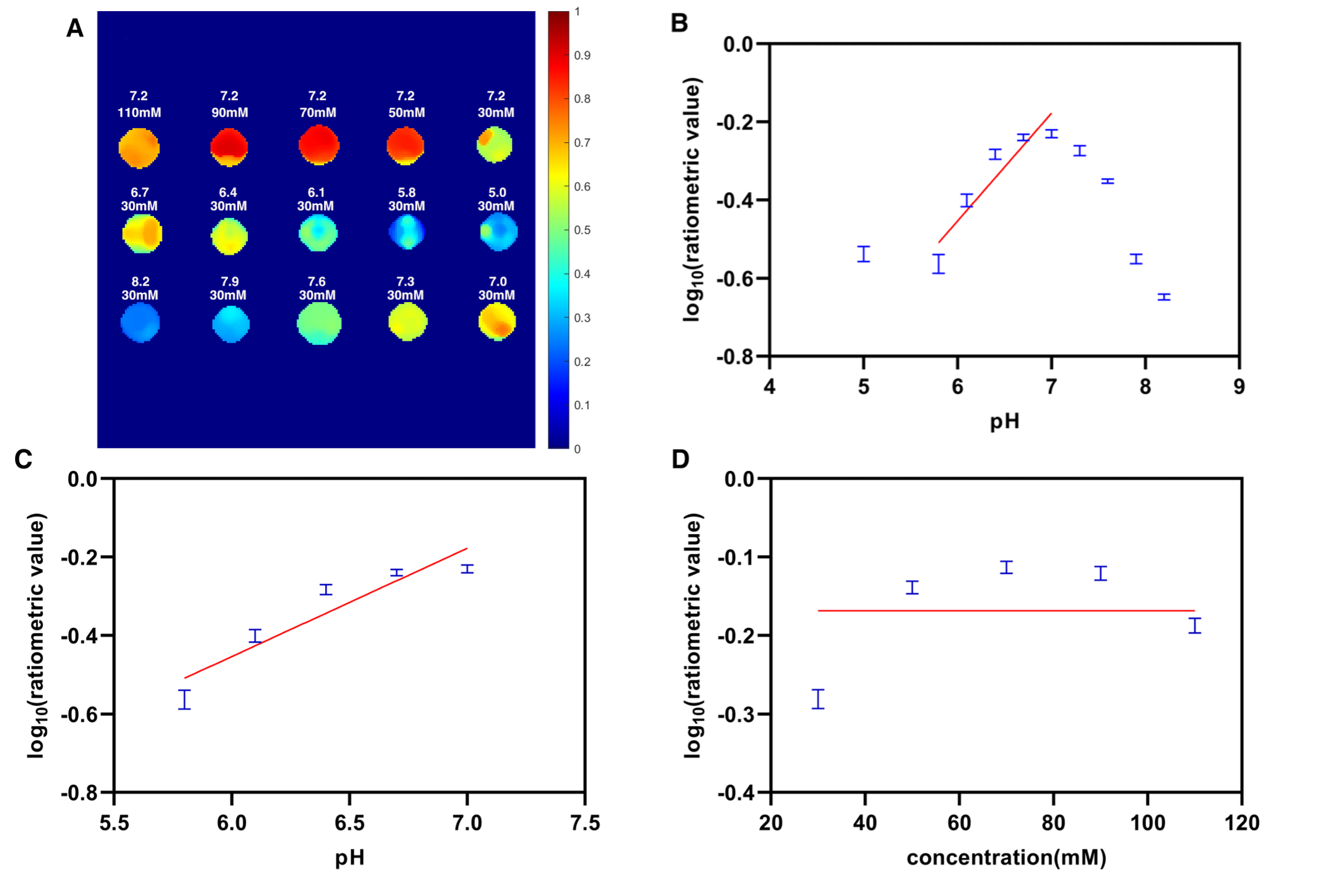
Figure 2: Experimental pH measurements in phantoms. (A) The calculated ratiometric map. The first row was tubes with different concentrations (30, 50, 70, 90, and 110 mM). The second and third rows were tubes of 30 mM ioversol at pH 5.0 to 8.2. (B) The log10 ratio of the CEST effect based on different pH phantoms. (C)The log10 ratio of the CEST effect was linearly correlated with the pH at the range of 5.8-7.0(R2=0.86, p<0.0001). (D)Mean log10 ratiometric values determined for several concentrations.
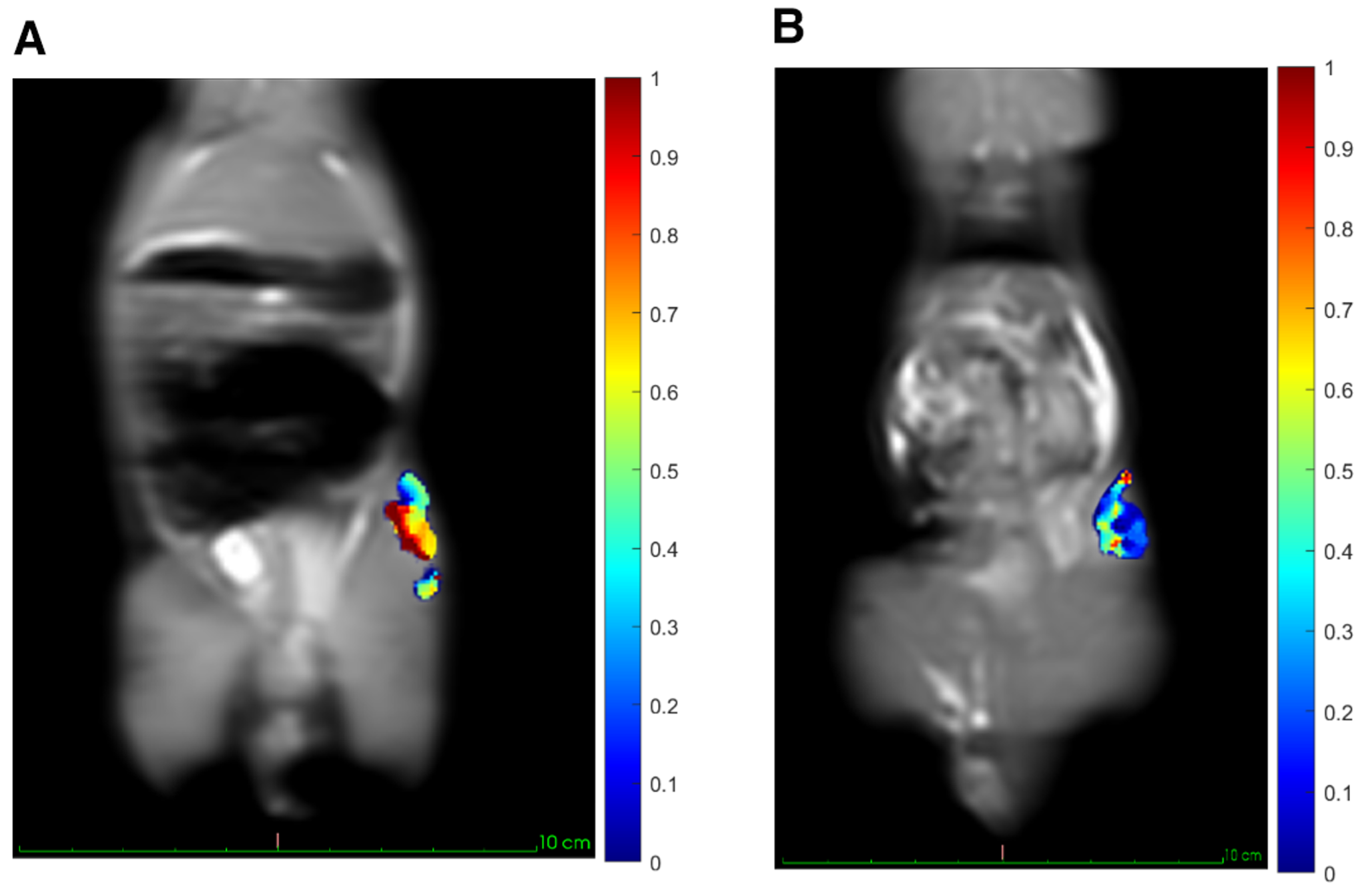
Figure 3: In vivo CEST imaging of osteosarcoma. T2 osteosarcoma image overlayed with CEST effect mask on tumor ROI. The ratiometric map calculated by using 1 μT and 3 μT CEST images. Length and width of the tumor(A) is 3cm*1.7cm. Length and width of the tumor(B) is 1.5cm*1.5cm.