1681
Direct traction device for clinical wrist MR imaging1Imaging and Interventional Radiology, The Chinese University of Hong Kong, Hong Kong SAR, China
Synopsis
Keywords: Whole Joint, New Devices
Motivation: Traction imaging helps visualize the wrist articular cartilage, intrinsic ligaments, and triangular fibrocartilaginous complex (TFCC). The current traction method is cumbersome to use limiting more widespread usage.
Goal(s): To design a direct wrist traction device that is practical, comfortable, effective, and versatile.
Approach: A new traction device was designed. One patient underwent wrist imaging before and after traction with this device. The degree of joint distraction and articular cartilage, ligament and TFCC visibility was evaluated as was the degree of subject discomfort.
Results: The traction device was easy to apply, achieved good traction with minimal subject discomfort.
Impact: This practical traction device can potentially improve the quality of wrist MR imaging, negating the need for wrist arthrography and improving the detection of articular cartilage, intrinsic ligament and TFCC injury.
Introduction
Articular distraction is quite widely utilized in wrist MRI with or, less commonly, without arthrography. Wrist traction effectively distracts the joint space and, in doing so, enhances cartilage visibility as well as the detection of intrinsic ligament and TFCC injuries1–5.Currently, wrist traction is typically performed using a pulley and cable system with weights. This approach is cumbersome, time-consuming, and lacks individualization. This current system of wrist traction also simultaneously distracts the ipsilateral elbow and shoulder. Regarding the traction weight used and the number of fingers to which it is applied varies among different studies. For example, one study used a 3 kg weight to apply traction to the index and ring fingers, while another utilized up to 10 kg to apply traction to all five fingers1,6. Also, using the pulley and cable traction system, patients need to be examined in the relatively uncomfortable “Superman” position, with the wrist positioned above the head. Traction is more effective following MR arthrography, as arthrography reduces inherent negative joint pressure. Arthrography does involve the intra-articular injection of contrast medium. As well as being invasive, arthrography is time-consuming and technically demanding.
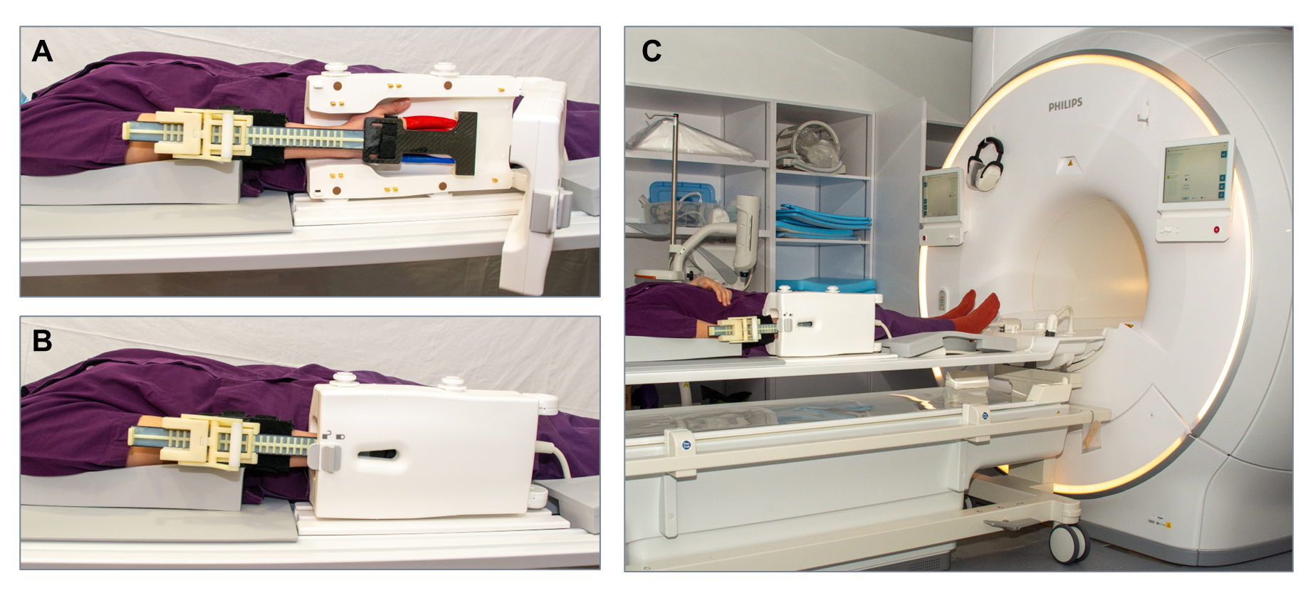
Therefore, we developed a novel wrist traction device that specifically distracts the wrist joint, excluding the elbow and shoulder joints. This device allows wrist MRI with traction to be performed with the patient lying in a supine position with their arm at their side. The aim was to develop a device which would improve non-arthrographic wrist MRI imaging whilst minimizing patient discomfort and being practical to use in an everyday clinical setting.
Methods
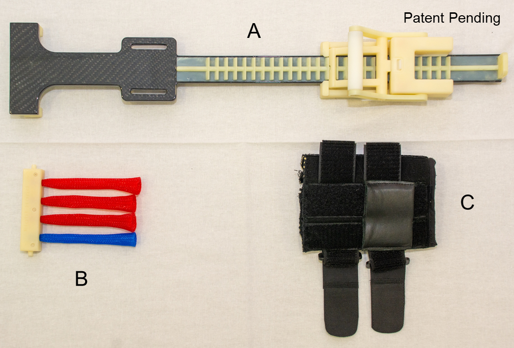
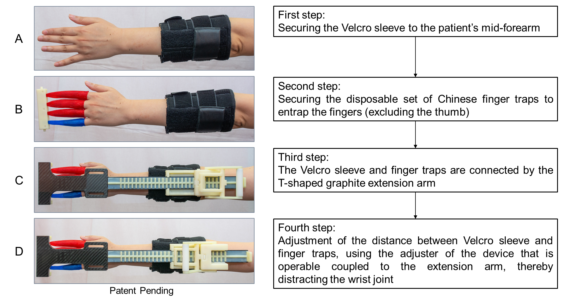
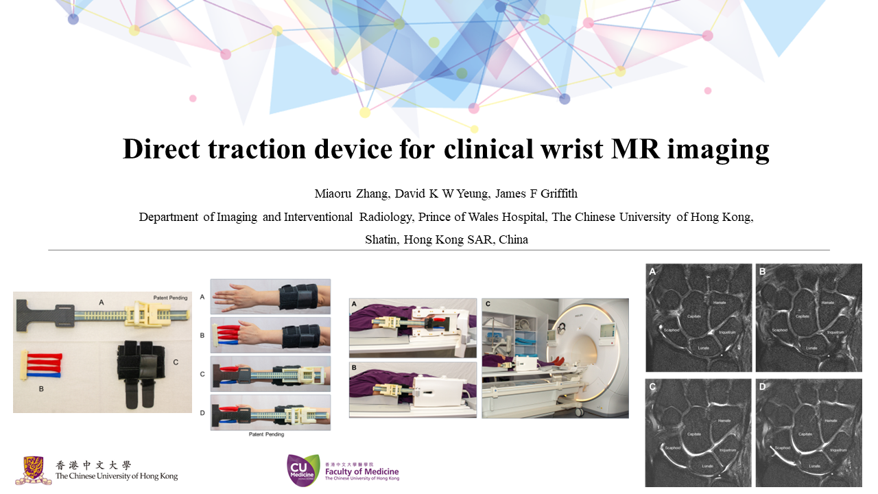
The wrist traction device has three main components: 1) a T-shaped graphite extension arm with a fluted segment for length adjustment using a movable lever arm; 2) a disposable set of Chinese finger traps applied to the index, middle, ring, and little fingers; and 3) a Velcro sleeve that secures the device on the mid-forearm (Fig. 1). All materials used in the wrist distraction device are MRI-compatible. The positioning procedure for the distraction device is shown in Figs. 2 and 3.One 27-year-old female patient was examined using the device. The examination was performed on a Philips Ingenia Elition 3.0T X MRI system. The degree of joint distraction was measured in millimeters and the visibility of the articular cartilage, intrinsic ligaments and TFCC were assessed on a three-point scale on proton-density and T2-weighted fat-saturated (T2-FS) coronal images, before and after traction5. The patient completed an in-house developed questionnaire utilizing a ten-point visual analog scale to assess the degree of wrist discomfort.
Results
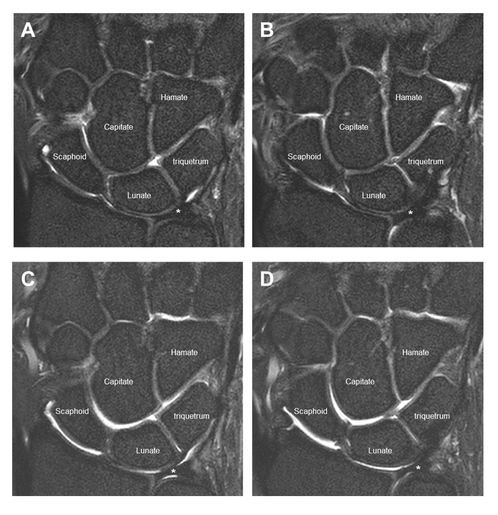
The patient reported her wrist discomfort at grade 3 before the application of traction. During traction, she experienced discomfort relief which was graded at level 2. The pain level remained unchanged after 10 minutes of traction. No discomfort was reported in any other body areas during the MRI traction examination.Figure 4 illustrates the positive effects of wrist distraction. There is widening particularly of the radioscaphoid, radiolunate, scaphocapitate, lunocapitate and triquetrohamate joints. The articular cartilage is more clearly seen, as are the scapholunate and lunotriquetral intrinsic ligaments, and the triangular fibrocartilage complex (TFCC).
Discussion
This feasibility study shows that the wrist distraction device is a wearable and user-friendly device that effectively distracts the wrist joint. It is practical to apply and causes minimal or no discomfort. It represents an improvement on previous methods of joint distraction.Conclusion
This newly developed wrist distractor is practical, effective, versatile, and comfortable. It is, as such, an improvement on previous methods of wrist distraction.Acknowledgements
The research was conducted in part at CUHK DIIR MRI Facility, which is jointly funded by Kai Chong Tong, HKSAR Research Matching Grant Scheme and the Department of Imaging and Interventional Radiology, The Chinese University of Hong Kong.References
1. Guntern D, Becce F, Richarme D, Palhais NS, Meuli R, Theumann N: Direct magnetic resonance arthrography of the wrist with axial traction: A feasibility study to assess joint cartilage. J Magn Reson Imaging 2011; 34:239–244.
2. Cerny M, Marlois R, Theumann N, et al.: 3-T direct MR arthrography of the wrist: Value of finger trap distraction to assess intrinsic ligament and triangular fibrocartilage complex tears. European Journal of Radiology 2013; 82:e582–e589.
3. Dallaudière B, Moreau-Durieux M-H, Larbi A, et al.: Effects of Axial Traction during Direct MR-Arthrography of the Wrist in Sports Injuries. Journal of the Belgian Society of Radiology 2016; 100:72.
4. Lee RKL, Griffith JF, Ng AWH, Nung RCH, Yeung DKW: Wrist Traction During MR Arthrography Improves Detection of Triangular Fibrocartilage Complex and Intrinsic Ligament Tears and Visibility of Articular Cartilage. American Journal of Roentgenology 2016; 206:155–161.
5. Lee RKL, Griffith JF, Tang WK, Ng AWH, Yeung DKW: Effect of traction on wrist joint space and cartilage visibility with and without MR arthrography. BJR 2017; 90:20160932.
6. Leventhal EL, Moore DC, Akelman E, Wolfe SW, Crisco JJ: Conformational changes in the carpus during finger trap distraction. J Hand Surg Am 2010; 35:237–244.
Figures