1678
Ventricular stroke volume is linked with thalamic volume in normal adult brains1Canon Medical Systems Corporation, Ellicott City, MD, United States, 2Johns Hopkins University, Baltimore, MD, United States, 3Canon Medical Systems Corporation, Tochigi, Japan, 4University of Wisconsin, Madison, WI, United States
Synopsis
Keywords: Myocardium, Brain, cardiac function
Motivation: Impaired cardiac function has been associated with cognitive deterioration. However, the correlation of cardiovascular function to cerebral structures remains unclear.
Goal(s): We studied whether the ventricular function is associated with measures of subcortical volumes in a general patient population.
Approach: All participants were imaged using a 3T MRI systems. Cardiac 2D SSFP CINE was acquired for cardiac function. Brain structural images were acquired using a 3D MPRAGE.
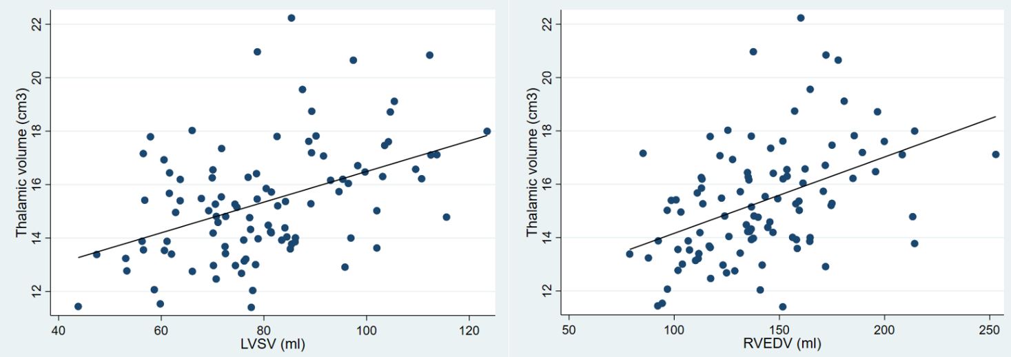
Results: Among seven subcortical structures examined in 101 healthy adults, only the thalamic volume demonstrated a positive association with left ventricular stroke volume and right ventricular end diastolic volume.
Impact: In healthy adults, the thalamic volume demonstrated a positive association with left and right ventricular stroke volume.
Introduction
Patients with heart failure (HF) exhibit greater structural brain alterations and higher dementia risks than the general population[1]. Neural atrophy in nearly every region of medical limbic circuit has been observed in HF patients. Reduction of cerebral blood flow has been suggested as the pathophysiological pathway linking HF and structural brain changes. Indeed, lower cardiac index levels were related to lower cerebral blood flow in older adults without stroke, dementia, or heart failure[2]. Thalamus is located near the center of the brain. As the brain’s relay station, almost all sensory information must pass through it before moving on to the cerebral cortex. Thalamus also assists with cognitive processes, such as memory and emotion. Small thalamic volumes were observed in older age and were linked to impaired cognition[3]. However, the link between the subcortical volumes and cardiac function is less studied. The aim of our study was to assess cross-sectionally the association between the thalamic volume and cardiac function in a general population with normal brain and cardiac function.Methods
All participants were imaged using a 3T MRI systems (Galan, Canon Medical Systems, Japan). Cardiac 2D SSFP CINE were acquired for the assessment of end diastolic volume (EDV), end systolic volume (ESV), mass (M), stroke volume (SV), and ejection fraction (EF) of the left ventricle (LV) and right ventricle (RV). Brain structural images were acquired in sagittal orientation using a 3D magnetization-prepared rapid-acquisition gradient echo with 1-mm isotropic resolution. Segmentation and parcellation of the cortical mantle were processed using default parameters in FreeSurfer v7.2. The subcortical volumes including thalamus, caudate, putamen, pallidum, hippocampus, amygdala, and accumbent were extracted from each set of images. Multivariable analysis adjusted for age, sex, BMI, heart rate, diabetes, and hypertension was performed to examine the relationship between cardiac CINE and brain structural measures. The significance threshold was Bonferroni-adjusted to 0.007 for the analysis of seven subcortical structures.Results
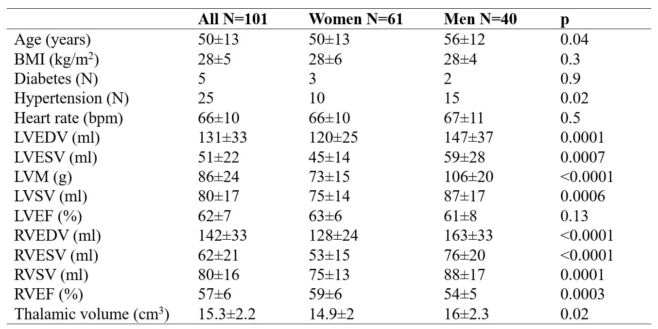
One hundred and one adult participants free from stroke and dementia were included in the study (mean age 53±12 years, range 22-79 years). Summary statistics stratified by sex is displayed in Table 1. There were significant differences in most cardiac measures between men and women. Among all seven subcortical measures, only thalamic volume remained related to cardiac parameters including LV and RV SV, and RVEDV in the multivariable analysis (figure 1, all P ≤ 0.004). Results were similar when participants with LV EF < 50 (N=6) were excluded.Discussion
Lower ventricular stroke volume corresponded to lower thalamic volume. Our finding is consistent with a study in 125 young adults (age 25±5 years) from Williamson et al.[4] Cardiac geometry and function were unrelated to brain volumes in other regions assessed (putamen, pallidum, hippocampus, amygdala, caudate, etc.) in our study which mostly comprised of volunteers with normal cardiac function. A few prior studies have examined the subcortical structural differences in patients with HF compared to controls. Brain volume loss (including putamen and hippocampal volumes) have been reported in patients with low EF[5, 6]. Future studies will investigate further the functional and structural cardiac-brain relations including HF patients for preventative measures of brain disorder.Acknowledgements
No acknowledgement found.References
[1] Leto L, Feola M. Cognitive impairment in heart failure patients. J Geriatr Cardiol 2014;11(4):316-28.
[2] Jefferson AL, Liu D, Gupta DK, Pechman KR, Watchmaker JM, Gordon EA, et al. Lower cardiac index levels relate to lower cerebral blood flow in older adults. Neurology 2017;89(23):2327-34.
[3] Woo MA, Macey PM, Fonarow GC, Hamilton MA, Harper RM. Regional brain gray matter loss in heart failure. J Appl Physiol (1985) 2003;95(2):677-84.
[4] Williamson W, Lewandowski AJ, Huckstep O, Visser E, Betts J, Jenkinson M, et al. Thalamic Volume is a Predictor of Cardiac Function at Rest and During Exercise in Young Adults. Circulation 2018;138(Suppl_1):A16361-A.
[5] Kumar R, Nguyen HD, Ogren JA, Macey PM, Thompson PM, Fonarow GC, et al. Global and regional putamen volume loss in patients with heart failure. Eur J Heart Fail 2011;13(6):651-5.
[6] Woo MA, Ogren JA, Abouzeid CM, Macey PM, Sairafian KG, Saharan PS, et al. Regional hippocampal damage in heart failure. Eur J Heart Fail 2015;17(5):494-500.
Figures