1677
Diagnostic Value of Three-Dimension Cine Enhanced Sensitivity Encoding by Static Outer-Volume Subtraction for Hypertrophic Cardiomyopathy1Department of Radiology, West China Hospital of Sichuan University, Chengdu, China, 2Clinical Science, Philips Healthcare, Chengdu, China
Synopsis
Keywords: Myocardium, Cardiovascular
Motivation: The two-dimensional (2D) cine balanced steady-state free precession (SSFP) sequence requires multiple breath-holds for cardiac imaging. A three-dimensional (3D) cine Enhanced sensitivity encoding (SENSE) by Static Outer-volume Subtraction (ESSOS) sequence was introduced to reduce the acquisition time.
Goal(s): To explore the diagnostic value of the 3D cine sequence in patients with hypertrophic cardiomyopathy (HCM).
Approach: All patients with HCM were scanned for cine imaging using conventional 2D SSFP and 3D ESSOS sequences. Cardiac structure and function parameters measured by 3D cine and 2D cine were compared.
Results: The 3D cine can rapidly obtain cardiac parameters with good diagnostic accuracy in patients with HCM.
Impact: The 3D ESSOS cine has good diagnostic accuracy for HCM. This sequence can acquire whole-heart images in a single breath hold, greatly improving efficiency and reducing patient discomfort. It should be used to screen people at high risk for HCM.
Introduction
Hypertrophic cardiomyopathy (HCM) is a common disease that causes heart failure, atrial fibrillation, ventricular arrhythmias, and sudden cardiac death, with high morbidity and mortality1. Quantitative analysis of cardiac structure and function is an important basis for the diagnosis, risk stratification and treatment decisions of HCM. The two-dimensional (2D) cine balanced steady-state free precession (SSFP) sequence is currently the preferred sequence for evaluating cardiac structure and function2, 3. However, the imaging time of traditional 2D cine is long. Patients need to cooperate with multiple breath-holds, and the amplitude of each breath-hold is required to be the same. Recently, a 3D cine Enhanced sensitivity encoding (SENSE) by Static Outer-volume Subtraction (ESSOS) acquisition has been proposed4, which can acquire isotropic 3D whole heart images in a single breath-hold. We sought to evaluate the clinical value of the 3D ESSOS cine sequence in the diagnosis of HCM by comparing the cardiac structure and function parameters measured by 3D ESSOS cine and conventional 2D SSFP cine sequences.Methods
This study was approved by the Local Ethics Committee and all subjects gave written informed consent. All participants diagnosed with HCM were scanned on a 3.0 T MR scanner (Ingenia Elition, Philips Healthcare, Best, The Netherlands) equipped with a 16-channel body matrix coil combined with a 12-channel spine matrix coil. Scanning sequences included conventional 2D cine SSFP and 3D cine ESSOS sequences with parameters listed in Table 1. According to the orientation and slice thickness of the 2D images, the two-chamber, three-chamber, four-chamber views and several (12-16) short-axis views covering the whole left ventricle from mitral valve to apex were reconstructed. Cardiac structure and function parameters were analyzed by cardiac analysis software CVI 42 version 5.14.2 (Circle Cardiovascular Imaging Inc., Canada). IBM SPSS Statistics version 26.0 (IBM Corp., Armonk, NK) was used for statistical analysis. The differences between the two sequences were compared using paired sample t-test or Wilcoxon signed rank test. Spearman correlation analysis and Bland-Altman analysis were used to evaluate the consistency of 3D and 2D cine sequences.Results
A total of 28 patients (mean age 50 ± 15 years, male/female: 20/8) with HCM were enrolled. The 3D cine images can clearly show the location and extent of myocardial thickening (Figure 1). The 28 patients had abnormal left ventricular wall thickening in different segments with different degrees, including ventricular wall thickening at anterior wall, inferior wall, lateral wall, septum and apex. Left ventricular ejection fraction (LVEF) and left ventricular end-diastolic mass index (LVEDMi) measured by 3D cine were smaller than those measured by 2D cine (all P < 0.05). There was no significant difference in right ventricular ejection fraction (RVEF) (P = 0.072) and left ventricular end-systolic mass index (LVESMi) (P = 0.127) between the two groups. There was a strong correlation between 3D and 2D cine in LVEF (r = 0.95, P < 0.001), LVEDMi (r = 0.71, P < 0.001), LVESMi (r = 0.93, P < 0.001) and RVEF (r = 0.91, P < 0.001).Discussion
Although the conventional 2D SSFP sequence is the clinical standard for measuring cardiac structure and function, some patients cannot tolerate its long examination time. The 3D ESSOS can complete the acquisition of cardiac cine images in a single breath-hold, which greatly improves the efficiency of examination. The image quality of 3D cine sequences is sufficient for accurate measurements of ventricular volume, mass and function in a single breath-hold5, 6. Although our study showed significant difference between 3D and 2D cine in measuring LVEF and LVEDMi, this difference was within the clinically acceptable range. The myocardial thickening and motion could be clearly observed on 3D cine images. The 3D cine sequence can rapidly evaluate cardiac structure and function in patients with HCM whose ventricular wall thickness is significantly different from normal people. And it might be used as a rapid and accurate screening method for the high-risk population susceptible to HCM.Conclusion
3D cine ESSOS can rapidly evaluate the cardiac structure and function in HCM patients with good diagnostic accuracy.Acknowledgements
No acknowledgement found.References
1. Ommen SR, Mital S, Burke MA, et al. 2020 AHA/ACC Guideline for the Diagnosis and Treatment of Patients With Hypertrophic Cardiomyopathy: Executive Summary. Circulation. 2020;142(25).
2. Guo J, Lu H, Chen Y, et al. Artificial intelligence study on left ventricular function among normal individuals, hypertrophic cardiomyopathy and dilated cardiomyopathy patients using 1.5T cardiac cine MR images obtained by SSFP sequence. The British Journal of Radiology. 2022;95(1133).
3. Elliott PM, Anastasakis A, Borger MA, et al. 2014 ESC Guidelines on diagnosis and management of hypertrophic cardiomyopathy The Task Force for the Diagnosis and Management of Hypertrophic Cardiomyopathy of the European Society of Cardiology (ESC). European Heart Journal. 2014;35(39):2733-2779.
4. Gomez-Talavera S, Fernandez-Jimenez R, Fuster V, et al. Clinical Validation of a 3-Dimensional Ultrafast Cardiac Magnetic Resonance Protocol Including Single Breath-Hold 3-Dimensional Sequences. JACC Cardiovasc Imaging. 2021;14(9):1742-1754.
5. Vincenti G, Monney P, Chaptinel J, et al. Compressed sensing single-breath-hold CMR for fast quantification of LV function, volumes, and mass. JACC Cardiovasc Imaging. 2014;7(9):882-892.
6. Okuda S, Yamada Y, Tanimoto A, et al. Three-dimensional cardiac cine imaging using the kat ARC acceleration: Initial experience in clinical adult patients at 3T. Magn Reson Imaging. 2015;33(7):911-917.
Figures
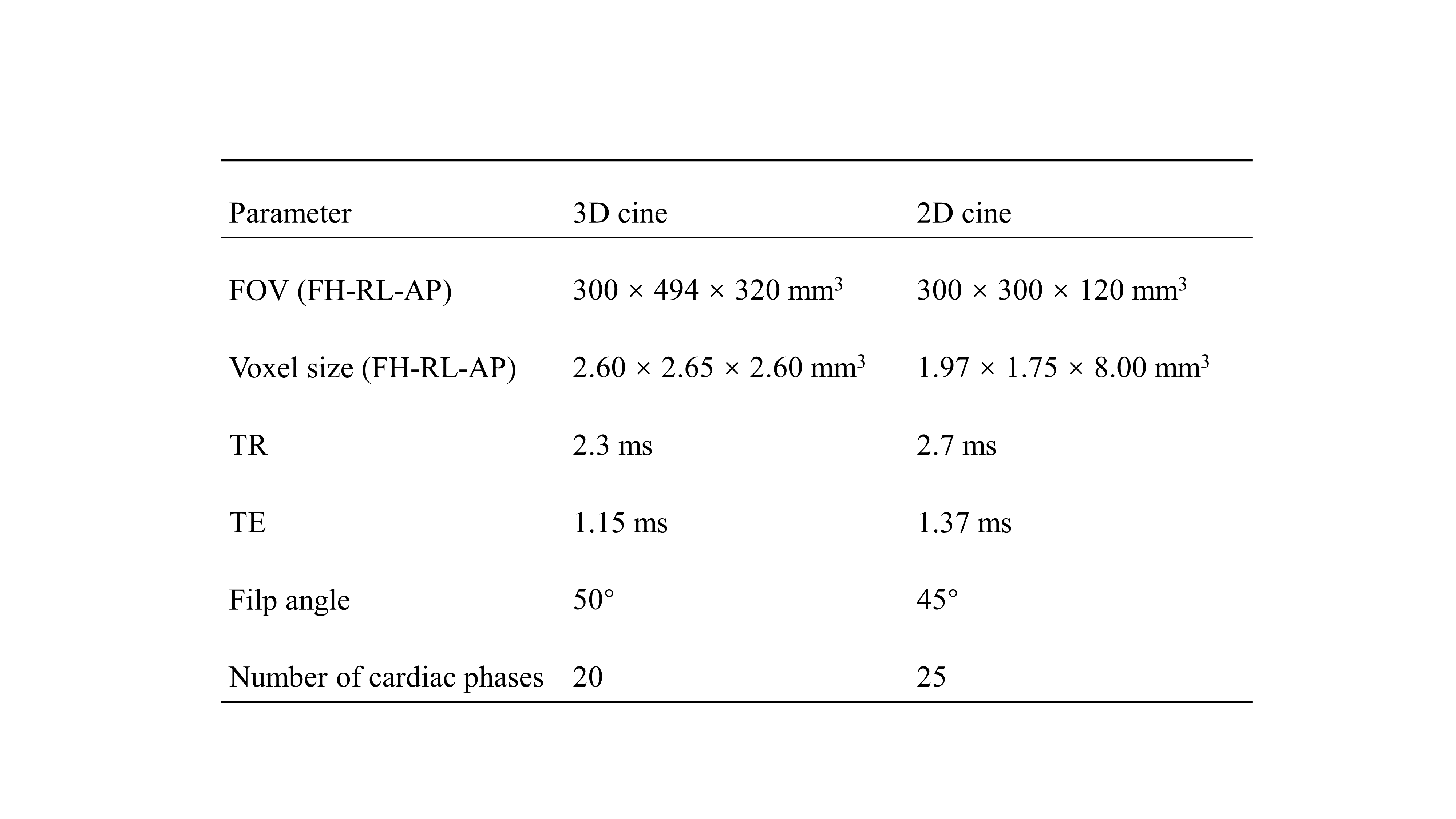
Table 1 Imaging parameters of 3D and 2D cine sequences. Note: 3D = three-dimensional; 2D = two-dimensional; field of view = FOV; repetition time = TR; echo time =TE.
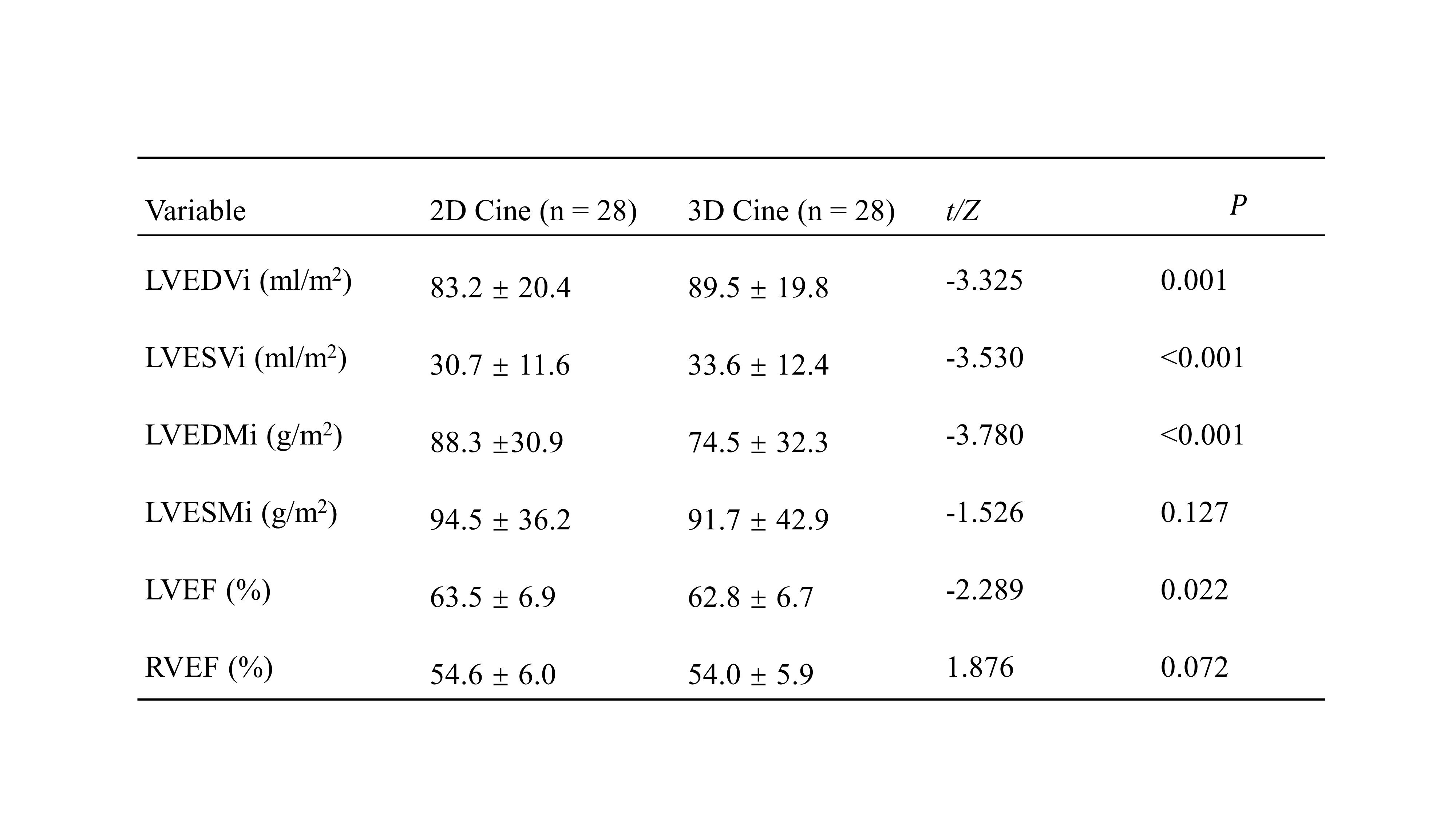
Table 2 Comparison of 3D cine and 2D cine in cardiac parameters in patients with HCM. Note: 3D = three-dimensional; 2D = two-dimensional; hypertrophic cardiomyopathy = HCM; left ventricular end-diastolic volume index = LVEDVi; left ventricular end-systolic volume index = LVESVi; left ventricular end-diastolic mass index = LVEDMi; left ventricular end-systolic mass index = LVESMi; left ventricular ejection fraction = LVEF; right ventricular ejection fraction = RVEF.
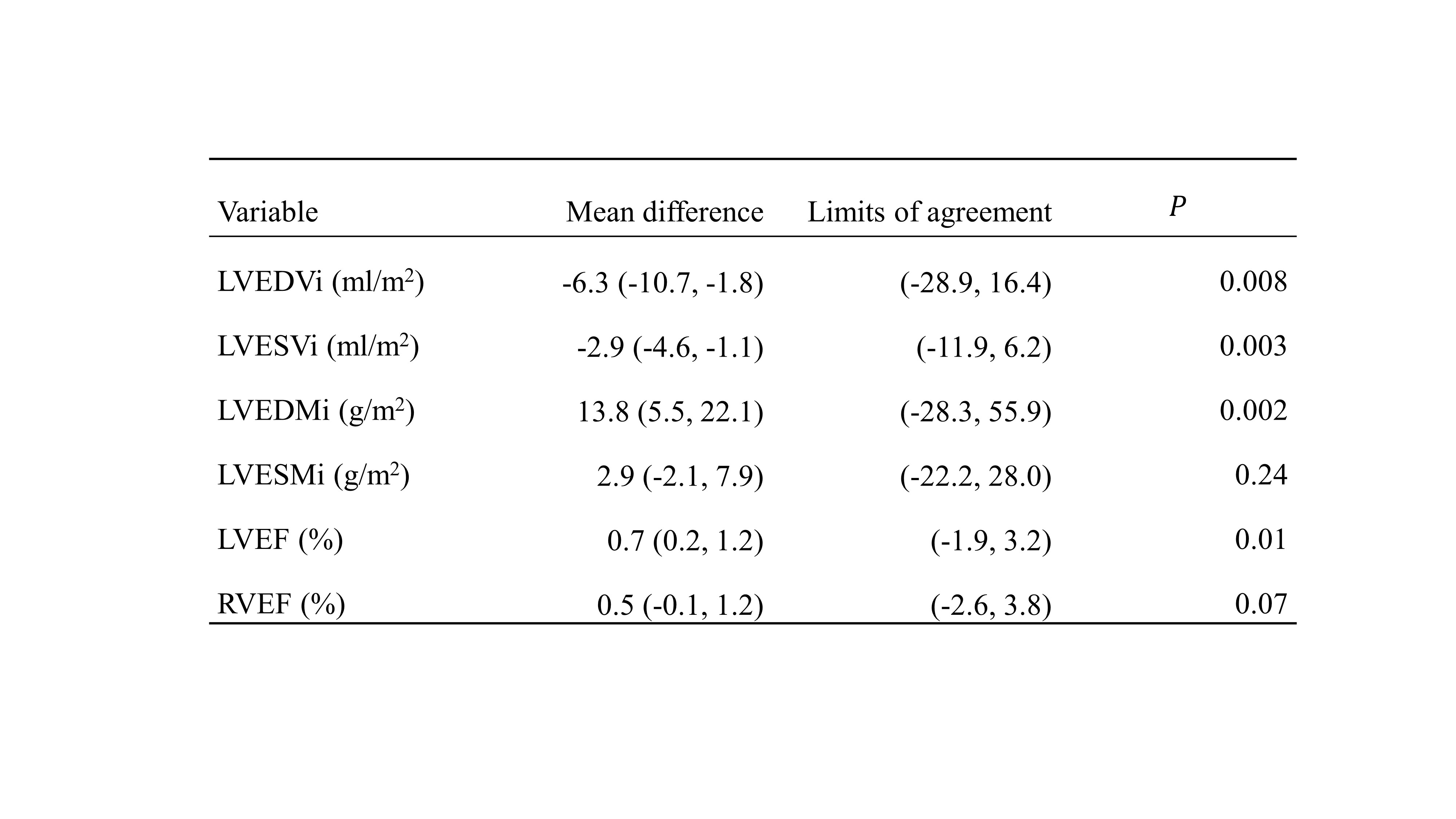
Table 3 Agreement of 3D cine measurements with 2D cine measurements in patients with HCM. Note: 3D = three-dimensional; 2D = two-dimensional; hypertrophic cardiomyopathy = HCM; left ventricular end-diastolic volume index = LVEDVi; left ventricular end-systolic volume index = LVESVi; left ventricular end-diastolic mass index = LVEDMi; left ventricular end-systolic mass index = LVESMi; left ventricular ejection fraction = LVEF; right ventricular ejection fraction = RVEF.
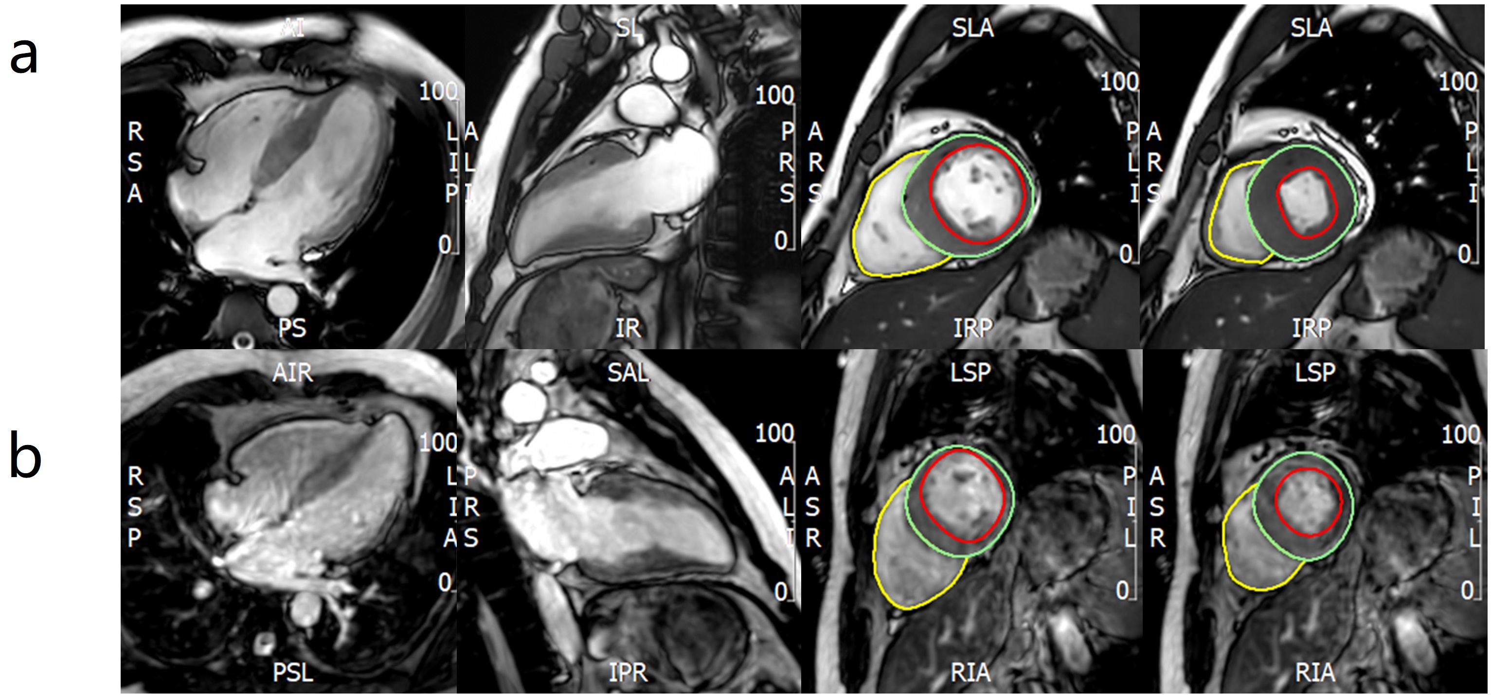
Figure 1 Two-dimensional (a) and three-dimensional (b) cine images of a patient with hypertrophic cardiomyopathy.