1670
3D Real-Time Whole Heart Cine MR Based on Spiral-In/Out bSSFP Sequence1United Imaging Healthcare, Houston, TX, United States, 2United Imaging Healthcare, Shanghai, China
Synopsis
Keywords: Myocardium, Cardiovascular, 3D real-time imaging
Motivation: To achieve high-temporal-resolution 3D whole-heart MR cine without the need for ECG, allow direct visualization of physiological motions, and assess cardiac function in clinical practice.
Goal(s): To attain 3D multi-slice whole-heart real-time imaging with <50 ms temporal resolution, while preserving adequate spatial resolution for cardiac functional analysis.
Approach: We employed golden-angle rotated spiral-in/out trajectory with 32× acceleration, and randomized variable density kz phase-encoding in the bSSFP sequence, and harnessed an iterative reconstruction algorithm.
Results: We attained a groundbreaking achievement in advancing MRI capabilities, obtaining an ECG-free 3D real-time cardiac cine with complete 12-slice whole-heart coverage and an impressive sub-50 ms temporal resolution.
Impact: We've employed advanced techniques to triumph over MRI's historical disadvantage of slower acquisition compared to other modalities. Pioneering a groundbreaking approach, we achieve high temporal resolution for 3D real-time imaging without ECG, breath-holding, or data segmentation, revolutionizing cardiac motion capture.
Introduction
Cardiovascular magnetic resonance imaging (CMR) assumes a pivotal role in evaluating cardiac function and morphology. Real-time imaging methods hold allure for their ability to effectively capture the dynamic movements of the heart. The most recent advancement in CMR cine techniques achieves an impressive temporal resolution of approximately 50 ms per 2D slice, markedly augmenting CMR's capability to elucidate the intricacies of cardiac motion. However, the pursuit of 3D real-time whole heart imaging presents a formidable challenge. This study stands as a pioneering effort, delving into the exploration of the feasibility of 3D real-time whole heart cine. This groundbreaking endeavor hinges on the innovative application of a spiral-in/out trajectory based on the balanced steady-state free precession (bSSFP) sequence, along with advanced reconstruction approach, representing a stride towards overcoming the significant hurdles associated with real-time visualization of the entire heart in 3D.Methods
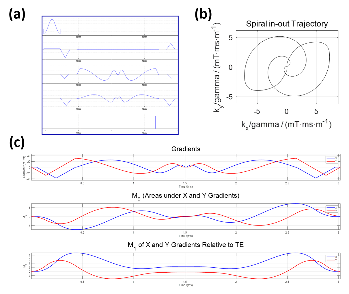
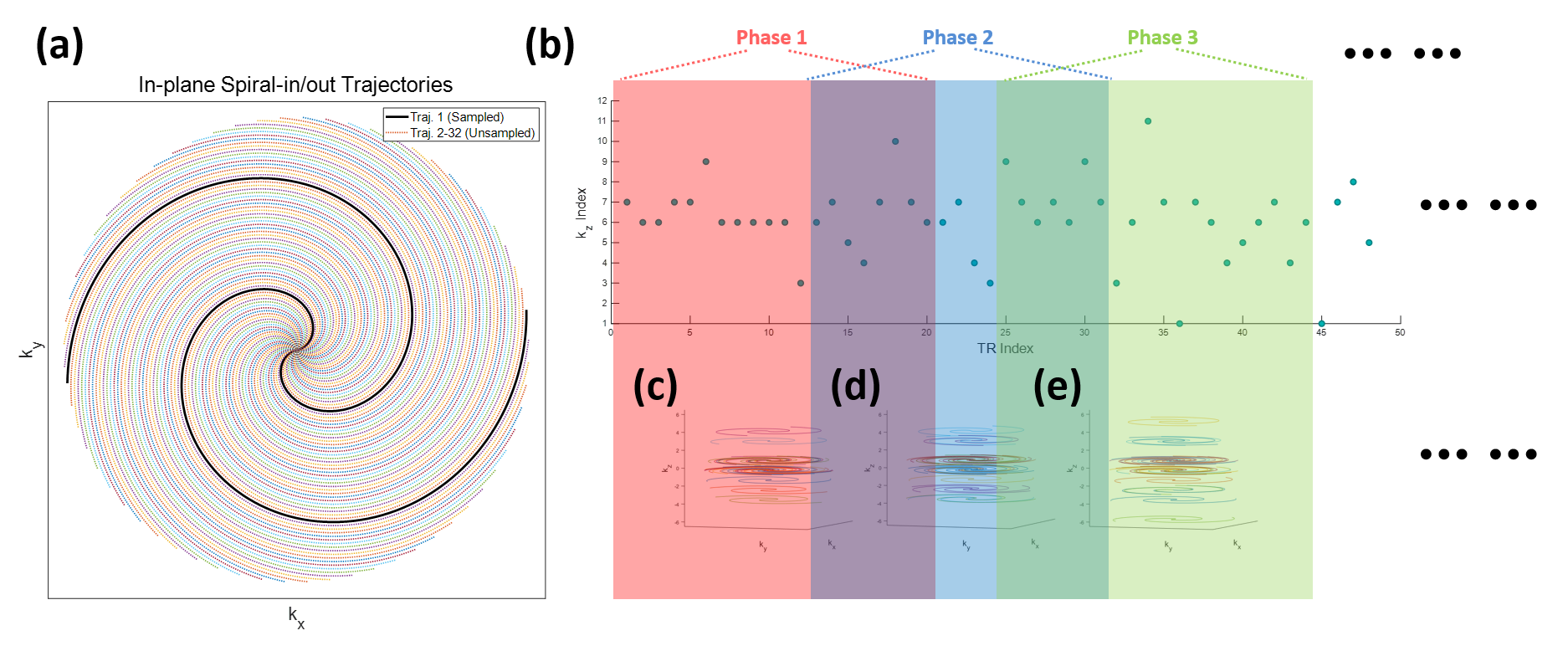
The spiral-in/out trajectory (Figure 1) features the nulling of both zeroth and first order gradient moments (M0 and M1). This trajectory finds integration into a bSSFP sequence (Figure 1a), finely tailored for advanced cardiac imaging applications.1,2 Figure 1b illustrates the complete in-plane k-space trajectory, while Figure 1c specifically details simulated field gradients and nulling behaviors of M0 and M1.For each successive repetition time (TR), the in-plane k-trajectory was designed to rotate at a golden angle. A configuration of 32 spiral-in/out interleaves was established for a fully sampled kx-ky plane. However, only 1 interleave was acquired, achieving a remarkable 32-fold in-plane acceleration (Figure 2a). A strategically randomized variable-density phase-encoding was applied along the slice-selection direction, and a temporal phase of a 3D volume was formed for every 12 acquired lines (i.e., 12 TRs), with an additional 8 acquisition lines data-shared from a previous phase (Figure 2b). This configuration yielded a temporal resolution of 49.2 ms (4.1 ms × 12), culminating in the completion of a 3D whole-heart acquisition.
A total of 200 phases of the 3D volume were acquired, with the initial 40 phases, encompassing dummy scans for achieving a steady state, discarded prior to reconstruction. Figures 2c-e illustrate the 3D k-space trajectories of the initial 3 phases, corresponding to the randomized variable density kz plot in Figure 2b. The duration of the cardiac cine spanned 6.89 seconds. Post-acquisition, a gradient-impulse response function (GIRF) correction was executed over the k-space trajectories,3 and iterative reconstruction unfolded under spatio-temporal constraints, detailed in reference.4
The sequence was implemented on a 1.5T MRI system (uMR 680, United Imaging Healthcare, Shanghai) equipped with a 24-channel super flexible coil. The gradient performance was 45 mT/m with a slew rate of 200 mT/m/s. Imaging parameters were: TR/TE = 4.10/2.05 ms, FA = 67°, FOV = 340×340 mm2, matrix size = 128×128, 8 mm slice thickness × 12 slices. Data was acquired from three healthy subjects.
Results
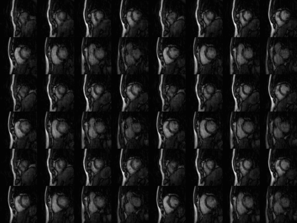
This abstract presents data from a single subject for simplicity. 3D real-time whole heart cine images were successfully acquired at a temporal resolution of 49.2 ms (Figure 3). For better presentation, the middle 8 slices out of a total of 12 are shown. This high temporal resolution enabled the precise visualization of cardiac motion across the entire cardiac cycle. The resulting images showcased outstanding contrast between blood and myocardium, coupled with exceptional spatial resolution, harnessing the advantages of bSSFP sequences within the realm of real-time cardiac MRI.Discussion and Conclusion
Spiral-in/out k-space trajectory offers the advantage of naturally nullifying both M0 and M1 gradient moments, making it an ideal choice for cardiac cine imaging. M0 nulling is a prerequisite for the bSSFP sequence, while M1 nulling renders the sequence insensitive to flow, eliminating flow artifacts and making it ideal for cardiac motion analysis. Additionally, the spiral-in/out trajectory features an efficient k-space sampling speed, enabling real-time imaging with higher temporal resolution.For the first time, our study presents a perfect solution for achieving 3D whole heart real-time cine MR using a spiral-in/out trajectory based on a bSSFP sequence. Spiral-in/out trajectory was particularly beneficial for capturing real-time cardiac events. The technique offers a flexible setting of temporal resolution by adjusting the number of data lines for each phase, albeit at the recognized trade-off of image quality. This technique offers the promise of high-quality, real-time cardiac imaging, making it a potential tool for comprehensive cardiac examinations, including those requiring high temporal resolution, such as arrhythmia assessments or stress tests. With advancements in spiral imaging and AI-assisted reconstruction, we can anticipate an upcoming era marked by a groundbreaking suite of 3D real-time sequences for CMR. The innovative approach will enhance applications like interventions and MR-guided therapies with prompt responsiveness.
Acknowledgements
No acknowledgement found.References
- Nayak, K. S., Hargreaves, B. A., Hu, B. S., Nishimura, D. G., Pauly, J. M., & Meyer, C. H. (2005). Spiral balanced steady‐state free precession cardiac imaging. Magnetic Resonance in Medicine, 53(6), 1468-1473.
- Feng, X., Salerno, M., Kramer, C. M., & Meyer, C. H. (2016). Non‐Cartesian balanced steady‐state free precession pulse sequences for real‐time cardiac MRI. Magnetic Resonance in Medicine, 75(4), 1546-1555.
- Campbell‐Washburn, A. E., Xue, H., Lederman, R. J., Faranesh, A. Z., & Hansen, M. S. (2016). Real‐time distortion correction of spiral and echo planar images using the gradient system impulse response function. Magnetic Resonance in Medicine, 75(6), 2278-2285.
- Zhao, Z., Lim, Y., Byrd, D., Narayanan, S., & Nayak, K. S. (2021). Improved 3D real‐time MRI of speech production. Magnetic Resonance in Medicine, 85(6), 3182-3195.
Figures