1651
Exploring the Reliability of Compressed Sensing Cardiac MR Cine in Heart Failure: Evidence from Varied MR Field Strengths and Vendors1Department of Radiology, Sir Run Run Shaw Hospital, Zhejiang University School of Medicine, Hangzhou, Zhejiang Province, China, Hangzhou, China, 2MR Research CollaborationTeam, Siemens-Healthineers Ltd, Shanghai, China, Shanghai, China
Synopsis
Keywords: Heart Failure, Heart
Motivation: The challenge of obtaining high-quality CMR cine images in patients with heart failure (HF) due to difficulties in breath-holding and arrhythmia.
Goal(s): Explore the feasibility of utilizing compressed sensing cardiac cine with varying levels of acceleration to acquire comparable cine images in HF patients.
Approach: We compare cardiac function, strain parameters, and image quality between bSSFP and CS images in both HF patients and healthy participants, utilizing MRI systems from different vendors.
Results: Our findings demonstrate a high level of consistency in most cardiac function and strain parameters between the two sequences. CS cardiac cine images exhibit comparable quality to bSSFP images.
Impact: This study offers prospective validation for the use of CS cardiac cine sequences in HF patients, addressing issues related to lengthy scanning durations and artifacts. It expands the eligible population for CMR scanning while optimizing image quality.
Introduction
Heart failure (HF) represents a significant global public health concern, with cardiac function indices playing a pivotal role in HF diagnosis and management1,2. HF patients often face challenges in maintaining repeated breath-holding during cardiovascular magnetic resonance (CMR) examinations, leading to suboptimal images and unexpected artifacts. Compressed sensing (CS) cardiac cine is a reliable imaging acceleration technique that can address these challenges effectively3. In this study, we aim to evaluate the reliability of CS-cine and its potential to efficiently replace conventional balanced steady-state free precession (bSSFP) cine images for clinical assessment of cardiac function and strain parameters in HF patients.Methods
We prospectively enrolled 28 HF patients and 33 healthy participants (HP) for cardiac magnetic resonance (CMR) examinations on a 3T MRI scanner (MAGNETOM Vida, Siemens Healthcare, Erlangen, Germany) and a subset of 38 HF patients on a 1.5T MRI scanner (uMR680, United Imaging, Shanghai, China). Inclusion criteria were consistent with established guidelines4. Each participant underwent cine imaging of the heart, acquiring images in four standard views (two-chamber, four-chamber, three-chamber, and short-axis) using both bSSFP and CS cine sequences. The crucial sequences parameters were as follows: a. 3T Siemens: bSSFP3.0 vs CS3.0: Temporal resolution = 39.2 vs 39.3 ms, Bandwidth= 970 vs 930 Hz/pixel, Acceleration factor (n = 3 vs 8.3), Scanning time = 58.85 vs 19.81 s. b. 1.5T uMR680: bSSFP1.5 vs CS1.5: TR = 3.12 vs 2.86 ms, TE=1.51 vs 1.34 ms, Temporal resolution = 31.2 vs 42.9 ms, Bandwidth= 1200 vs 1200 Hz/pixel, Acceleration factor (n = 2 vs 11.4), Scanning time = 119.58 vs 18.54 s. Subsequently, the cine images were imported into CVI42 software (Circle Cardiovascular Imaging Inc., Calgary, Canada) for volume function and strain analysis. Statistical analyses were performed using the Wilcoxon matched-pairs signed-rank test or paired t-test. To assess the correlation and agreement of quantitative parameters between the different sequences, we conducted linear regression and Bland–Altman analysis. Furthermore, intra- and inter-observer consistency were evaluated using the intra-class correlation coefficient (ICC).Results
The CS cine sequences exhibited significantly shorter acquisition times compared to the bSSFP cine sequences. In the HF group, left ventricular ejection fraction (LVEF) consistently showed a slight underestimation with the CS cine sequence in comparison to the conventional cine sequence under the 3T scanner (HF 3.0T bSSFP vs CS: 39.37% vs 37.32%, p < 0.006), but not under the 1.5T scanner (HF 1.5T bSSFP vs CS: 30.24% vs 29.8%, p = 0.126). There were no statistically significant differences in right ventricular ejection fraction (RVEF) parameters among these three groups. The correlation between global radial strain (GRS) and global circumferential strain (GCS) values in the biventricular chambers remained consistently high (R > 0.75, p < 0.001) across all three groups, while global longitudinal strain (GLS) exhibited lower R values compared to the above two. Global strain values derived from conventional and CS cine sequences indicated that CS cine typically yielded lower values (p < 0.05). Furthermore, CS-cine-based biventricular functional and strain parameters exhibited a strong correlation with those of conventional cine sequences, with more than 90% of data points falling within the limits of agreement. Inter- and intra-observer agreements were consistently strong (ICC = 0.80 to 0.99, p < 0.001) for CS cine in both HF groups (3.0T and 1.5T).Discussion
CS cine sequences offer the advantage of reducing scanning time while maintaining image quality in HF patients, regardless of MR field strengths and vendors. Cardiac parameters derived from CS cine exhibit a high level of agreement with those obtained from conventional cine sequences, except for specific strain parameters related to the right ventricle (RV). This difference may be attributed to the inherent characteristics of the RV, such as its thin wall, irregular shape, and contour, which present challenges in accurately assessing its function5. The slight reduction in LVEF when assessed using CS cine images, particularly in the RV and at the 1.5T field strength, may be due to the reduced spatial and temporal resolution in CS images, potentially leading to an overestimation of end-systolic volume (ESV)6. Similarly, strain parameters obtained from CS images consistently tend to yield lower measurements, consistent with findings from previous studies7.Conclusions
This study has affirmed the practical utility of CS cardiac cine sequences in patients with HF who face difficulties in maintaining breath-hold, as it significantly reduces scan times without compromising image quality, irrespective of varying field strengths and MR vendors. Additionally, the assessment of cardiac function and strain using CS cine demonstrates strong concordance with conventional cine analysis.Acknowledgements
This work was supported by the National Natural Science Foundation of China (Grant No. 81873908, for HJH).References
1. Metra, M. et al. Developed by the task force for the diagnosis and treatment of acute and chronic heart failure of the European Society of Cardiology (ESC).
2. Contaldi, C. et al. Role of Cardiac Magnetic Resonance Imaging in Heart Failure. Heart Fail. Clin. 17, 207–221 (2021).
3. Feng, L. et al. Compressed sensing for body MRI. J. Magn. Reson. Imaging 45, 966–987 (2017).
4. Roifman, I. et al. Utilization and impact of cardiovascular magnetic resonance on patient management in heart failure: insights from the SCMR Registry. J. Cardiovasc. Magn. Reson. 24, 65 (2022).
5. Tadic, M. Multimodality Evaluation of the Right Ventricle: An Updated Review. Clin. Cardiol. 38, 770–776 (2015).
6. Li, Y. et al. Cardiac cine with compressed sensing real-time imaging and retrospective motion correction for free-breathing assessment of left ventricular function and strain in clinical practice. Quant. Imaging Med. Surg. 13, 2262–2277 (2023).
7. Chen, Y. et al. Feasibility of single-shot compressed sensing cine imaging for analysis of left ventricular function and strain in cardiac MRI. Clin. Radiol. 76, 471.e1-471.e7 (2021).
Figures
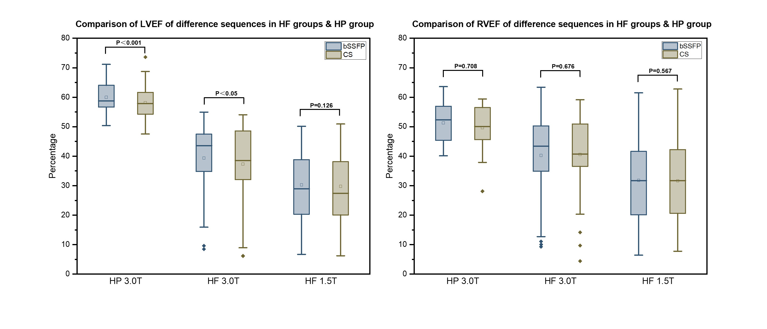
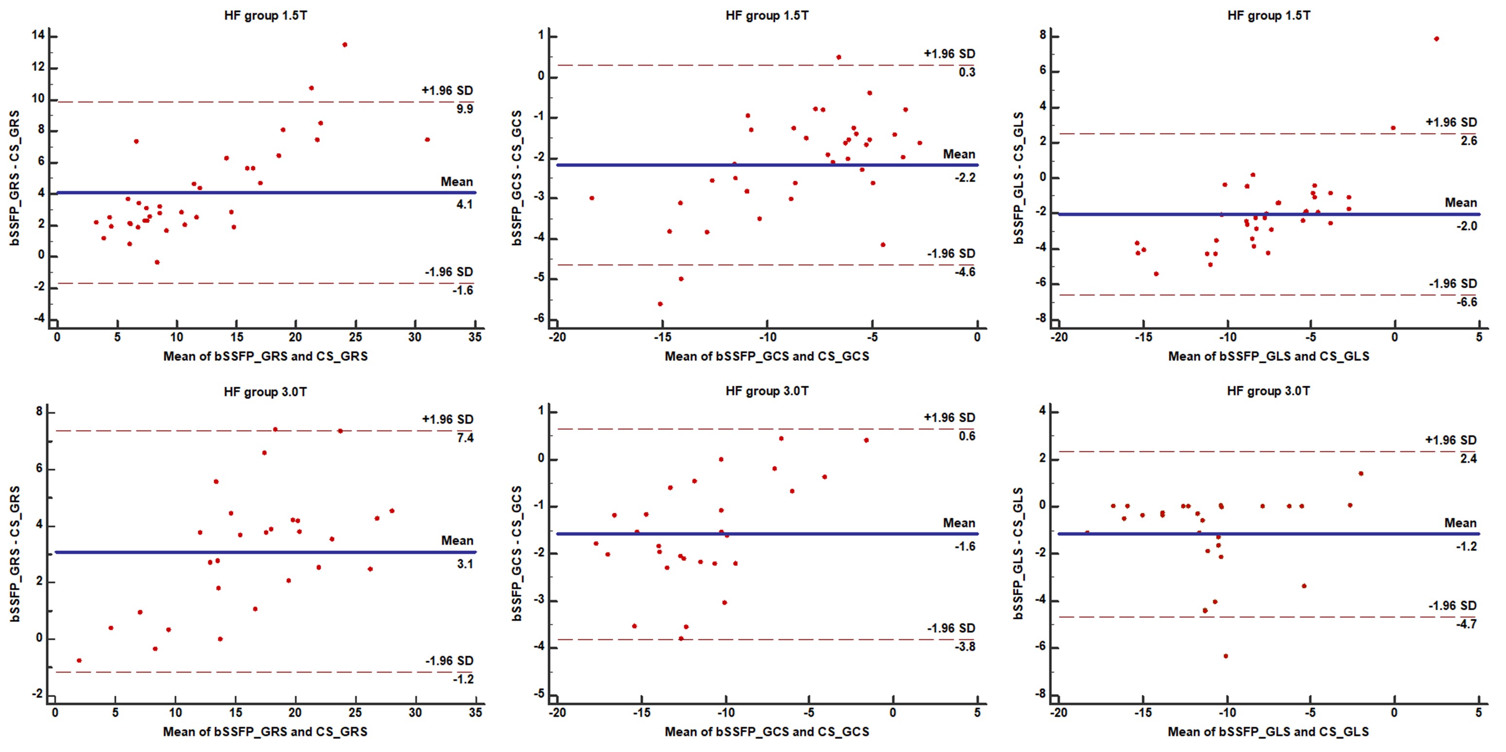
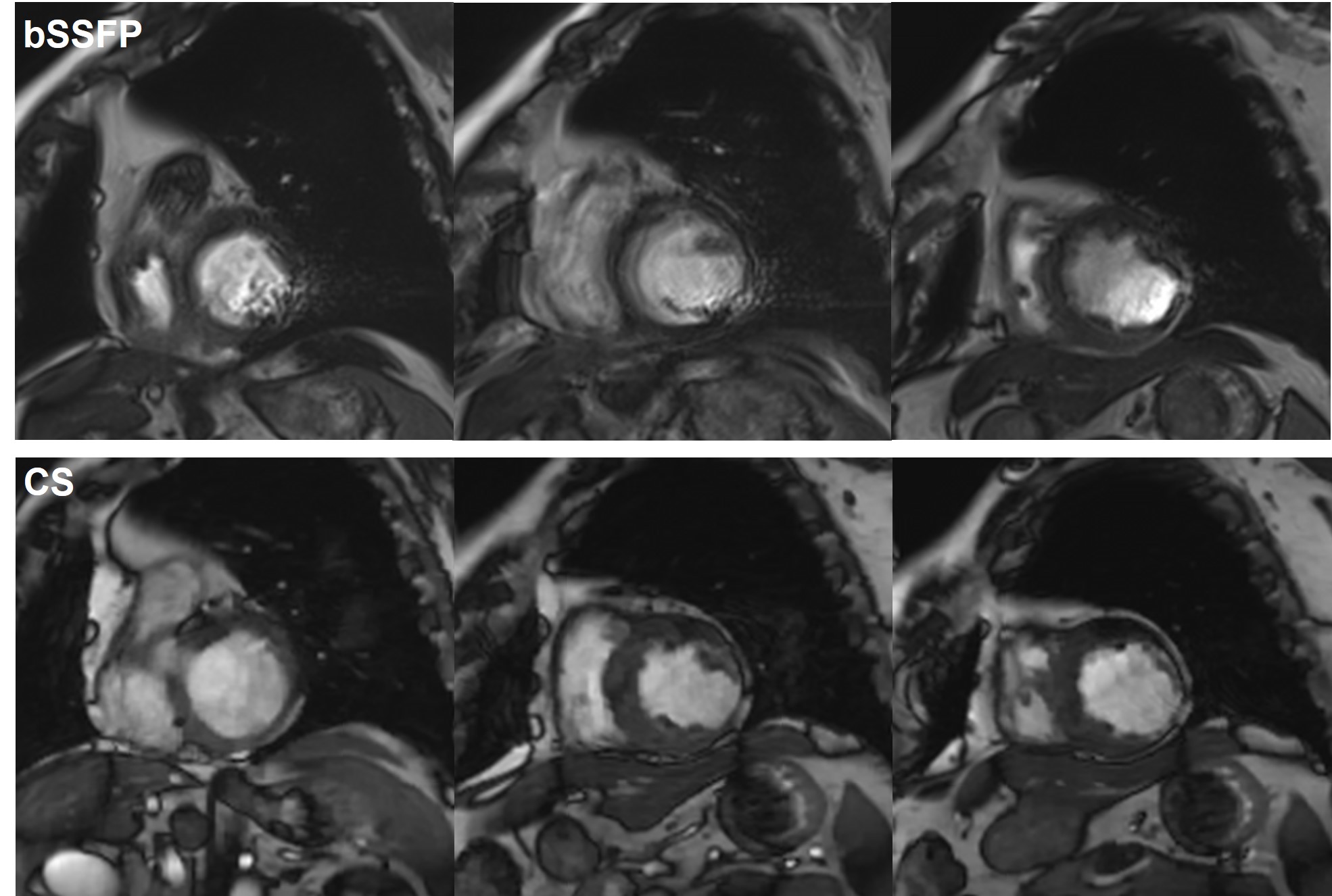
Figure 3: 3T scanner (MAGNETOM Vida, Siemens Healthcare, Erlangen, Germany) bSSFP vs CS cine images.
The image portrays a 72-year-old male patient who is grappling with HF and encountering difficulties in maintaining breath-hold during the examination. Notably, the conventional cine images revealed prominent respiratory artifacts. In stark contrast, the CS cine images displayed significantly enhanced image quality.