1644
Left ventricular blood flow characteristics in children with premature ventricular contractions by 4D flow CMR1Shengjing Hospital of China Medical University, Shenyang, China, 2Siemens Healthcare GmbH, Erlangen, Germany, 3Siemens Healthineers, Beijjing, China, 4Circle Cardiovascular Imaging Inc, Calgary, AL, Canada, 5Siemens Healthineers, Shenyang, China
Synopsis
Keywords: Arrhythmia, Arrhythmia, CMR,4D FLOW
Motivation: A novel imaging method was developed to diagnose children with premature ventricular contractions (PVCs), thereby preventing heart failure or cardiomyopathy.
Goal(s): To determine the changes in the blood flow composition of LV in children with PVCs, a free breathing 4D flow CMR was performed. The relationship of PVC load with LV function was explored.
Approach: The children with PVCs underwent 24-hour dynamic ECG and CMR examination within 3 days. Thus, we determined their cardiac function and hemodynamic parameters.
Results: Children with PVCs had reduced direct blood flow in LV, which showed a significant negative correlation with PVC load.
Impact: In this study, we analyzed the characteristics of the left ventricular blood flow in children with premature ventricular contractions (PVCs). Thereafter, we elucidated the pathophysiological mechanism of PVCs, which provided the basis for clinical treatment and prognostic evaluation.
Introduction
Premature ventricular contractions (PVCs), which is a common arrhythmia, originates from the right ventricle or the left ventricle’s outflow tract. This condition occurs in healthy children, with a normal heart structure. The incidence of isolated PVCs is 18-21% of children. However, PVCs cause changes in electrical-mechanical coupling, which triggers complex hemodynamic changes in ventricles. In children with frequent PVCs, the clinical outcomes are poor. A detailed diagnosis is performed to identify potential risk factors. Our aim is to determine the characteristics of blood flow in the left ventricle of children with PVCs. Therefore, a four-dimensional flow cardiovascular magnetic resonance (4D flow CMR) technique was performed. Thus, we evaluated the relationship between the following parameters: the composition of the left ventricular blood flow, the efficacy of left ventricular function, and the burden of PVCs.Method
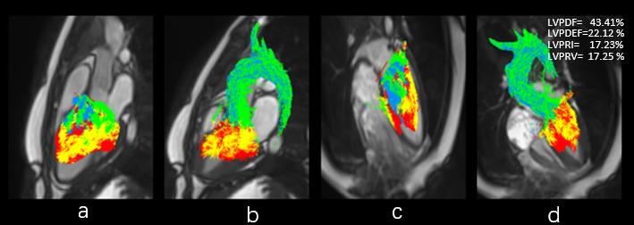
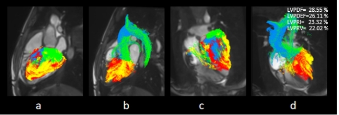
In our hospital, 20 children (age: 8 ± 2.9,female: 13cases) were diagnosed with PVCs in the period extending from June 2023 to September 2023. The prospective study was performed on the PVCs group. Furthermore, 15 children did not have cardiopulmonary diseases, and they were subjected to CMR examination. These children constituted the control group (age: 8 ± 3.8, male: 2 cases). A 24-hour Holter electrocardiogram and CMR scan were performed within a span of three days. To perform a CMR examination of 4D flow, we used a 3T scanner (MAGNETOM Prisma, Siemens Healthineers AG, Erlangen, Germany) . A written informed consent letter was signed by all the subjects. A 4D flow research sequence was used with ECG-triggering and navigator-gating. The parameters of the scan were as follows: Temporal resolution = 38.56 ms, TE=2.28 ms, excitation angle =7 degrees, FOV = 360 mm×280 mm, matrix = 144×78, voxel size = 2.5 mm×2.5 mm×2.5 mm, venc = 120-150 cm/s, 25 cardiac phases. The total scan time was 6 to 8 minutes. The CMR data were transferred to cvi42 (Version 5.10.7, Circle Cardiovascular Imaging, Calgary Alberta, Canada.). The ventricular function included the LV end-diastolic and end-systolic volumes, and the LV ejection fraction. All the flow parameters were measured by a cardiovascular radiologist, who had two years of experience in operating 4D flow software. Thus, the background phase correction, the noise filer and the velocity aliasing correction, and the endocardial segmentation were performed. The positions of LV pathlines were divided into the following four functional flow components: the percent direct flow in the left ventricle (LVPDF), the percent delayed ejection flow in the left ventricle (LVPDEF), the percent retained inflow in the left ventricle (LVPRI), and the percent residual volume in the left ventricle (LVPRV) (Figure 1 and 2). Each component constituted a portion of the total end-diastolic volume. Statistical analyses were performed using SPSS 20 software IBM, Armonk,USA). T test or Mann-Whitney U test were used to compare the differences in hemodynamic parameters and ventricular function parameters. Spearman correlation analysis was performed to assess the correlations between the following parameters: the composition of LV blood flow, the left ventricular function, and the PVC load of the experimental group. All the tests were 2-sided, and P < 0.05 was considered to be a statistically significant difference.Result
In the PVCs group, the LVPDF, LVPDEF, LVPRI, and LVPRV were 34.45%± 5.91%, 23.46%±4.51%, 23.41%±3.41% and 18.67%±4.75%, respectively, In the control group, the aforementioned parameters were 44.41%± 3.07%, 20.41%± 4.71%,22.60% ± 4.00%,12.57% ± 2.51%, respectively (Table 1). The LVPDF of the PVC group was significantly lower than that of the control group (p < 0.01). The LVPRV of the PVCs group was significantly greater than that of the control children(p=0.01). The LVPDF and LVPDEF of the PVCs group showed a negative correlation ((r=−0.434, P=0.159). The LVPRI and LVEF of the PVCs group also showed a negative correlation (r=−0.529, P=0.077). The combination of LVPDF+LVPDEF was (59.19±4.69)%, and it showed a significantly positive correlation with LVEF (r=0.905, P<0.001). There was no statistically significant difference between LVEF and (LVPDF+LVPDEF) (P=0.348). The LVPDF of PVCs group showed a significantly negative correlation with the PVC burden (r= -0.671, P=0.048).Discussion and Conclusion
A 4D flow CMR technique was used to assess the left ventricular function of children with PVCs in free-breathing. In these children, the direct blood flow decreased and the residual volume to the left ventricle increased. This implies that the direct blood flow to the left ventricle is correlated with the left ventricular function and the PVC load.Acknowledgements
No acknowledgement found.References
[1] KAMIŃSKA H, MAŁEK Ł A, BARCZUK-FALĘCKA M, et al. The Role of Cardiac Magnetic Resonance in Evaluation of Idiopathic Ventricular Arrhythmia in Children [J]. Journal of Clinical Medicine, 2021, 10(7).
[2] ALFONSO SESTITOA M P, GREGORY A. SGUEGLIAA, LUIGI NATALEC,ANGELICA DELOGUB, FABIO INFUSINOA, GABRIELLA DE ROSAB, FULVIO BELLOCCIA,FILIPPO CREAA AND GAETANO A. LANZA. Cardiac magnetic resonance of healthy children and young adults with frequent premature ventricular complexes [J]. Journal of Cardiovascular Medicine 2007.
Figures