1637
MRI of tendinopathy of the common extensor tendon and common flexor tendon and its application for injury classification1Department of Radiology, Beijing Jishuitan Hospital, Capital Medical University, Beijing, China
Synopsis
Keywords: Tendon/Ligament, Tendon/Ligament
Motivation: We analyzed magnetic resonance imaging (MRI) features of the elbow tendons to provide information for early diagnosis and treatment.
Goal(s): To analyze the injury classification of the common extensor tendon (CET) and common flexor tendon (CFT) tendinopathy by MRI.
Approach: 62 patients with elbow tendinopathy as the case group and 20 healthy subject as the control group. All subjects underwent elbow MR examination. The MRI of tendinopathy were read by 2 senior radiologists independently, and the consistency was tested by the κ test. The MRI manifestations for injury classification were discussed.
Results: MRI can be used to diagnose tendinopathies and classify injuries.
Impact: Tendinopathy of CFT and CET of elbow joint has its characteristic MRI manifestations, indicating that MRI can be used for the diagnosis injury classification of tendinopathy. In addition, MRI can also be used for the injury classification.
Introduction
Elbow tendinopathy is common in clinical practice, especially common extensor tendon (CET) and common flexor tendon (CFT) tendinopathy, which often lead to elbow pain and dysfunction, affecting patients' quality of life. Early determination of the location and extent of the injury is an important guide to improving treatments and improving patient prognosis[1]. X-ray and CT examinations are preferred for elbow trauma to clarify whether there are fractures and dislocations, but they cannot show changes in the subtle anatomical structures such as tendons[1, 2]. Magnetic resonance imaging (MRI) has good soft tissue resolution and multiplanar imaging characteristics, which can clearly show the anatomical structure of elbow tendons and the site, range and extent of tendons injuries. In this study, we analyze the MRI findings of tendinopathy of the common extensor tendon (CET) and common flexor tendon (CFT) and to explore its application for injury classification.Methods
Sixty‑two patients with elbow tendinopathy of CET and/or CFT from August 2019 to April 2022 were enrolled as the case group, and 20 healthy subjects (40 elbows) were recruited as the control group. All subjects underwent elbow MR examination.The MRI of tendinopathy of CFT and CET were read by 2 senior radiologists independently, and the consistency was tested by the κ test. The MRI manifestations for injury classification were discussed.Results
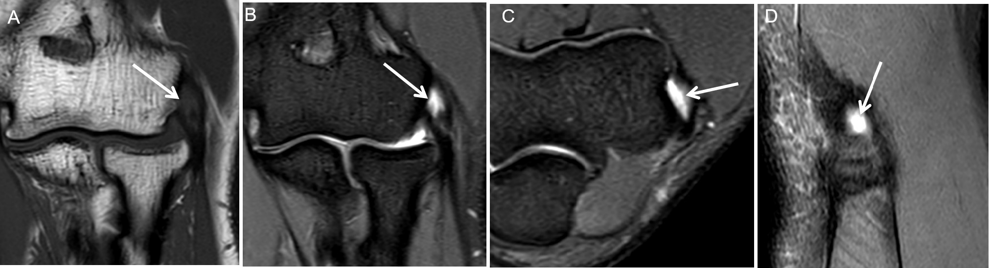
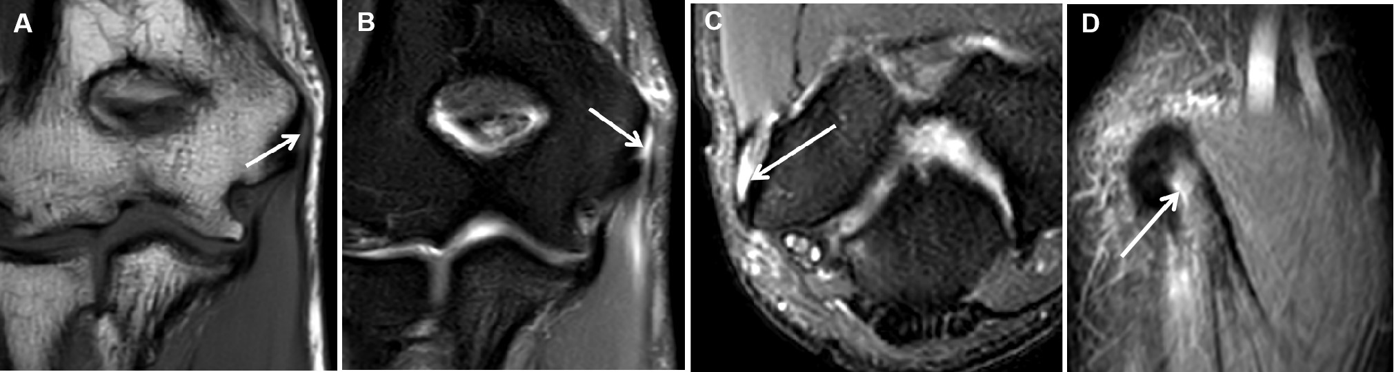
The κ values of CFT, CET, medial collateral ligament complex and lateral collateral ligament complex were 0.645, 0.657, 0.615 and 0.653 respectively. MRI clearly showed the anatomical structure of CFT, CET and surrounding soft tissue. The CFT and CET of the elbow of 20 normal volunteers (40 sides) in the control group showed thin line or banded low signal in each MRI sequence, and the muscle fibers were continuous. In 27 patients with classification Ⅰ injury of CFT and/or CET of elbow joint, MRI showed thickening or thinning of tendons and increased signal intensity on fat suppression T2‑weighted imaging. In 27 cases of classification Ⅱ injury, MRI showed that the fluid signal involved 20% to 80% of the tendon thickness. In 8 cases of classification Ⅲ injury, MRI showed that the fluid signal involved more than 80% of the tendon thickness.Discussion
Elbow tendinopathy is a common condition affecting upper limb motion and function, usually caused by specific occupational or sporting activities. Previous studies have shown that CET injuries usually involve the radial collateral ligament and lateral ulnar collateral ligament[2-4]. In this study, the incidence of grade I, II, and III CET tendinopathy with lateral collateral ligament complex injuries was 55%, 77%, and 100%, respectively, which suggests that the more severe the CET injury, the higher the incidence of ipsilateral collateral ligament injury. CFT tendinopathy is often associated with medial collateral ligament complex injuries. In this study, the incidence of medial collateral ligament complex injuries accompanying grade I, II, and III CFT tendinopathy was 33%, 57%, and 33%, respectively, which suggests that the incidence of medial collateral ligament complex injuries did not significantly increase with the severity of CFT tendinopathy, which is inconsistent with previous studies[5, 6], and thus further large-sample studies are needed.Conclusions
Tendinopathy of CFT and CET of elbow joint has its characteristic MRI manifestations, indicating that MRI can be used for the diagnosis of tendinopathy. In addition, MRI can also be used for the injury classificationAcknowledgements
No acknowledgement found.References
[1] Dewan AK, Chhabra AB, Khanna AJ, Anderson MW, Brunton LM. MRI of the elbow: techniques and spectrum of disease: AAOS exhibit selection. J Bone Joint Surg Am. 2013. 95(14): e99 1-13. [2] Acosta Batlle J, Cerezal L, López Parra MD, Alba B, Resano S, Blázquez Sánchez J. The elbow: review of anatomy and common collateral ligament complex pathology using MRI. Insights Imaging. 2019. 10(1): 43. [3] Qi L, Zhu ZF, Li F, Wang RF. MR imaging of patients with lateral epicondylitis of the elbow: is the common extensor tendon an isolated lesion. PLoS One. 2013. 8(11): e79498. [4] Cha YK, Kim SJ, Park NH, Kim JY, Kim JH, Park JY. Magnetic resonance imaging of patients with lateral epicondylitis: Relationship between pain and severity of imaging features in elbow joints. Acta Orthop Traumatol Turc. 2019. 53(5): 366-371. [5] Amin NH, Kumar NS, Schickendantz MS. Medial epicondylitis: evaluation and management. J Am Acad Orthop Surg. 2015. 23(6): 348-55. [6] Ciccotti MC, Schwartz MA, Ciccotti MG. Diagnosis and treatment of medial epicondylitis of the elbow. Clin Sports Med. 2004. 23(4): 693-705, xi.Figures