1636
Enhancing MRI Image Quality and Meniscus Injury grading in Knee Joints with Deep Learning1Department of MRI, the First Affiliated Hospital of Zhengzhou University, Zhengzhou, China, 2MR Research China, GE Healthcare, Beijing, China
Synopsis
Keywords: Whole Joint, Bone, deep learning; knee joint; image quality; meniscus injury; MRI
Motivation: The need for accurate meniscus injury diagnosis and the limitations of traditional MRI.
Goal(s): To enhance knee MRI through deep learning algorithms.
Approach: A cohort of 46 patients underwent MRI with and without deep learning reconstruction (DLR). Radiologists subjectively assessed image quality and evaluated meniscus damage.
Results: DLR improved image quality, reducing noise and artifacts. Radiologists consistently rated DLR images higher. DLR excelled in detecting subtle meniscus injuries compared to traditional MRI.
Impact: This study demonstrates that deep learning-based MRI reconstruction substantially improves image quality and the detection of subtle meniscus injuries, offering enhanced diagnostic accuracy in knee joint assessments.
Introduction
The meniscus is a crucial fibrocartilage structure in the knee joint, serving to absorb pressure, stabilize the knee, and reduce friction during leg movement [1]. Twisting or rotating the knee can lead to meniscus damage, causing persistent knee pain and the risk of osteoarthritis. Timely diagnosis and grading of meniscus injuries are essential for effective treatment. Arthroscopy is the gold standard for diagnosis but is invasive, making MRI the preferred choice due to its excellent soft tissue resolution and non-invasiveness. However, conventional MRI has limitations, such as suboptimal image quality, lengthy acquisition times, and susceptibility to operator skill.Advancements in deep learning algorithms have led to the integration of artificial intelligence in medical image analysis, enhancing image quality, automatic detection, and segmentation. Deep learning-based MRI reconstruction pipelines [2, 3], when applied to knee MRI, maintain or even improve image quality while significantly reducing scanning time. These deep learning algorithms have shown promise in improving the evaluation of meniscus injuries in knee MRI images, potentially revolutionizing the diagnostic process and patient care. This study aims to test the hypothesis that deep learning-based knee MRI can enhance overall image quality and speed, further enhancing the assessment of meniscus injuries in knee MR images.Material and methods
This prospective cohort study involved 46 adult patients who underwent knee joint MRI examinations at the First Affiliated Hospital of Zhengzhou University from December 2022 to April 2023. The participants, consisting of 23 males and 23 females with an average age of 46.35, were included based on specific criteria, including the presence of meniscus injury and age over 18 years, with exclusion criteria involving MRI contraindications or an inability to complete the required scans. A 3T GE MRI scanner (SIGNA Premier, GE Healthcare, Waukesha, WI) was used for the scanning. The study assessed the image quality subjectively using three sets of images: conventional reconstructed images without deep learning reconstruction (DLR), accelerated MRI images without DLR, and accelerated MRI images with DLR. Two radiologists independently analyzed the images, rating artifacts, noise, and overall image quality on 5-point scales. Meniscus injury was evaluated based on McNab grading by the same two radiologists.Statistical analysis was conducted using SPSS 26.0 software, assessing image quality differences between various image sets and the consistency of image results between the two physicians using weighted kappa values. The results were interpreted with specific kappa value ranges indicating the level of consistency. The significance level was set at P<0.05 for statistical significance.Results and discussion
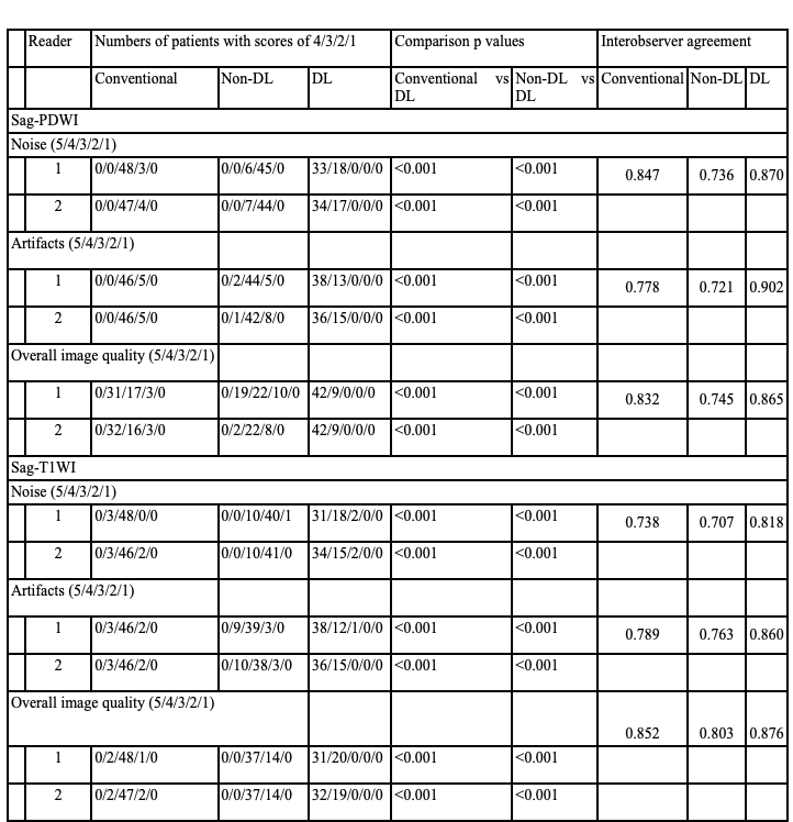
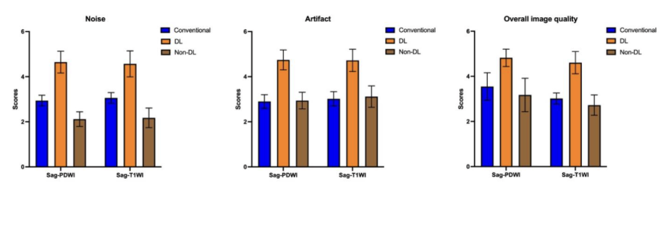
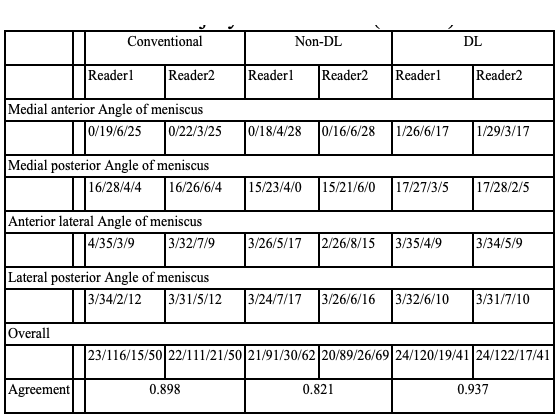
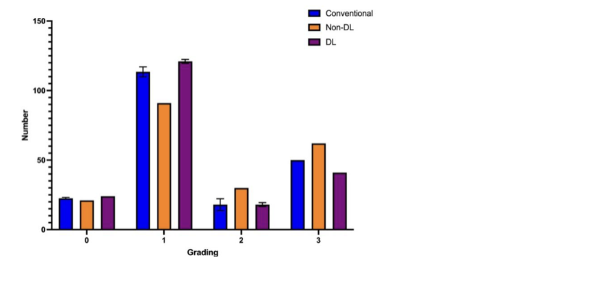
The study's image quality evaluation revealed that two radiologists rated the overall image quality, subjective noise, and artifacts of images processed with deep learning reconstruction (DLR) higher compared to conventional and non-DLR images. In table 1 and Figure 1, DLR images mostly received scores of 5 and 4 for overall image quality, subjective noise, and artifacts, while traditional images had generally lower scores than non-DLR images. Significant differences were observed in subjective noise, artifacts, and overall quality between DLR images and conventional images, as well as between DLR and non-DLR images (P<0.05). The two radiologists showed good consistency in their subjective evaluations of image quality, with kappa values ranging from 0.721 to 0.902.Regarding the assessment of meniscus damage, two radiologists evaluated meniscus injuries in knee joint images processed with and without DLR (Table 2 and Figure 2). The diagnostic results demonstrated good consistency, with kappa values between 0.898, 0.821, and 0.937 (P<0.05). The use of deep learning algorithms in image processing aided in the evaluation of mild meniscus injuries, with higher values for level 1 and level 2 meniscus injuries compared to traditional magnetic resonance images and non-DLR images. This suggests that DLR images can better capture subtle meniscus damage.Conclusions
In summary, the implementation of the DLR algorithm effectively mitigates image noise and eliminates undesirable artifacts, all while maintaining the speed of MRI image reconstruction. This enhancement in image quality results in an improved spatial resolution during scanning, ensuring the provision of intricate details essential for the accurate assessment of meniscus damage or lesions. This capability to discern even minor meniscus injuries is of paramount importance in clinical practice, facilitating the early identification and diagnosis of subtle structural anomalies within the meniscus.Acknowledgements
No acknowledgment.References
1. Hung TNK, Vy VPT, Tri NM, et al. Automatic Detection of Meniscus Tears Using Backbone Convolutional Neural Networks on Knee MRI. J Magn Reson Imaging. Mar 2023;57(3):740-749.
2. Hahn S, Yi J, Lee HJ, et al. Image Quality and Diagnostic Performance of Accelerated Shoulder MRI With Deep Learning-Based Reconstruction. AJR Am J Roentgenol. Mar 2022;218(3):506-516.
3. Yasaka K, Tanishima T, Ohtake Y, et al. Deep learning reconstruction for 1.5 T cervical spine MRI: effect on interobserver agreement in the evaluation of degenerative changes. Eur Radiol. Sep 2022;32(9):6118-6125.
Figures