1635
Meniscus tear detection on knee magnetic resonance images with U-net model1The Center for Artificial Intelligence in Diagnostic Medicine, University of California Irvine, Irvine, CA, United States
Synopsis
Keywords: Other Musculoskeletal, Machine Learning/Artificial Intelligence, Meniscus
Motivation: Making an accurate diagnosis of meniscus tears from knee magnetic resonance imaging (MRI) is difficult.
Goal(s): To evaluate the accuracy of the 3D/2D deeply supervised U-net model for meniscus tears detection on knee MRI.
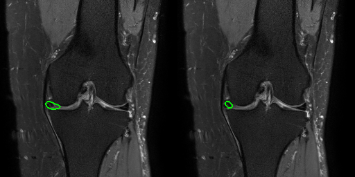
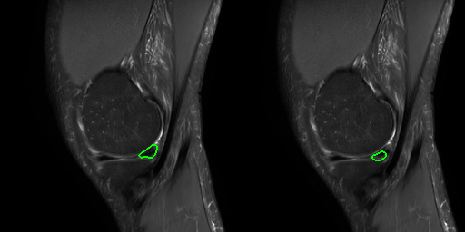
Approach: A total of 391 adult knee MRI scans were annotated in the tear regions of the menisci in coronal and sagittal images as ground truth. Tear detection was performed as a segmentation within the exam in the 679 test dataset.
Results: The accuracy of the coronal model and sagittal model were 0.76 and 0.74, respectively.
Impact: The diagnostic model for meniscus tears on knee magnetic resonance imaging might be useful as a screening tool and diagnostic aid for radiologists but further improvement is required for more accurate detection.
Introduction
Making an accurate diagnosis of meniscus tears from knee magnetic resonance imaging (MRI) is time-consuming and requires specialized knowledge and skills. We develop a 3D/2D deeply supervised U-net model that can make a detection of meniscus tears in coronal and sagittal views of knee MRI scans. The purpose of this study was to evaluate the accuracy of this meniscus tears detection algorithm on knee MRI.Methods
A total of 1070 adult knee MRI scans that were taken at our facility with both coronal and sagittal fat-suppressed proton density-weighted sequences were enrolled in this study. All scans were performed on a 3T scanner (Achieva, Philips Healthcare, Netherlands) with an 8-channel knee coil and had their radiology reports that were written by the board-certified musculoskeletal radiologists. Of these, 391 cases were assigned to training dataset as a ground-truth and the residual 679 cases to test dataset. Three-dimensional (slice-by-slice) segmentation masks were generated to demarcate tear regions of the medial and lateral menisci both in coronal and sagittal volume acquisitions manually and independently based on their radiology report findings. Tears were defined as high signal in the meniscus reaching the articular surface on either coronal or sagittal images. Fraying, blunting, truncation, maceration, or contusion without tear were not considered tear. All annotations were reviewed and finalized by an expert musculoskeletal radiologist with over 30 years of experience. Two separate fully convolutional contracting-expanding (U-Net) models were trained for meniscal tear segmentation on coronal and sagittal volumes, respectively. Each model is implemented as a 19-layer 3D/2D deeply supervised U-Net composed of 106,915 trainable parameters. For any given single slice prediction, the 3D/2D approach utilizes a total of five slices (two adjacent slices before and after) to generate predictions. The model is optimized using deep supervision with auxiliary loss functions at each resolution within the expanding layers defined using a binary cross-entropy loss function with a smoothing value of 0.2. Training is implemented with the Adam optimizer, learning rate of 1e-3, learning rate decay of 0.001 per epoch, batch size of 8, and a total of 30,000 iterations. During inference, a threshold based on total number of positive meniscal tear prediction voxels across both coronal and sagittal volumes is used to determine global presence or absence of tear. Based on this approach, model performance is evaluated using overall sensitivity, specificity, and accuracy.Results
Using only coronal volume datasets, overall model sensitivity and specificity is 0.765 and 0.795, respectively. Using only sagittal volumes datasets, overall model sensitivity and specificity is 0.808 and 0.677, respectively. After aggregating results in both coronal and sagittal volumes, overall model accuracy is 0.76 and 0.74, respectively.Conclusion
The current results were relatively inferior compared to previous reports, which had the sensitivity and specificity in the 80% range1, 2, 3. The combination of the coronal and sagittal model and data augmentation techniques to provide the models with more data would be important to improve the overall model. We anticipate future improvements in model performance may be achieved by additional preprocessing including cropping the large field-of-view MR scan to include only regions containing menisci near the center of the acquisition.Acknowledgements
No acknowledgement found.References
1. Pedoia V, Norman B, Mehany SN, Bucknor MD, Link TM, Majumdar S. 3D convolutional neural networks for detection and severity staging of meniscus and PFJ cartilage morphological degenerative changes in osteoarthritis and anterior cruciate ligament subjects. J Magn Reson Imaging. 2019 Feb;49(2):400-410.2. Fritz B, Marbach G, Civardi F, Fucentese SF, Pfirrmann CWA. Deep convolutional neural network-based detection of meniscus tears: comparison with radiologists and surgery as standard of reference. Skeletal Radiol. 2020 Aug;49(8):1207-1217.3. Shin H, Choi GS, Shon OJ, Kim GB, Chang MC. Development of convolutional neural network model for diagnosing meniscus tear using magnetic resonance image. BMC Musculoskelet Disord. 2022 May 30;23(1):510.Figures