1634
Comparison of lesion detection performance between 3D high-resolution isotropic MRI using MIXTURE and conventional 2D-TSE in knee joints1Radiology, Eastern Chiba Medical Center, Chiba, Japan, 2Faculty of Health Sciences, Institute of Medical, Pharmaceutical and Health Sciences, Kanazawa University, Ishikawa, Japan, 3Philips Japan, Tokyo, Japan, 4Philips Healthcare, Hamburg, Germany, 5Diagnostic Radiology and Radiation Oncology, Graduate School of Medicine, Chiba University, Chiba, Japan, 6Radiology, Chiba University Hospital Comprehensive Radiology Center, Chiba, Japan
Synopsis
Keywords: Cartilage, MSK
Motivation: Cartilage degeneration can be evaluated non-invasively by quantitative T2 mapping MRI. However, clinical knee joint MRI requires multiple contrasts to diagnose various diseases, and T2 mapping for quantitative evaluation must be additionally performed.
Goal(s): To evaluate the lesion detection performance between 2D-TSE and 3D MIXTURE in clinical knee MRI.
Approach: MIXTURE is a 3D TSE that can set arbitrary echo times using the T2 preparation pulses, and enables several image contrasts and T2 mapping by acquiring at least two echo time images.
Results: With regard to the lesion depiction, 3D MIXTURE and conventional 2D TSE were considered comparable with equivalent diagnostic confidence.
Impact: MIXTURE can acquire morphological and quantitative images in a single scan. We evaluated the diagnostic accuracy of the morphological images. We will clarify the diagnostic accuracy of MIXTURE by comparing it with the conventional 2D-TSE method for knee joints.
Introduction
The progression of knee osteoarthritis has a close relation to cartilage degeneration. Cartilage degeneration can be evaluated non-invasively by quantitative T2 mapping MRI1. However, clinical knee joint MRI requires multiple contrasts to diagnose various diseases2-5, and T2 mapping for quantitative evaluation must be additionally performed. Thus, we proposed a new sequence called MIXTURE (Multi-Interleaved X-prepared TSE with inTUitive RElaxometry). MIXTURE is a 3D TSE that can set arbitrary echo times using the T2 preparation pulses, and enables several image contrasts (such as PDW, T2W) and T2 mapping by acquiring at least two echo time images (Fig.1). In a previous study, MIXTURE has demonstrated that it can simultaneously perform anatomic and pathologic evaluations in knee imaging6,7. Although 2D-TSE is commonly used in knee MRI examinations, the lesion detection performance of MIXTURE is unknown. The purpose of this study was to evaluate the lesion detection performance between 2D-TSE and 3D MIXTURE in clinical knee MRI.Methods
Twenty patients with knee joint pain (64.6±14.1 years old, 12 women and 8 men) were examined on 3.0T whole-body clinical systems (Ingenia CX, Philips Healthcare, Best, the Netherland) with a 16ch knee coil. The study was approved by the local IRB. Both conventional 2D TSE and 3D MIXTURE (PDW and fat suppressed T2W) were performed. Conventional 2D TSE consisted of five sequences or scans that were routinely done at our institute for patient knee joint MRI (Fig.2). MIXTURE images were reconstructed by multi-planar reconstruction (MPR), producing 2 mm coronal and axial images, for a total of 6 images. Two radiologists (16 and 6 years of experience, respectively) evaluated all images for the presence of 12 common imaging findings in internal derangements of the knee, subdivided into categories of ligaments, meniscus, bone, synovium, cartilage and tendons. The readers recorded the presence of all 12 imaging findings (Yes/No) and scored the diagnostic confidence of the sequences used (0 = No Confidence, 1 = Low Confidence, 2 = Moderate Confidence, and 3 = High Confidence). Clinical information was not made available to the readers to ensure comparisons were made using imaging findings alone. Equivalent diagnostic confidence, positive percentage agreement (PPA) and negative percentage agreement (NPA), and Cohen's Kappa statistic were calculated for each imaging method.Results and Discussions
The overall diagnostic confidence was found comparable for all six regions between the conventional 2D TSE and 3D MIXURE. While four regions showed equivalently results (diagnostic confidence percentage above 70%) for ligaments, meniscus, bone, and synovium, differences were found for cartilage and tendon (Fig. 3). 3D MIXTURE had a higher PPA and NPA for all regions except synovium, compared to 2D TSE. This is mainly attributed by much thinner slices in 3D imaging, which not only lead to a smaller partial volume effect, but also allow for a smooth visual inspection of the tissue structures through all consecutive slices, particularly useful for ligaments, meniscus, and cartilage. The inter-reader rating by Cohen's Kappa statistic for all regions ranged from 0.247 to 0.485 for 2D TSE and 0.248 to 0.724 for MIXTURE, respectively (Fig. 4). A similar Cohen's Kappa statistic for both methods in most regions indicates good agreement and comparable performance between 3D MIXTURE and conventional 2D TSE. Figure 5 shows representative clinical cases.Conclusion
With regard to the lesion depiction, 3D MIXTURE and conventional 2D TSE were considered comparable with equivalent diagnostic confidence.Acknowledgements
No acknowledgement found.References
1. Xia Y, Farquhar T, Burton‐Wurster N, Ray E, Jelinski LW. Diffusion and relaxation mapping of cartilage‐bone plugs and excised disks using microscopic magnetic resonance imaging. Magn Reson Med. 1994;31(3):273-282. doi:10.1002/mrm.1910310306
2. Bredella MA, Tirman PF, Peterfy CG, et al. Accuracy of T2-weighted fast spin-echo MR imaging with fat saturation in detecting cartilage defects in the knee: comparison with arthroscopy in 130 patients. AJR Am J Roentgenol. 1999 Apr;172(4):1073-80.
3. Cheung LP, Li KC, Hollett MD, et al. Meniscal tears of the knee: accuracy of detection with fast spin-echo MR imaging and arthroscopic correlation in 293 patients. Radiology. 1997 May;203(2):508-12.
4. Mirowitz SA, Shu HH. MR imaging evaluation of knee collateral ligaments and related injuries: Comparison of T1-weighted, T2-weighted, and fat-saturated T2-weighted sequences—correlation with clinical findings. J Magn Reson Imaging. 1994 Sep-Oct;4(5):725-32.
5. Sonin AH, Pensy RA, Mulligan ME, et al. Grading Articular Cartilage of the Knee Using Fast Spin-Echo Proton Density–Weighted MR Imaging Without Fat Suppression. AJR Am J Roentgenol. 2002 Nov;179(5):1159-66.
6. Yoneyama M, Sakai T, Zhang S, et al. MIXTURE: A novel sequence for simultaneous morphological and quantitative imaging based on multi-interleaved 3D turbo-spin echo MRI. Proc Intl Soc Mag Res Med. 2021;29:4203.
7. Sakai T, Yoneyama M, Watanabe A, et al. Simultaneous anatomical, pathological and T2 quantitative knee imaging with 3D submillimeter isotropic resolution using MIXTURE. In: Proc Intl Soc Mag Reson Med. Vol 29. 2021:0845.
Figures
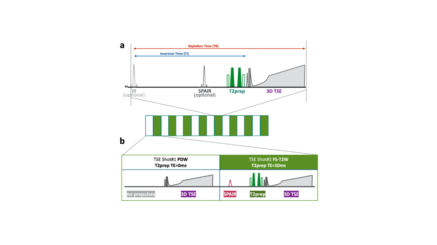
Figure 1. Scheme of the MIXTURE (Multi-Interleaved X-prepared tse with inTUitive RElaxometry).
(a) T2-mapping was performed using T2-prepared 3D segmented turbo spin- echo (TSE) with variable refocusing pulse trains.
(b) Two images with different TE (TE = 0 and 50ms) were acquired with interleaved acquisition. To obtain the compatible contrasts with routine TSE images, TSE shot#1 did not apply any pre-pulses (as “PDW) and shot#2 applied both SPAIR and T2pre (as “fat-suppressed T2W”).
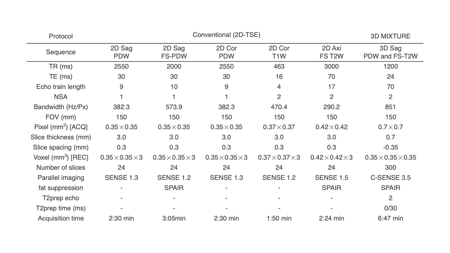
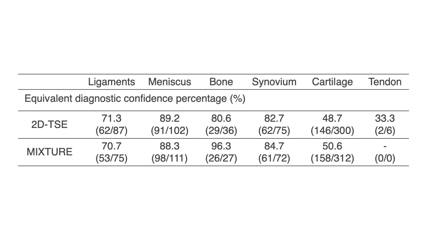
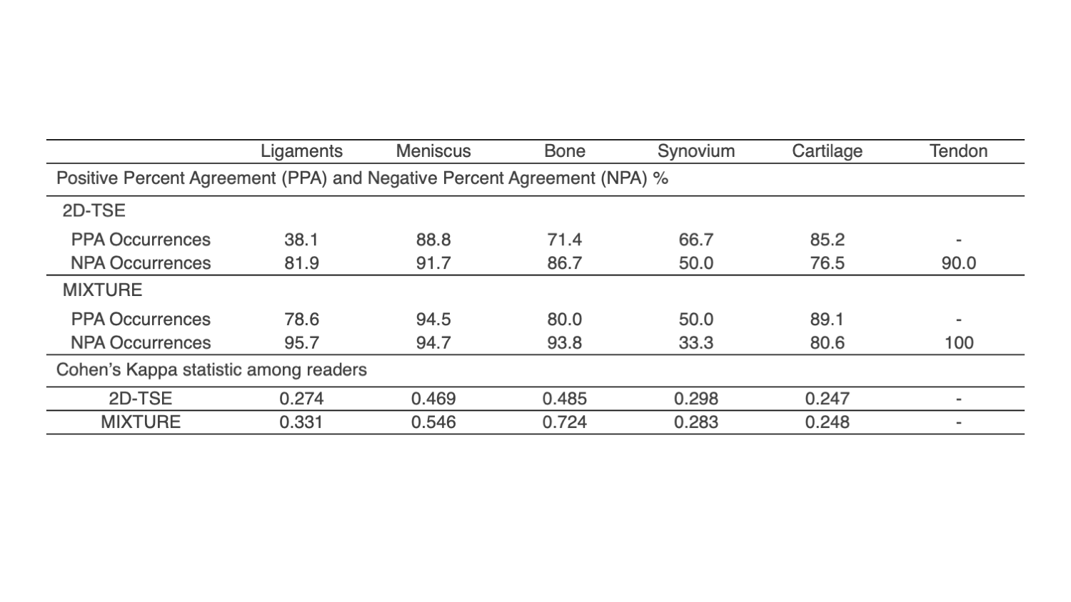
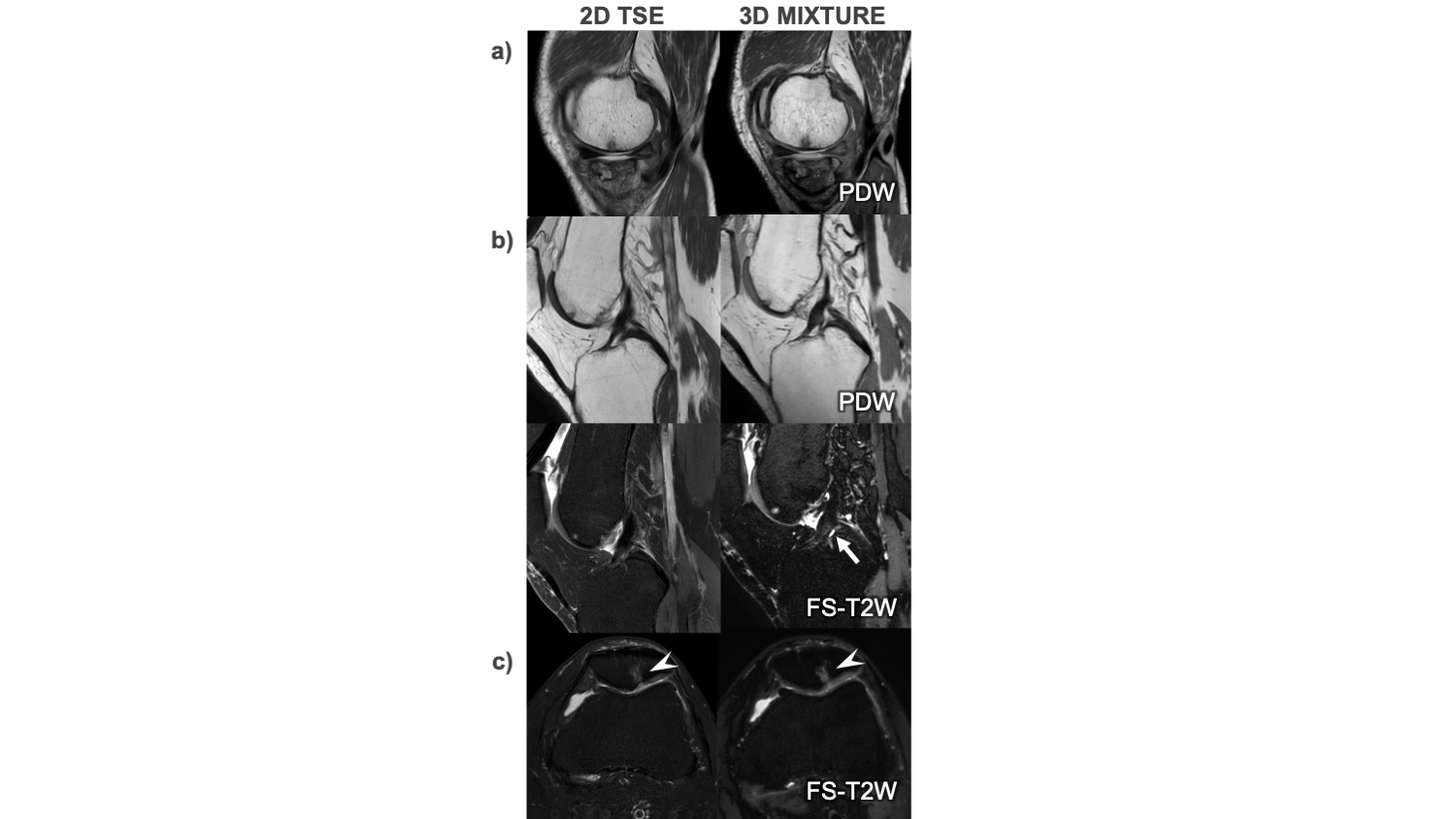
Figure 5. Representative clinical cases of conventional 2D TSE and 3D MIXTURE in knee MRI.
a) Bone marrow edema of the femur and osteonecrosis of the tibia b) Chronic ACL tear was seen in sagittal PDW image from 3D MIXTURE, but not visible in 2D TSE. In the same patient, edema/degeneration of ACL can be seen in FS-T2W images in 3D MIXTURE (arrow) but was unclear in 2D TSE.c) Bone marrow edema of the patella and irregularities of the patellar cartilage (arrow heads)