1633
Evaluating Muscle Health with Multiparametric MRI after FES-Bike Exercise Training in People with Spinal Cord Injury1Istituto di Sistemi e Tecnologie Industriali Intelligenti per il Manifatturiero Avanzato, Consiglio Nazionale delle Ricerche, Milano, Italy, 2Istituto di Tecnologie Biomediche, Consiglio Nazionale delle Ricerche, Segrate, Italy, 3Neuroimaging Unit, Scientific Institute IRCCS “Eugenio Medea”, Bosisio Parini (LC), Italy, 4Department of Mechanical Engineering, Politecnico di Milano, Milano, Italy, 5Department of Electronics, Information and Bioengineering, Politecnico di Milano, Milano, Italy
Synopsis
Keywords: Muscle, Muscle, Multi parametric MRI; Quantitative MRI; Rehabilitation
Motivation: Our research is driven by the need for advanced, non-invasive tools to assess the rehabilitation effectiveness in people with Spinal Cord Injury (SCI).
Goal(s): To determine the effects of Functional Electrical Stimulation (FES)-bike exercise training on muscle health in SCI patients using multiparametric MRI (mpMRI).
Approach: Four males with SCI conducted a 12-week FES-bike exercise training program. mpMRI was used to assess muscle health comprehensively considering muscle volume, fat fraction, diffusion parameters.
Results: Our study demonstrated significant increase in muscle volume and decrease in fat infiltration in the thigh muscles of people with SCI after 12-weeks of FES-bike training.
Impact: Our study highlights the potential of mpMRI for assessing the effectiveness of FES-bike training in people with SCI. This paves the way for more comprehensive evaluation methods, fostering precise monitoring of muscle health during rehabilitation and promoting tailored treatment strategies.
Introduction
Spinal cord injury (SCI) leads to muscle deterioration, weakness, and metabolic dysfunction, that compromise the patients’ quality of life and increase health complications. One way to counteract these effects is Functional Electrical Stimulation (FES), a technique using low level electrical impulses to induce muscle contractions of paralyzed muscles1. FES-bike exercise training, which combines cycling with lower limb stimulation, already demonstrated to improve muscle function and health in people with SCI2. Morphological Magnetic Resonance Imaging (MRI) is commonly used as a non-invasive method to evaluate changes in muscle volume (MVol) and cross-sectional areas (CSA)3, but it does not provide information on other aspects of muscle health, such as tissue composition, perfusion, and microstructure. Multiparametric MRI (mpMRI) is a more comprehensive approach that can assess these aspects of muscle health4,5 and its use, still overlooked in this specific field, can be a valuable tool to investigate muscle changes occurring after FES training. Therefore, the main aim of this work was to evaluate the effect of FES-bike exercise training on muscle health in SCI patients using mpMRI.Methods
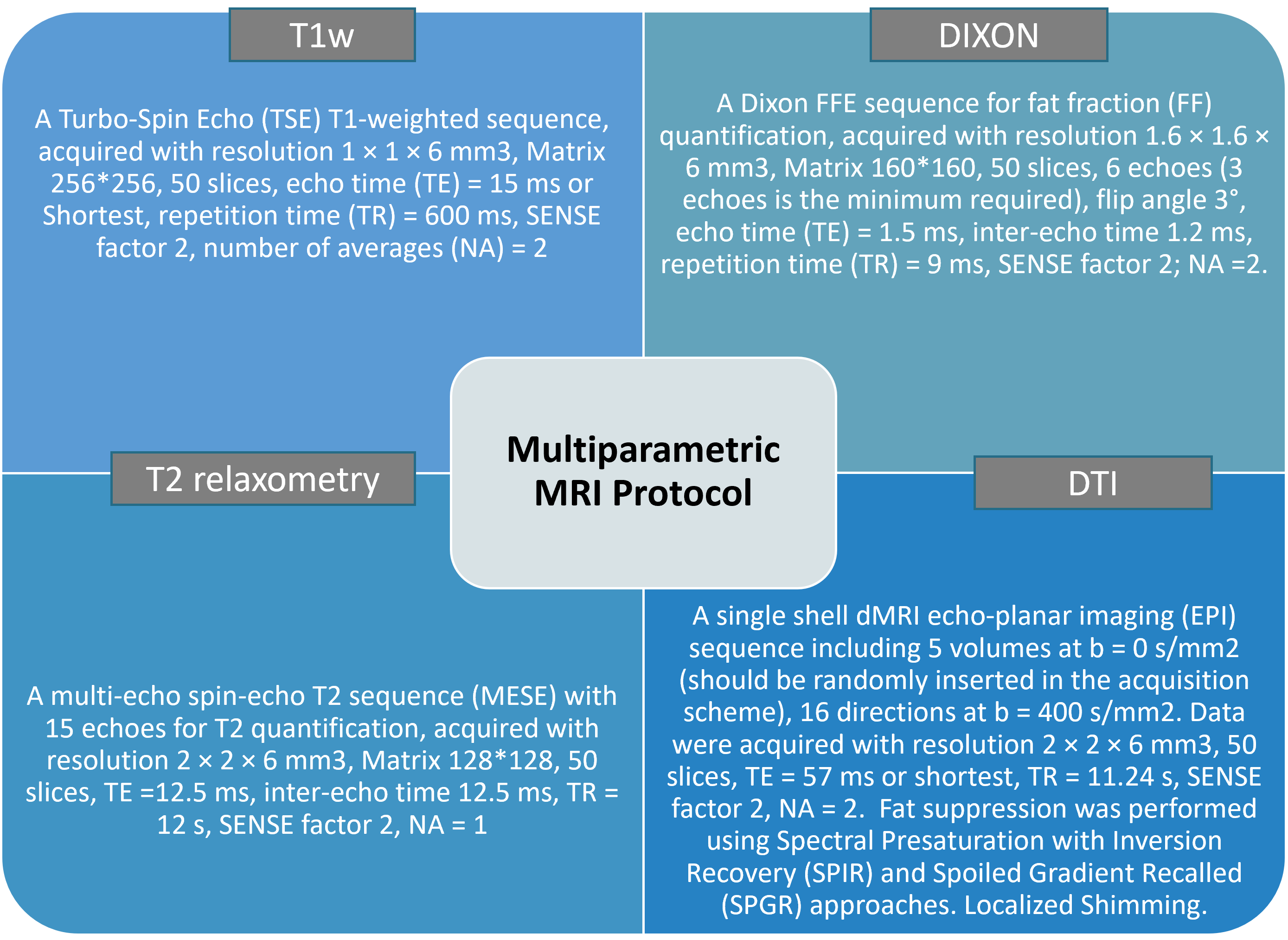
Four male with SCI with complete loss of motor function (age: 30±8 yo; ASIA A; time since injury: 2.7 years) were enrolled in this study. Subjects completed a 12-week FES-bike training of two 30-minute sessions per week cycling at 40 rpm, by stimulating 4 muscles per leg (quadriceps, hamstrings, gluteus maximum and gastrocnemius) with biphasic squared pulses (maximum current amplitude=140mA, stimulation frequency=40 Hz, pulse width=500 ms). The dominant leg thigh muscles were scanned using a 3T scanner (Philips-Achieva) at two time-points: at the beginning (T0) and at the end (T1) of the training program. The MRI protocol included a T1-w Turbo Spin Echo (TSE), a 6 points Fast-Field-Echo (FFE) Dixon, a 15 echoes Multi-Echo-Spin-Echo (MESE) and a diffusion tensor imaging (DTI) sequence with a single shell (b=400 s/mm2) of 16 non-collinear gradient directions. See Figure 1 for more details. Fat Fraction (FF) was estimated from the Dixon Images using a bicompartmental exponential model considering B0 field and T2* relaxation times. T2 relaxation times were estimated from the MESE images using the Extended Phase Graph approach, whereas the DTI parameters (Fractional Anisotropy (FA), Mean Diffusivity (MD), Radial and Axial Diffusivity (RD, AD)) were calculated using Explore DTI tool (https://www.exploredti.com/). ROIs were semiautomatically drawn on T1-w images using Dafne tool (https://dafne.network/) considering the twelve muscles composing the thigh. The MVol, maximum CSA, FF, T2 relaxation time, and DTI parameters were measured and compared between the different time points.Results
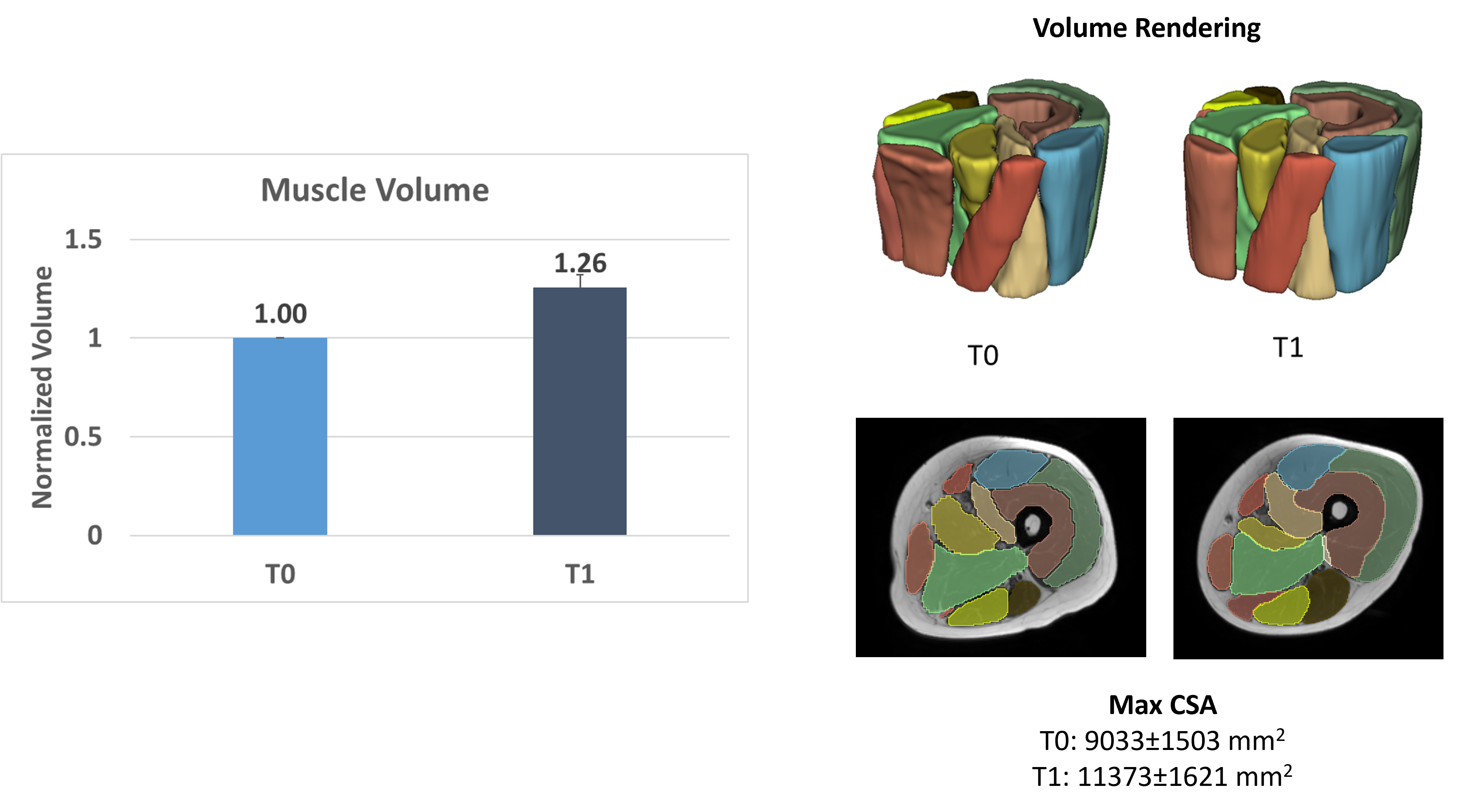
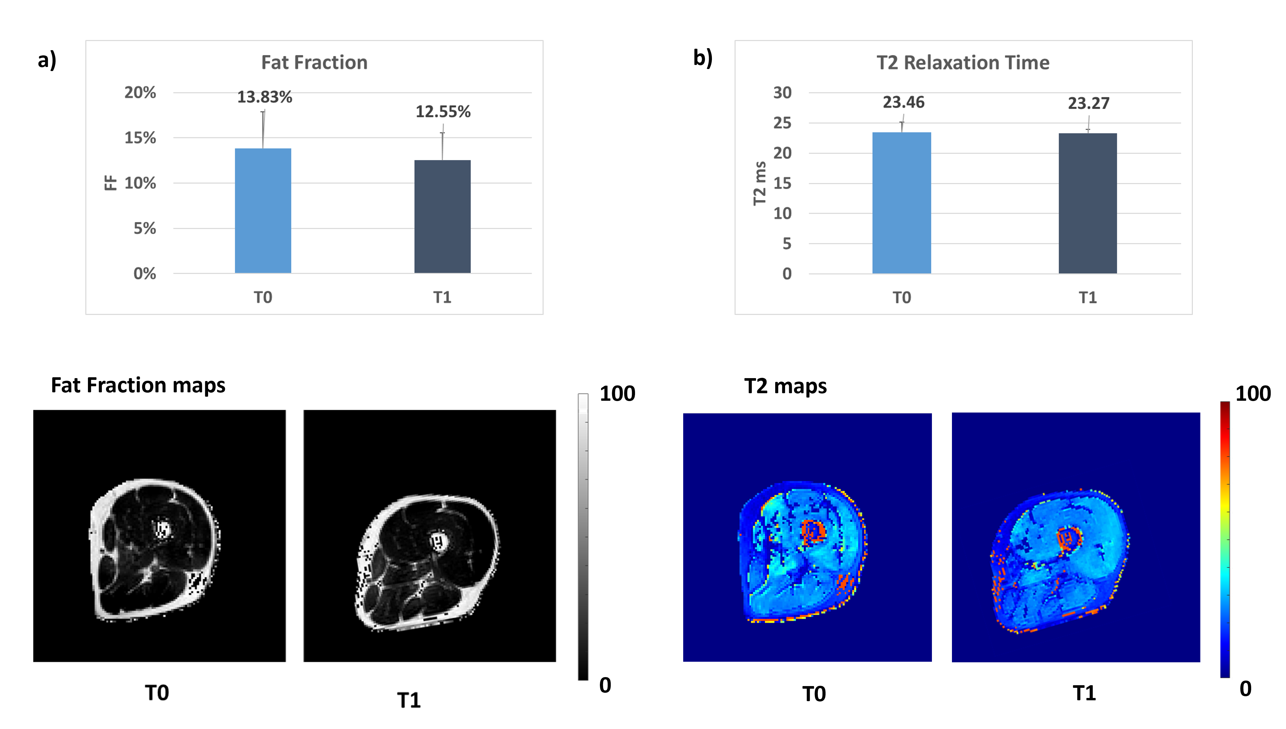
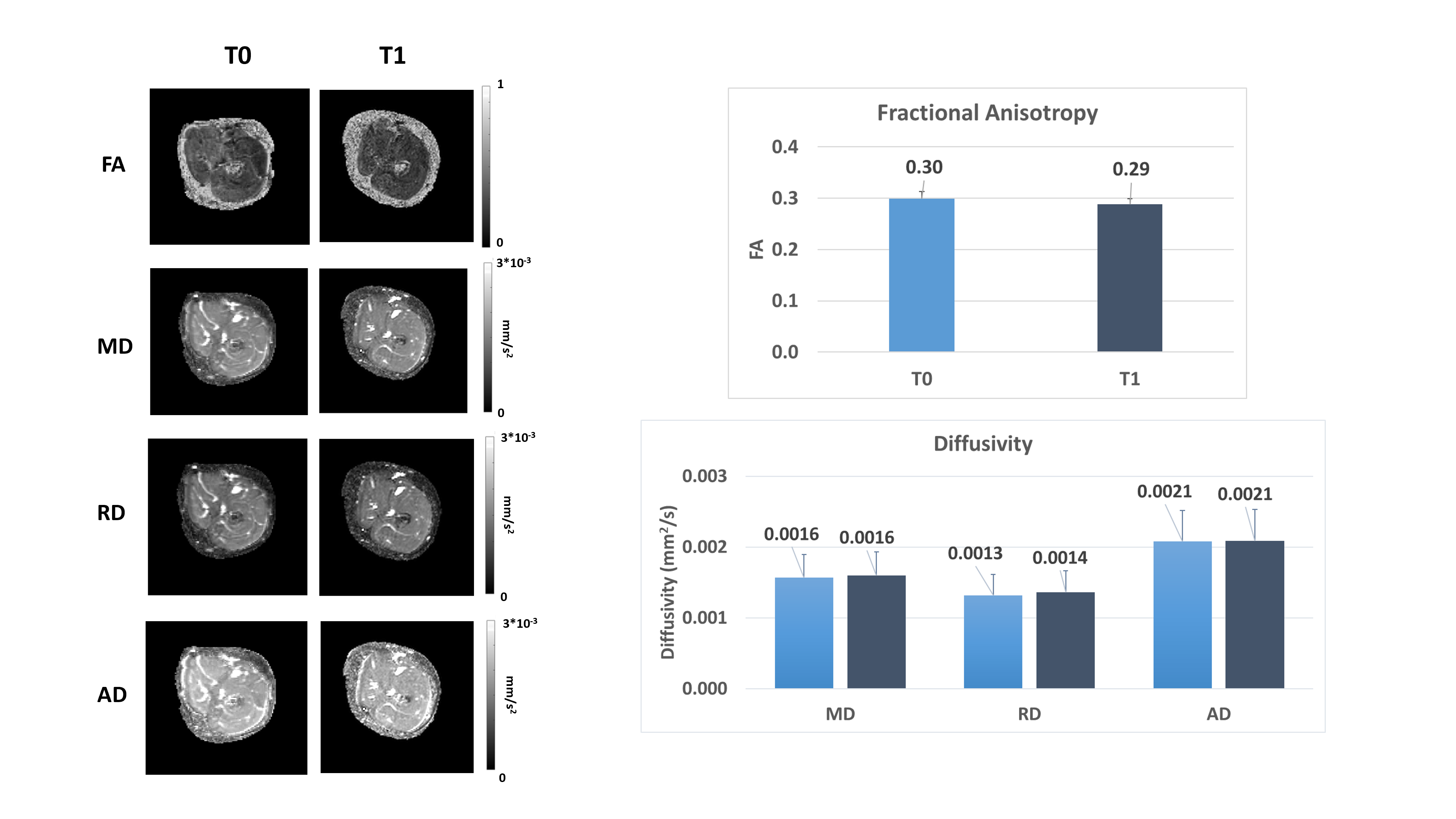
FES-bike exercise training significantly increased the overall muscle volume of about +25% and analogously the maxCSA (T0= 9033±1503 mm2; T1= 11373±1621 mm2) of about 26%, as shown in Figure 2. Similarly, FF, a parameter related to fat infiltration, decreased of about 9% (T0=13.83±4%; T1=12.55±3%), as shown in Figure 3a, in the thigh muscles after 12 weeks of training. Conversely, T2 relaxation time, a parameter related to "disease activity" and edema, remained unchanged after the training (T0=23.45±1.71 ms; T1=23.3±0.63 ms), as shown in Figure 3b. As to DTI parameters, which are related to muscle microstructure, FA showed a slight decrease of about 3.5% (T0=0.30±0.01; T1=0.29±0.01) and RD showed a slight increase of about 8% (T0=0.0013±9*10-5 mm2/s; T1=0.0014±3.8*10-5), whereas MD (T0=0.0016±1.1*10-4 mm2/s; T1=0.0016±4*10-5) and AD (T0=0.0021± 1.3*10-4 mm2/s; T1=0.0021± 7*10-5) didn’t show any relevant changes. Further details are displayed in Figure 4.Discussion
Our findings confirm that FES-bike training induces thigh muscle hypertrophy and reduces fat infiltration in people with SCI. These results align with prior literature, underlining the potential of this rehabilitation strategy for improving muscle health. The unchanged T2 relaxation time suggests that edema was not present in the thigh muscles, even before the initiation of the intervention. Notably, the changes in DTI parameters, specifically the decrease in FA and increase in RD, may indicate a transition from Type I to Type II muscle fibers. However, further confirmation through muscle biopsy is warranted. It is crucial to acknowledge that our study's primary limitation is the small sample size, but the results underscore the potential of mpMRI as a robust tool for assessing muscle health following FES-bike training.Conclusion
Multiparametric MRI offers a comprehensive and informative approach for evaluating muscle health after FES-bike exercise training in people with SCI. This technique allows for precise and non-invasive assessment of muscle volume, fat infiltration, and microstructural changes, providing valuable insights into the effectiveness of rehabilitation strategies. Our research highlights the potential of FES-bike exercise training in preventing muscle atrophy, fat infiltration, and fiber degeneration in individuals with SCI.Acknowledgements
This research was supported by INAIL (Istituto Nazionale per l’assicurazione contro gli Infortuni sul Lavoro, Italy), with PR19-RR-P5 – FESleg project.References
1. Gorgey, A. S., Dolbow, D. R., Dolbow, J. D., Khalil, R. K., & Gater, D. R. (2015). The effects of electrical stimulation on body composition and metabolic profile after spinal cord injury–Part II. The journal of spinal cord medicine, 38(1), 23-37.
2. van der Scheer, J. W., Goosey-Tolfrey, V. L., Valentino, S. E., Davis, G. M., & Ho, C. H. (2021). Functional electrical stimulation cycling exercise after spinal cord injury: a systematic review of health and fitness-related outcomes. Journal of neuroengineering and rehabilitation, 18(1), 1-16.
3. Sadowsky, C. L., Hammond, E. R., Strohl, A. B., Commean, P. K., Eby, S. A., Damiano, D. L., ... & McDonald, J. W. (2013). Lower extremity functional electrical stimulation cycling promotes physical and functional recovery in chronic spinal cord injury. The journal of spinal cord medicine, 36(6), 623-631.
4. Li, K. E., Dortch, R. D., Welch, E. B., Bryant, N. D., Buck, A. K., Towse, T. F., ... & Park, J. H. (2014). Multi‐parametric MRI characterization of healthy human thigh muscles at 3.0 T–relaxation, magnetization transfer, fat/water, and diffusion tensor imaging. NMR in Biomedicine, 27(9), 1070-1084.
5. Berry, D. B., Rodriguez‐Soto, A. E., Englund, E. K., Shahidi, B., Parra, C., Frank, L. R., ... & Ward, S. R. (2020). Multiparametric MRI characterization of level dependent differences in lumbar muscle size, quality, and microstructure. JOR spine, 3(2), e1079.
Figures