1631
Longitudinal Texture Analysis of the Infrapatellar Fat Pad in ACL-Injured Knees1Hospital for Special Surgery, New York, NY, United States, 2University of California, San Francisco, San Francisco, CA, United States
Synopsis
Keywords: Other Musculoskeletal, MSK
Motivation: Anterior cruciate ligament (ACL) injury is associated with development of posttraumatic osteoarthritis and infrapatellar fat pad (IPFP) abnormalities.
Goal(s): Our goal was to quantify texture features of the IPFP in patients with ACL injury (pre- and post-ACLR) and compare to healthy contralateral knees.
Approach: Image texture features were calculated from bilateral knee MRIs acquired at baseline (TP0), 6-months post-ACLR (TP1), and 1-year post-ACLR (TP2) from patients with acute, unilateral ACL tears.
Results: The IPFP of ACL-injured knees had greater texture variability as compared to healthy contralateral knees up to 1-year post-ACLR.
Impact: Texture heterogeneity of the IPFP of ACL-injured knees were higher up to 1-year post-ACLR compared to intact contralateral knees. MRI-based texture analysis can be used to differentiate between injured and contralateral knees.
Introduction
Anterior cruciate ligament (ACL) injury is associated with development of posttraumatic osteoarthritis (PTOA) [1–3]. The mechanisms behind cartilage degeneration and subsequent OA development following ACL injury are multifactorial, involve multiple compartments within the knee, and are not fully understood. The infrapatellar fat pad (IPFP) has been implicated as a source of OA development due to its inflammatory phenotype and high innervation of substance-P containing fibers [4]. Image texture analysis is a quantitative analysis technique that characterizes the spatial variations in pixel intensity within imaged tissues. Previous work has demonstrated that MRI-based IPFP texture features show greater discrimination for knee OA than clinical scores [5]. Further, ACL injury and surgery has been linked to IPFP abnormalities [6,7]. However, it is unclear how MRI-based IPFP texture features may change over time post ACL-injury and reconstruction (ACLR). Therefore, the objectives of this study were to: 1) Quantify texture features of the IPFP in patients with ACL injury pre- and post-ACLR, and 2) Compare texture features between injured and contralateral knees.Methods
This study had IRB approval with informed consent obtained. Bilateral MRI images were acquired at baseline (TP0), 6-months post-ACLR (TP1), and 1-year post-ACLR (TP2) from patients with acute, unilateral ACL tears. This cohort was previously evaluated for morphologic and quantitative changes to the articular cartilage [8]. Patients were treated with 1 of 4 graft types: hamstring tendon, tibialis posterior, bone–patellar tendon–bone (BTB), or Achilles tendon. The full volume of the IPFP in the injured and contralateral knees was automatically segmented using custom semantic segmentation software (Mathworks, Natick, MA) and manually edited as needed using ITK-Snap. Pixel intensity data within the ROIs was processed using maZda software [9] to calculate the gray level co-occurrence matrix (GLCM) to quantify the spatial organization of voxel intensity values. The texture features derived from the GLCM included: entropy (disorder), inverse difference moment (homogeneity), correlation (gray level linear dependence between neighboring pixels), contrast (variability), and angular second moment (homogeneity). Paired t-tests were performed to compare differences in texture features between injured and contralateral knees at each timepoint, and texture features within each knee between timepoints. T-tests were performed to compare texture features between those who received bone–patellar tendon–bone (BTB) grafts vs. soft-tissue grafts at each timepoint. A Bonferroni correction was applied for multiple comparisons.Results
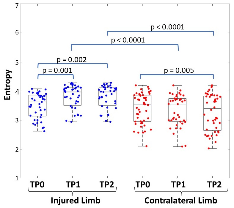
48 patients were included in the analysis (18 female, 30 male; Age: 28.2 ± 11.7 yrs; BMI: 24.5 ± 2.4). Graft tissues included 20 BTB grafts and 28 soft-tissue grafts. ACLR vs Contralateral Knees: At baseline, GLCM texture features did not differ between injured and contralateral knees. At 6 months post-ACLR, the injured knees had higher entropy (+12%, p<0.0001), lower inverse difference moment (-42%, p<0.0001), and lower contrast (-18%, p=0.003). 1-year post-ACLR, injured knees had higher entropy (+15%, p < 0.0001), lower inverse difference moment (-53%, p<0.0001), lower contrast (-13%, p=0.005), and lower angular second moment (-76%, p<0.001). Within ACLR Knees: 6 months post-ACLR, contrast was lower (-23%, p=0.005) but entropy was higher (+7%, p=0.001) as compared to baseline. Between 6 months post-ACLR and 1-year post-ACLR, texture features did not differ within ACLR knees. At 1-year post-ACLR, entropy remained higher (+6%, p=0.002) and correlation was higher (+15%, p = 0.01) compared to baseline. Texture features did not differ among graft types at any timepoint. Within Contralateral Knees: Texture features did not differ between baseline and 6-months or between 6-months to 1-year. Angular second moment was increased (+34%, p=0.001), inverse difference moment was increased (+20%, p=0.003), and entropy was decreased (-6%, p=0.005) 1-year post-ACLR compared to baseline (Figure 1).Discussion
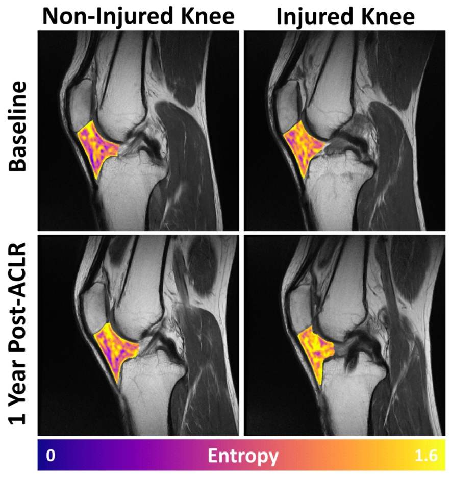
The IPFP of ACL-injured knees had greater texture variability as compared to healthy contralateral knees up to 1-year post-ACLR (Figure 2). These results agree with previous findings that heterogeneous signal alterations of the IPFP indicate pathologic changes [5,10]. Within injured knees, heterogeneity of the IPFP increases 6 months post-ACLR but appeared to stabilize by 1-year post-ACLR. However, even at 1-year post-ACLR the IPFP was more heterogeneous than at baseline, indicating long-term changes. Within healthy contralateral knees, homogeneity appears to increase in the IPFP with time; however, these differences were not apparent until 1-year later. Unexpectedly, the finding of decreased contrast in injured knees is inconsistent with the other results and requires further examination. Our results suggest that IPFP heterogeneity is increased in injured knees post-ACLR and texture analysis can differentiate between injured and contralateral knees.Conclusion
Texture heterogeneity of the IPFP of ACL-injured knees were higher up to 1-year post-ACLR as compared to healthy contralateral knees. Differences of MRI-based texture features exist between injured and contralateral knees.Acknowledgements
The authors would like to acknowledge funding from the Arthritis Foundation and the Arthritis Foundation ACL Consortium who contributed data to this study and the support of Drs. Scott Rodeo, Kimberly Amrami, Aaron Krych, and Benjamin Ma for their clinical contributions to this study.References
1. Shelbourne et al, Sport. Traumatol. Arthrosc 1997; 2. Nakata et al, J. Arthrosc. Relat. Surg. 2008; 3. Friel et al, Clin Sports Med 2013; 4. Dragoo et al, Sport. Med 2012; 5. Li et al, Radiology 2022; 6. Heilmeier et al, Osteoarthr. Cartil. 2020; 7. Murakami et al, Am. J. Sports Med. 1997; 8. Davidson et al., Sports Health 2023, 9. Szczypinski et al., Comp Meth Prog Biomed. 2017; 10. Han et al, Annals Rheumatic Diseases 2016.Figures