1629
Multi-parametric Diffusion MRI and Multiple Linear Regression Predict Regional Spinal Cord Damage1Department of Radiology and Radiological Sciences, Vanderbilt University Medical Center, Nashville, TN, United States, 2Vanderbilt University Institute of Imaging Science, Vanderbilt University Medical Center, Nashville, TN, United States, 3Department of Biomedical Engineering, Vanderbilt University, Nashville, TN, United States
Synopsis
Keywords: Large Animals, Nonhuman Primates, Spinal Cord, Multiple Linear Regression
Motivation: Diffusion MRI provides several quantitative parameters for evaluating structural alterations in spinal cord resulting from injuries.
Goal(s): Our primary objective is to comprehensively assess the sensitivity and specificity of measures derived from both the spherical mean technique and diffusion tensor imaging for assessing regional damage in the cervical spine of non-human primates after a targeted injury to a unilateral dorsal column.
Approach: We acquired diffusion MRI data and obtained silver-stained histological sections for validation.
Results: Our results suggest that diffusion MRI can detect and characterize axonal and cell body damage and assess the severity of spinal cord injury from multiple linear regression.
Impact: SMT and DTI offer sensitive and specific metrics to spinal cord injury (SCI). Vax correlates strongly with histologic assessments of SCI, independent of axonal orientation. Multiple linear regression provides better estimates of tissue damage after SCI than single linear regression.
Purpose
This study aims to delineate regional changes in diffusion parameters obtained from diffusion tensor imaging (DTI) and the multi-compartment microscopic diffusion imaging with the spherical mean technique (SMT) and to assess their sensitivity and specificity for detecting structural changes in cervical spinal cord injury (SCI). Our final goal is to develop a multiple linear regression (MLR) model to be able to predict regional spinal cord damage based on these diffusion measures.Methods
MRI scans of anesthetized squirrel monkeys were conducted at 9.4T (N = 10), before and after a unilateral dorsal column lesion in the cervical spinal cord. The diffusion data were acquired using a spin-echo diffusion sequence with echo-planar imaging readout. Three b-shells with different b values (750, 1000, and 2000 s/mm2) were sampled in 30 directions each. The apparent axonal volume fraction Vax, intrinsic axonal diffusivity Dax, and extra‐axonal transverse diffusivity Dex were derived from SMT. Conventional DTI parameters, including fractional anisotropy (FA), axial diffusivity (AD), radial diffusivity (RD), and mean diffusivity (MD), were also obtained. In histology validations, the regional mean values of lightness (LGT) and fraction of normal tissue (FNT) were extracted from corresponding silver-stained tissue sections. Receiver operating characteristic (ROC) analysis was used to evaluate the sensitivity and specificity by calculating the true positive and false positive rates for different measures, respectively. The detectability for each measure was evaluated by the area under the ROC curve (AUC). Regional correlations were estimated between diffusion and histological measures using the Pearson correlation. MLR employed seven diffusion measures as regressors to predict regional damage detected by histological measures.Results
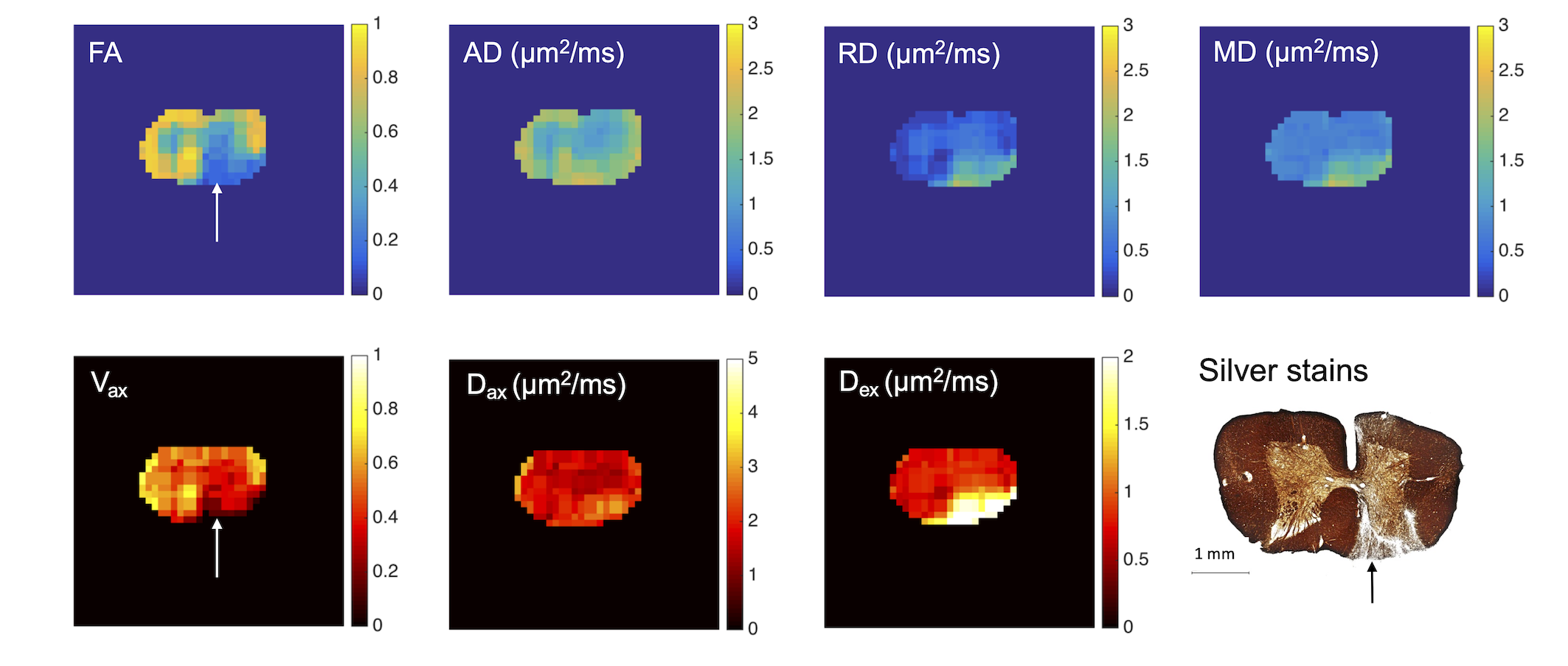
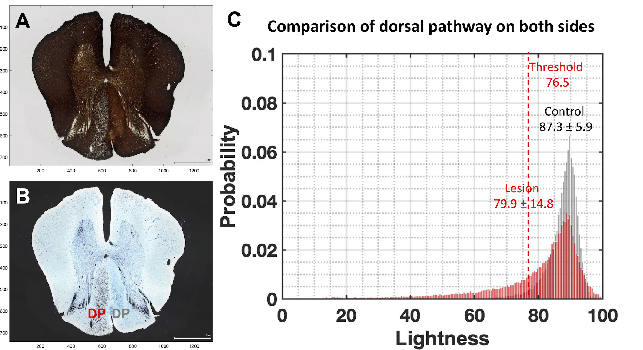
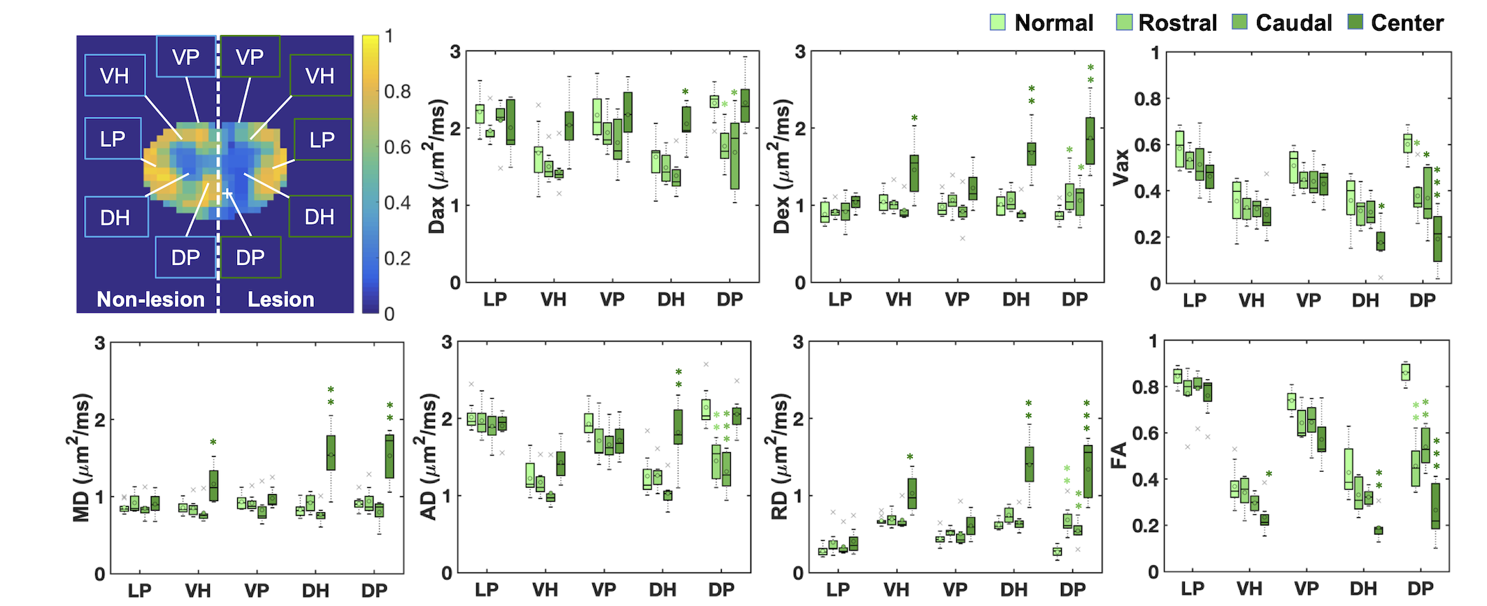
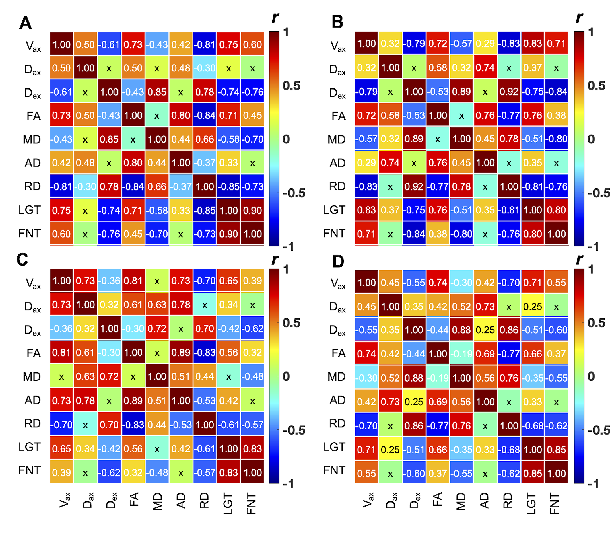
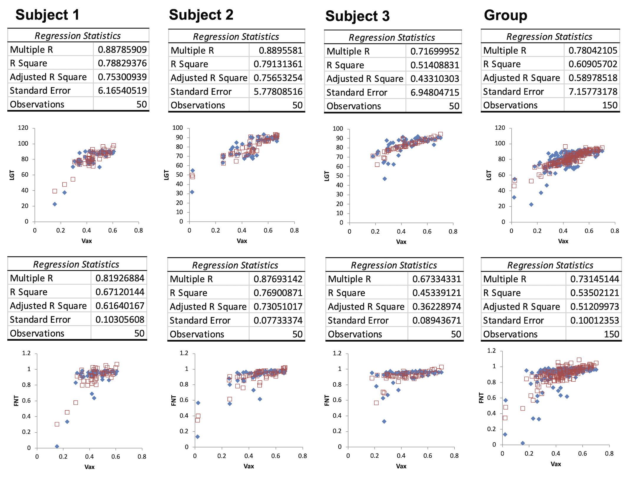
Both DTI and SMT parametric maps effectively differentiated normal white matter, gray matter, and abnormal tissues in the cervical spinal cord following unilateral dorsal column lesions (Fig. 1). The regional tissue damage was validated by histological results (Fig. 1). Figure 2 displays the quantification of histological indices in dorsal pathway (DP). The averaged LGT in DP on the lesion side was significantly lower than that on the non-lesion side (Fig. 2). When comparing the mean and standard deviation (SD) across pixels of LGT in DP, pixels with significantly higher LGT (above the respective regional threshold of LGT) were identified to quantify FNT (Fig. 2). For this histological section, the FNT values for DP on the control and lesion sides were 0.961 and 0.733, respectively, when a threshold of 76.5 (LGT Mean-1.96SD of DP region) was applied. Among SMT and DTI parameters, FA exhibited the highest sensitivity to column lesions (Fig. 3). Vax showed comparable detectability for severe tissue damage at the lesion site (AUC = 1) as FA but showed slightly lower detectability for tissue damage in the dorsal pathway rostral or caudal to the lesion site (AUC = 0.97 and 0.96, respectively). While both Vax and FA were positively correlated with histologic measures, Vax showed significantly higher correlations with LGT and FNT compared to FA (Fig. 4). Although Dex and RD displayed higher sensitivity to SCI than Dax and AD at the lesion site, Dax and AD had higher detectability for damaged tissues in segments rostral and caudal to the lesion. Notably, Dax and AD increased at the lesion site where cysts formed but decreased in the dorsal pathway either rostral or caudal to the lesion site. The regional Dex and RD had a stronger negative correlation with LGT and FNT than Dax and AD. MLR, incorporating seven diffusion parameters as regressors, reliably predicted regional tissue damage following SCI, with higher accuracy for tissue compared to cysts (Fig. 5). MLR provides model that approximates the regional tissue damage assessed by histological indices better than any single diffusion measure (Figs. 4-5), indicated by higher adjusted R2. From the statistics of MLR, Vax and AD provided the most significant contribution to predict LGT, with p-value 8.57 x 10⁻⁴ and 9.83 x 10⁻⁴ respectively using 7 MRI measures as regressors. FA and Vax played the most significant role in predicting FNT, with p-value 8.00 x 10⁻⁵ and 1.15 x 10⁻³ respectively using 7 MRI measures as regressors.Conclusion
Both SMT and DTI offer sensitive measures to detect and characterize axonal and cell body damage post-injury and assess the severity of SCI. SMT derived Vax exhibits the strongest association with histologic assessments LGT and FNT of SCI, suggesting Vax could potentially be used as an imaging biomarker for enhancing SCI characterization independently of axonal orientation.Acknowledgements
We thank Mrs. Chaohui Tang and Mr. Fuxue Xin of the Vanderbilt University Institute of Imaging Science for their assistance in animal preparation and care during MRI data collection. We also thank Dr. Ming Lu and Dr. Xinqiang Yan for customizing coils for cervical spinal cord imaging. This study is supported by DOD grant W81XWH-17-1-0304, and NIH grant NS092961.References
No reference found.Figures