1625
Quantitative assessment of rotator cuff injury using zero echo time magnetic resonance imaging (ZTE-MRI) in combination with Synthetic MRI1Department of Radiology, Taihe Hospital, Hubei University of Medicine, Shiyan 442000, China, shiyan, China, 2Department of Radiology, The Central Hospital Of Wuhan,Wuhan 430000, China, wuhan, China
Synopsis
Keywords: Tendon/Ligament, Bone, Magnetic resonance imaging
Motivation: The use of ZTE-MRI and Synthetic MRI technologies is increasing in musculoskeletal MRI, showing significant clinical symptoms in patients with limited shoulder joint mobility caused by supraspinatus tendon injuries
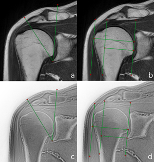
Goal(s): ZTE technology offers superior bone display capabilities, making it ideal for studying the relationship between CSA and AI and rotator cuff injuries
Approach: Prospective clinical study on the combined application of novel magnetic resonance technology for assessing supraspinatus tendon injuries
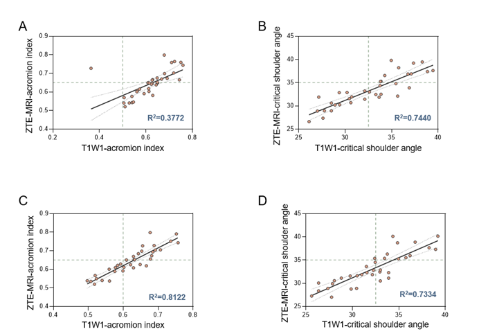
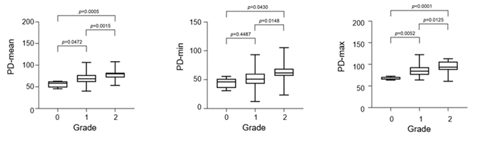
Results: The PLCC indicated a positive correlation between ZTE-MRI and T1WI measurements, with high consistency within and between groups. Boxplot statistics revealed a significant difference in the PD value of Synthetic-MRI quantitative values.
Impact: We used advanced magnetic resonance technology and relevant parameters to assess rotator cuff injury, employing a quantitative approach for classification and providing precise reference indicators for clinical imaging diagnosis and surgical treatment.
Introduction
Shoulder tendon injuries1 can cause pain, decreased function, and limited range of motion. Radiographic findings in patients with rotator cuff injuries include inflammatory edema and structural lesions. Magnetic resonance imaging (MRI) technology provides superior contrast resolution, enabling non-invasive assessment of challenging soft tissue structures that are difficult to assess with traditional radiography or computed tomography (CT). Although conventional qualitative magnetic resonance imaging (MRI) is widely considered the noninvasive gold standard for evaluating rotator cuff tears, it has limited efficacy in evaluating tendinopathy. Rotator cuff injury has a certain impact on Critical shoulder Angle(CSA)2 and Acromial index(AI)3, so the new technology ZTE-MRI 4was used to study the correlation between the angle change and the injured rotator cuff, and the new technology Synthetic MRI 5was used to quantitatively grade rotator cuff injuries.Methods
Patients with suspected rotator cuff injury who underwent MRI examination at Taihe Hospital from September 2022 to July 2023 were prospectively recruited. After applying the inclusion and exclusion criteria, 35 patients were included in the study and scanned using conventional scanning technology and ZTE-MRI combined with Synthetic MRI technology. Two radiologists analyzed and post-processed the images.Results
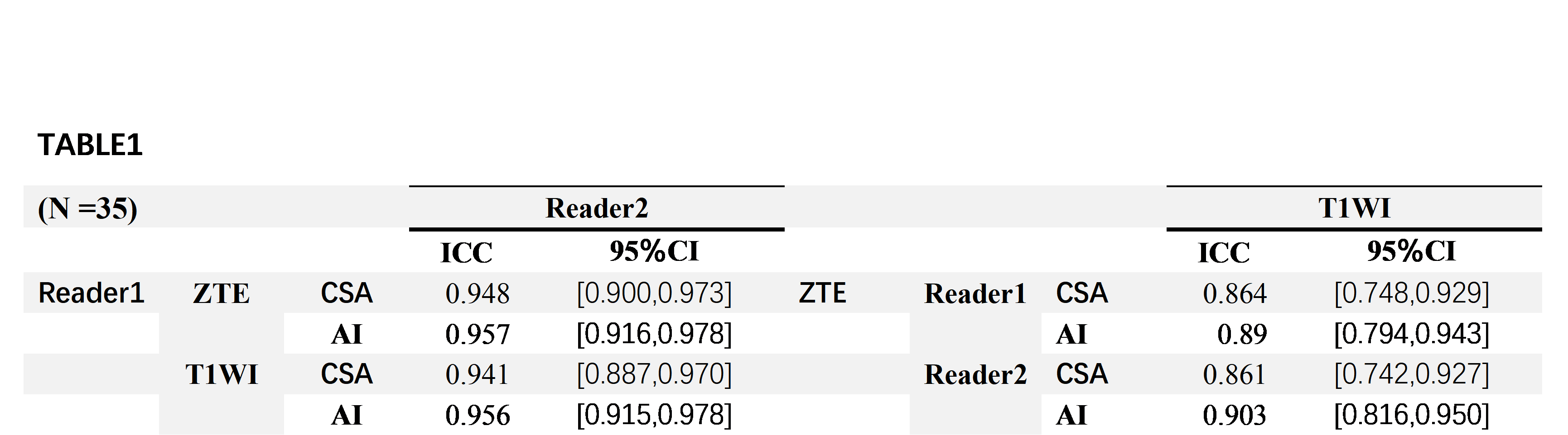
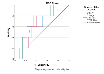
SPSS22 was used for statistical analysis of all data, and the Shapiro–Wilk test was used to test the normality of all measurement data. When the ZTE and T1WI weighted correlation angle measurement parameters met the normality test, the Pearson linear correlation coefficient was used to indicate that the parameters did not meet the normality. The Spearman rank-order correlation coefficient is used to test the consistency, and the inter-group correlation coefficient (ICC) is used to test the consistency between the test raters and within the raters. Spearman rank correlation test was used to calculate the correlation between different levels of different T1, T2, and PD values. In terms of angle evaluation between ZTE-MRI and conventional T1WI sequences: Pearson's linear correlation coefficient shows that there is a positive correlation between ZTE-MRI AI angle and T1WI AI angle measurement (reader1 R2=0.3772, reader2 R2=0.8122, Figure 4A, 4C). The ICC range is 0.861~0.903. Inter-reader agreement ranged from 0.945 to 0.957. (Table 1) Evaluation of ZTE-MRI and conventional T1WI sequence control group and experimental group: The ROC curve in the small sample evaluation showed a certain degree of differentiation between the normal group and the control group, with the ROC range being 0.646~0.711 (Figure 5) Evaluation between Synthetic MR and Zlakin Grading: There is a statistical difference between the quantitative PD value and the grade. The p values in PDmean and PDmax are both less than 0.05. The PD value has certain reference value in the classification of rotator cuff injury grades.Discussion
Magnetic resonance is the gold standard for non-invasive examination of rotator cuff injuries, and its TIWI sequence in routine scans has a high reference value. ZTE-MRI has the advantage of zero echo time imaging and is superior to clinical conventional T1WI sequences in displaying bone cortex. Our research results show that ZTE-MRI and T1WI have a very high consistency in CSA and AI measurements. ZTE- MRI can be used to measure clinical rotator cuff injury indicators to a certain extent. At the same time, in the control group and the experimental group, the area under the curve of CSA in ZTE-MRI was 0.711, which has a good effect. Considering that the small amount of data may cause data bias, it is necessary to increase the amount of data for research. In synthetic MRI, the PD value data is relatively stable and can distinguish the differences between grades very well. Among them, the damage to the lateral subregion and middle subregion of the supraspinatus tendon is more serious than that of the medial subregion. Especially in the area 1cm distal to the supraspinatus muscle with less blood supply, the degeneration is more serious.Conclusion
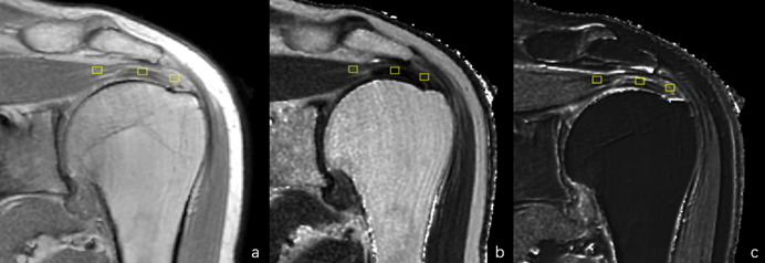
ZTE-MRI has higher resolution than conventional T1WI in bone applications. CSA angle measurement in ZTE-MRI has good accuracy. Synthetic MRI quantitative PD value has certain reference value in grading supraspinatus tendon injury.Acknowledgements
nothingReferences
Murrell, G. A. C.; McInnes, I. B.; Rodeo, S. A. Tendinopathy. Nat. Rev. Dis. Primer 2021, 7 (1), 1. https://doi.org/10.1038/s41572-020-00234-1.
(2) Spiegl, U. J.; Horan, M. P.; Smith, S. W.; Ho, C. P.; Millett, P. J. The Critical Shoulder Angle Is Associated with Rotator Cuff Tears and Shoulder Osteoarthritis and Is Better Assessed with Radiographs over MRI. Knee Surg. Sports Traumatol. Arthrosc. 2016, 24 (7), 2244–2251. https://doi.org/10.1007/s00167-015-3587-7.
(3) Kim, J. R.; Ryu, K. J.; Hong, I. T.; Kim, B. K.; Kim, J. H. Can a High Acromion Index Predict Rotator Cuff Tears? Int. Orthop. 2012, 36 (5), 1019–1024. https://doi.org/10.1007/s00264-012-1499-4.
(4) Fujisaki, A.; Tsukamoto, J.; Narimatsu, H.; Hayashida, Y.; Todoroki, Y.; Hirano, N.; Takeda, K.; Shin, S.; Ota, S.; Anai, K.; Fukumitsu, S.; Yoshimatsu, Y.; Kono, Y.; Ueno, M.; Ide, S.; Murakami, Y.; Aoki, T. Zero Echo Time Magnetic Resonance Imaging; Techniques and Clinical Utility in Musculoskeletal System. J. Magn. Reson. Imaging 2023, jmri.28843. https://doi.org/10.1002/jmri.28843.
(5) Ji, S.; Yang, D.; Lee, J.; Choi, S. H.; Kim, H.; Kang, K. M. Synthetic MRI : Technologies and Applications in Neuroradiology. J. Magn. Reson. Imaging 2022, 55 (4), 1013–1025. https://doi.org/10.1002/jmri.27440.
(6) Aktas, E.; Sahin, B.; Arikan, M.; Ciledag, N.; Buyukcam, F.; Tokgoz, O.; Caglar, E.; Aribas, B. K. MRI Analysis of Coracohumeral Interval Width and Its Relation to Rotator Cuff Tear. Eur. J. Orthop. Surg. Traumatol. 2015, 25 (2), 281–286. https://doi.org/10.1007/s00590-014-1490-4
Figures