1624
Comparison of MRI FRACTURE sequence and CT for three-dimensional bone imaging in patients with shoulder dislocation1Department of Radiology, Sun Yat-sen Memorial Hospital, Sun Yat-sen University, Guangzhou, China, 2Department of Orthopaedics, Sun Yat-sen Memorial Hospital, Sun Yat-sen University, Guangzhou, China, 3Philips Healthcare, Guangzhou, China, 4Philips Healthcare, Shenzhen, China, 5Philips Healthcare, Nanchang, China
Synopsis
Keywords: Whole Joint, Multimodal, FRACTURE, CT, shoulder
Motivation: It is highly desirable to evaluate both soft tissue and bone in a single examination. FRACTURE could be used as a viable auxiliary method of conventional MRI to allow evaluating soft tissue injury and quantifying bone defect simultaneously.
Goal(s): To evaluate the equivalence of FRACTURE and 3D-CT in quantifying bone loss in patients with shoulder dislocation and measuring the morphological parameters of the shoulder.
Approach: Fifty-six patients with shoulder dislocation underwent both MRI (including FRACTURE) and CT scanning. Paired t-test was utilized to assess the difference between them.
Results: There was no statistical difference between the measurements of 3D-CT and FRACTURE.
Impact: FRACTURE, a radiation-free and reliable examination, could serve as an effective auxiliary method to conventional MRI. It allows for simultaneous evaluation of soft tissue injury and accurate quantification of bone defect.
Introduction
Accurate assessment of glenoid defect is crucial for pre-operative planning in patients with shoulder dislocation [1]. In most cases, 3% glenoid defect is less than 1mm. Therefore, the precise imaging modality is essential for the accurate evaluation of glenoid defect. In most clinical settings, patients with shoulder dislocation are commonly required both MRI and CT to assess soft tissue injury and bone defect, respectively. Thus, it is highly desirable to evaluate both soft tissue and bone in a single examination. However typical MRI sequences are limited in the ability to image bone, especially cortical bone and trabecular bone. 3D MRI Fast field echo Resembling A CT Using Restricted Echo-spacing (FRACTURE) sequence could be used as an effective auxiliary method to conventional MRI. It allows for simultaneous evaluation of soft tissue injury and accurate quantification of bone defect. In this study, we aimed to evaluate the equivalence of FRACTURE and 3D-CT in quantifying bone loss in patients with shoulder dislocation and measuring the morphological parameters of the shoulder.Methods
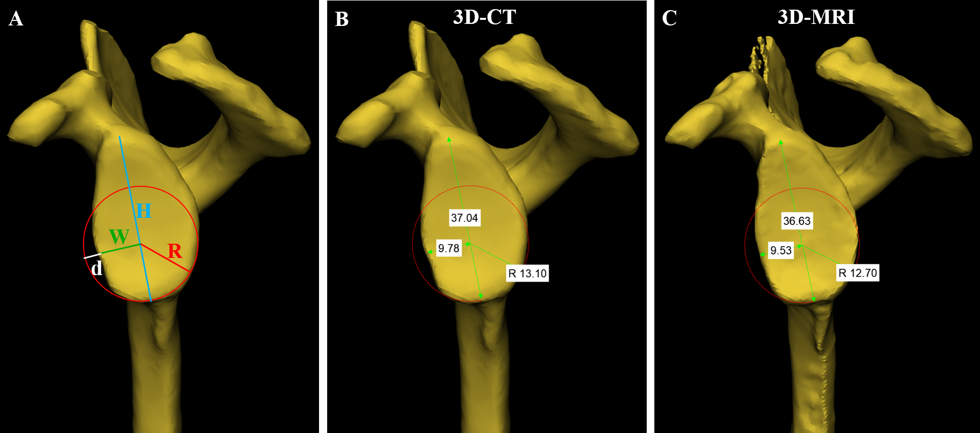
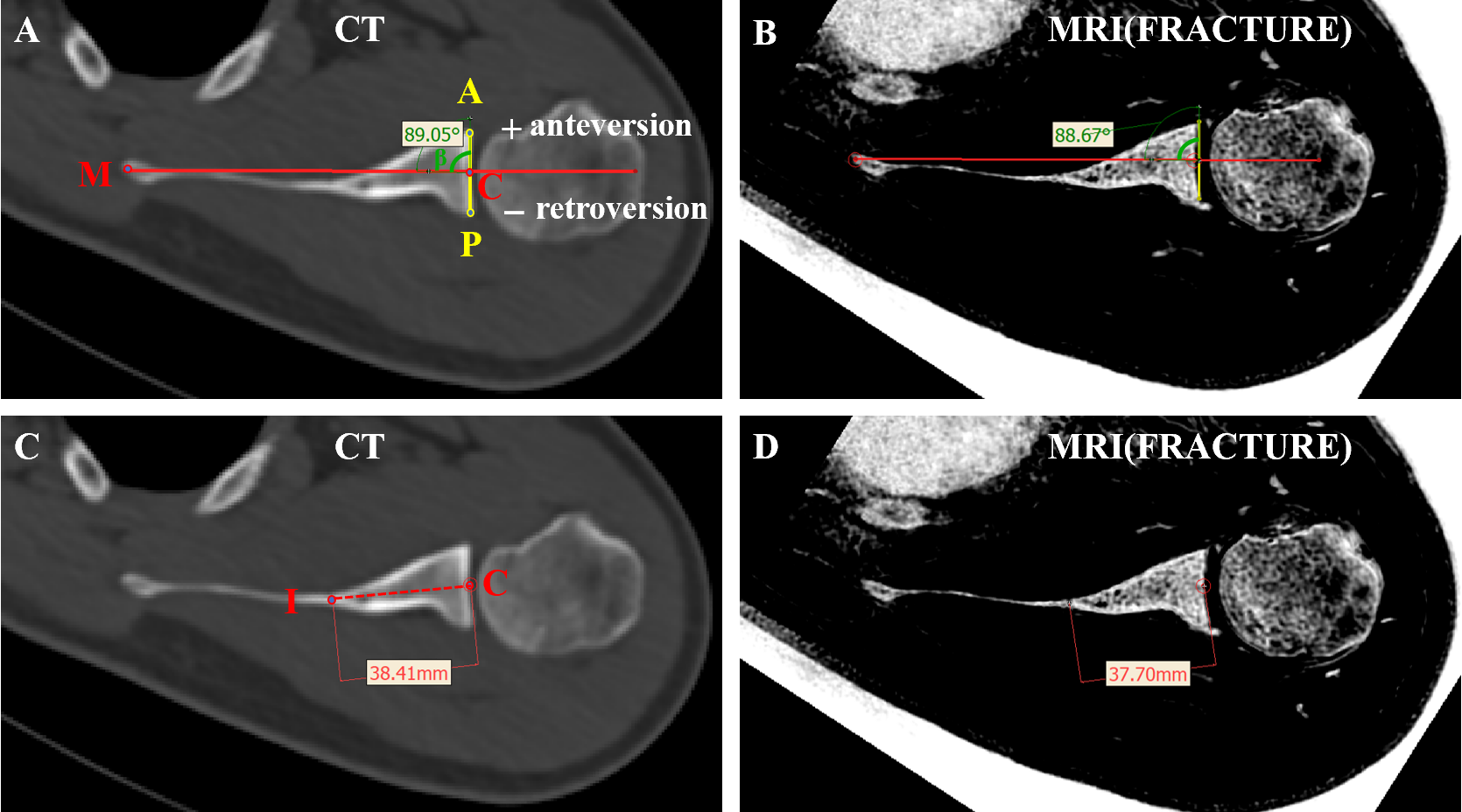
A total of 56 patients with shoulder dislocation (16 bipolar bone defect, 5 only Hill-Sachs lesion, and 35 without bone defect) who underwent both MRI(included FRACTURE) and CT within 1 week were finally enrolled in the study from July 01, 2022 to June 30, 2023. MRI scanning was acquired on a 3T scanner (Ingenia CX, Philips, Best, The Netherlands) with 8-channel shoulder array coil. FRACTURE sequence was acquired with the following parameters: TR,15ms; echoes,4; TE first, in-phase 4.6ms; echo-spacing, 4.6ms; FOV, 180x180mm; voxel size, 0.8x0.8x0.8mm3. For bone defect cases(N=21), two independent observers measured and evaluated glenoid defect, percentage of glenoid defect, glenoid track (GT), Hill-Sachs Interval (HSI), on-track/off-track on the 3D model. For all cases (N=56), two independent observers measured glenoid width, glenoid height, humeral head fitting sphere radius and Critical Shoulder Angle (CSA) on 3D model, while glenoid version and vault depth were measured on axial image. Inter-observer and intra-observer agreement was evaluated by Interclass Correlation Coefficient (ICC). Paired t-test was utilized to assess the difference between CT and FRACTURE. Bland-Altman plot was constructed to evaluate the consistency between CT and FRACTURE. Paired chi-square test and Cohen’s kappa statistic were used for Binary variable(on-track/off-track). In addition, post-processing time and costs of FRACTURE and CT were also recorded.Results
The measurements (glenoid defect, percentage of glenoid defect, GT, HSI) of 21 bone defect cases showed no statistical difference between 3D-CT and 3D-MRI. No significant difference was found between 3D-CT and 3D-MRI in the diagnosis of "on-track/off-track"(P> 0.05), and the diagnostic agreement was perfect (Kappa=1.00, P < 0.05). The measurements (glenoid height, glenoid width, humeral head fitting sphere radius, glenoid version, vault depth, CSA) of all 56 cases revealed no significant difference between 3D-CT and 3D-MRI. For all parameters, the Bland-Altman plots demonstrated weeny measurement error between 3D-CT and 3D-MRI, and the ICCs showed excellent inter-observer and intra-observer consistency. The post-processing time of 3D-MRI was significantly higher than that of 3D-CT (P < 0.05).Discussion & Conclusion
The HSI and GT of bone defect cases were evaluated between 3D-CT and 3D-MRI for the first time, and the diagnosis of "on-track/off-track" was performed. 3D-MRI (FRACTURE) is equivalent to 3D-CT in quantifying bone loss in patients with shoulder dislocation and measuring shoulder morphological parameters. As a radiation-free and reliable examination, FRACTURE could be used as an effective auxiliary method to conventional MRI to allow simultaneously evaluating soft tissue injury and accurately quantifying bone defect. Not only avoiding the radiation exposure risk and additional cost of CT examination, but also simplifying the preoperative process for patients and surgeons. It would be worthy and easy to be popularized in clinical practice.Acknowledgements
No acknowledgement found.References
[1] Kubicka, A.M., Stefaniak, J., Lubiatowski, P. et al. Reliability of measurements performed on two dimensional and three dimensional computed tomography in glenoid assessment for instability. International Orthopaedics (SICOT) 40, 2581–2588 (2016).
[2] Johnson B, Alizai H, Dempsey M. Fast field echo resembling a CT using restricted echo-spacing (FRACTURE): a novel MRI technique with superior bone contrast. Skeletal Radiol. 2021 Aug;50(8):1705-1713.
Figures