1623
White and gray matter abnormalities in active young boxers after sports-related concussions1Lanzhou University Second Hospital, Lanzhou, China, 2GE Healthcare MR Research, Lanzhou, China
Synopsis
Keywords: Traumatic Brain Injury, Traumatic brain injury
Motivation: The neurological effects of active young boxers with sport-related concussions (SRC) are still unclear.
Goal(s): To reveal the micro- and macrostructural changes in active young boxers with SRC.
Approach: Using 3D-T1WI and neurite orientation dispersion and density imaging (NODDI) in young boxers with SRC (n=57) were compared to matched healthy control groups (HCs) (n=72).
Results: In the gray and white matter of concussed boxers, T1WI and NODDI measures showed lower white matter volume, greater CSF and CSP, and significant white matter abnormalities than in the controls.
Impact: This study demonstrated the significant effects of repeated concussions on the micro- and macrostructural in active young boxers, suggesting that we should monitor and maintain the neurological health of these athletes over the long term.
Introduction
Sport-related concussion (SRC) is a major public health concern that affects millions of individuals every year1. Boxing is one of the common types of SRC, and boxers often suffer countless concussions and sub-concussions during their careers, causing great damage to neurological function2. The pattern and extent of damage caused by SRC to the grey and white matter of the brain is unknown. Given the small sample size and inconsistent results of previous studies, this study explores SRC macro - and micro-structure alteration patterns in a larger population of boxers.Methods
57 boxers with a history of SRC and 72 healthy exercise-loving controls with non-SRC (NSRC) were recruited. Athletes were interviewed to determine their years in sports and the number of concussions. All subjects underwent the same Neuropsychological assessment before the MRI scan. All MRI examinations were performed on a 3T MR scanner (SIGNATM Premier; GE Healthcare, Waukesha, WI, USA) with a 48-channel head coil. The NODDI data was obtained with TR = 5,705 ms, TE = 68.8 ms, FOV = 240 mm, matrix = 120 × 120, 78 slices, slice thickness = 2 mm. The NODDI protocol consisted of acquiring diffusion-weighted images along three b-values: twenty directions at b = 1,000 s/mm2, forty directions at b=1,800 s/mm2, and sixty directions at b=2,500 s/mm2. The T1-weighted images with 1-mm isotropic resolution were acquired: FOV = 25.6 × 25.6 cm2, 392 slices, TR/TE = 2632/3 ms, TI = 1000 ms. Using the "recon-all" process in FreeSurfer version 7.1, the cerebral cortex and brain segment volume are reconstructed according to a standardized pipeline. The incidence of cavum septi pellucidi (CSP), the length of CSP and cavum, and the ratio of CSP length to septum length were calculated by two raters independently. NODDI fitting was performed using the CUDA Diffusion Modelling Toolbox (cuDIMOT, https://users.fmrib.ox.ac.uk/~moisesf/cudimot/) running on Graphics Processing Units (GPUs). TBSS was employed to investigate group differences in DTI and NODDI metrics using TFCE and 5,000 permutation testing.Results
Neuropsychological testingAll participants' demographic and clinical characteristics are summarized in Table 1. Compared to the control group, the SRC group performed less well in terms of attention, information processing speed, and language learning ability. There were significant differences in anxiety scores between the two groups.
White Matter Microstructure
Compared with the HCs group, the FA, NDI, and ISOVF of the SRC group were reduced, and MD, AD, and RD values were significantly increased (PFWE < 0.01). NDI decreased widely and significantly in the white matter of SRC group athletes (Fig. 1).
Gray and white matter Volumetric Analyses
Volumetric Analyses of between-group revealed the ratio of gray matter volume to total intracranial volume (TIV) and the ratio of cerebral spinal fluid (CSF) volume to TIV increased, and the ratio of white matter volume to total intracranial volume (TIV) reduced((P < 0.05) (Table 2).
Cavum Septum Pellucidum
The presence of CSP was 57.1% in the SRC group and 38.9% in the HCs group (P=0.04). The length of the CSP and the ratio of CSP length to septum length were significantly greater in boxers with SRC (Table 3).
Discussion
This study found significant abnormalities in micro- and macrostructural in active young boxers with SRC compared to healthy controls. The result was consistent with previous study3, and the white matter abnormalities in boxers were more significant than reported in previous studies, suggesting that repetitive direct contact sport is more damaging to the brain. For boxers with recurrent chronic SRC, the histopathological features of white matter were characterized by axonal degeneration and demyelination, as well as reversible axonal remyelination and irreversible axonal breaks4, which may be associated with diminished NDI. The myelin protein of white matter is a lipid-rich substance that insulates axons and coordinates neural communication functions5. Boxers who suffer repeated concussions may involve multifocal damage to myelin proteins in the subcortical white matter, resulting in diffuse changes and reduced white matter volume. A study of 164 active boxers also found CSP to be present in 49% and to continue to progress at subsequent follow-up6. Formation of CSP may involve shear forces leading to immediate fenestration or secondary injury, cerebral atrophy, and ventricular dilatation leading to progressive formation7. Further studies are needed to investigate the pathologic mechanisms underlying the development of CSP after repetitive head impacts.Conclusion
This study demonstrated the effects of repeated concussions on the micro- and macrostructural in active young boxers with SRC. The brains of active boxers have shown significant changes, suggesting that we should monitor and maintain the neurological health of these athletes over the long term.Acknowledgements
This work was supported by the National Natural Science Foundation of China (No. 81960309), the Gansu Province Clinical Research Center for Functional and Molecular Imaging (No. 21JR7RA438), and the Science and Technology Project of Gansu Province (No. 21JR7RA403).References
1. Gessel LM, Fields SK, Collins CL, Dick RW, Comstock RD. Concussions among United States high school and collegiate athletes. J Athl Train 2007;42:495-503.
2. Hirad AA, Bazarian JJ, Merchant-Borna K, Garcea FE, Mahon BZ. A common neural signature of brain injury in concussion and subconcussion. Science Advances 2019;5.
3. Palacios E, Owen J, Yuh E, et al. The evolution of white matter microstructural changes after mild traumatic brain injury: A longitudinal DTI and NODDI study. Science advances 2020;6:eaaz6892.
4. Armstrong RC, Mierzwa AJ, Marion CM, Sullivan GM. White matter involvement after TBI: Clues to axon and myelin repair capacity. Exp Neurol 2016;275 Pt 3:328-333.
5. Joseph JR, Swallow JS, Wilisey K, et al. Elevated markers of brain injury as a result of clinically asymptomatic high-acceleration head impacts in high-school football athletes. Journal of Neurosurgery 2019;130:1642-1648.
6. Aviv RI, Tomlinson G, Kendall B, Thakkar C, Valentine A. Cavum Septi Pellucidi in Boxers. Can Assoc Radiol J 2010;61:29-32.
7. Hwang J, Kim JE, Kaufman MJ, et al. Enlarged Cavum Septum Pellucidum as a Neurodevelopmental Marker in Adolescent-Onset Opiate Dependence. Plos One 2013;8.
Figures

Table 1 Demographic and clinical characteristics of all participants.
Note: BMI = PCS = Post-Concussion Scale; MoCA = Montreal Cognitive Assessment; PCS = SAS = Hamilton Anxiety Scale; SDS = Hamilton Depression Scale; RAVLT = Rey Auditory Verbal Learning Test; NA = not available; SD = standard deviations; IQR = interquartile range.a two-sample t-test; b chi-square test.
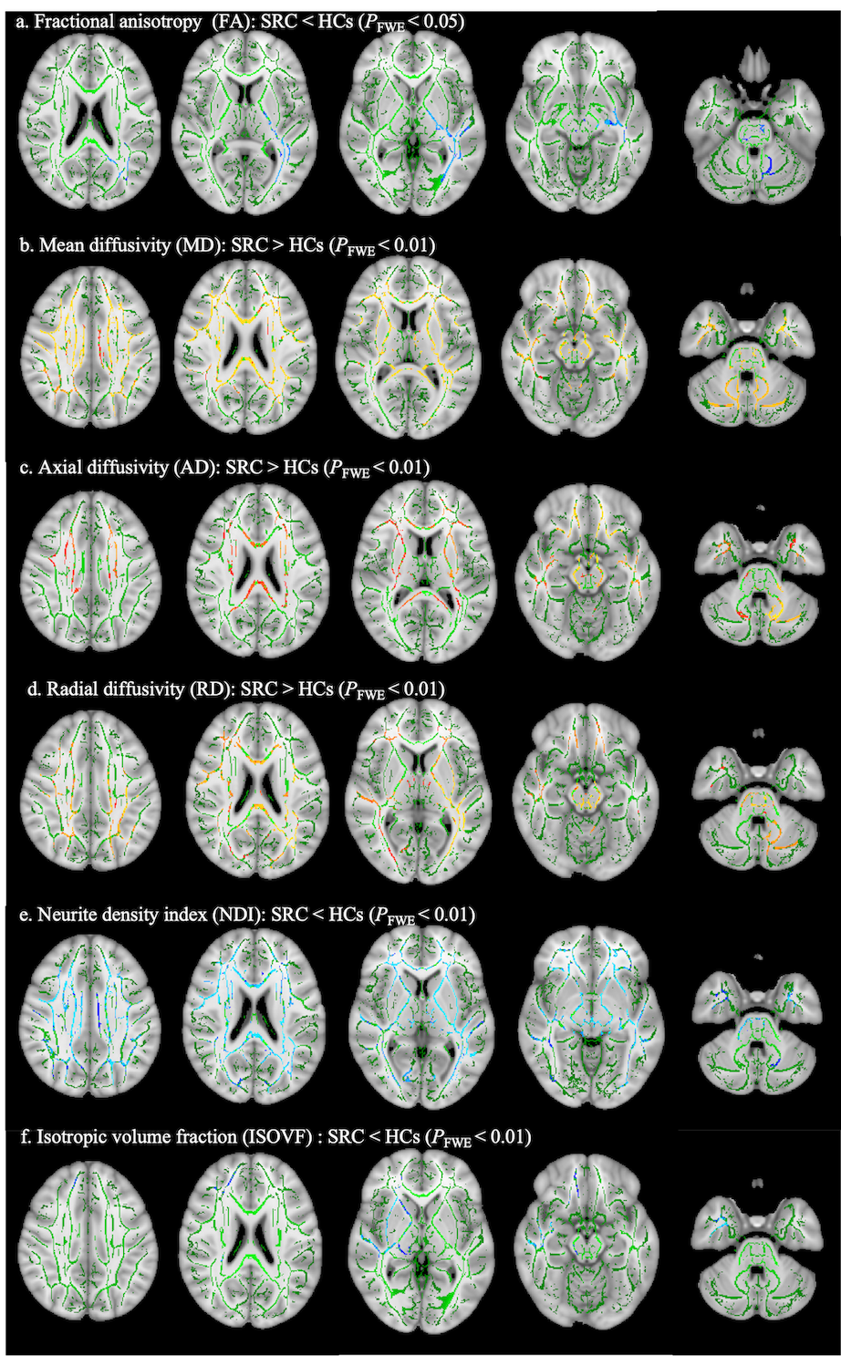

Table 2 Distributions of different MRI measurements of SRC and HCs group.
Note: Data are mean distribution ± standard deviation. TIV = Total intracranial volume. CSF = cerebral spinal fluid.

Table 3 Distributions of different MRI measurements of SRC and HCs group.
Note: Data are mean distribution ± standard deviation. CSP = cavum septi pellucidi. a two-sample t-test; b chi-square test.