1621
Structural connectome disruption and gray matter morphologic changes in active young boxers1Lanzhou University Second Hospital, Lanzhou, China, 2GE Healthcare MR Research, Beijing, China
Synopsis
Keywords: Traumatic Brain Injury, Traumatic brain injury
Motivation: The effects of repeated concussions on the structural covariance network of active young boxers are unknown.
Goal(s): To reveal the effects of sport-related concussion (SRC) on the brain morphology of boxers and to construct a structural covariance network to investigate global and local property changes in SRC networks.
Approach: The cortical thickness and structural covariance network properties were calculated using 3D-T1WI of 57 SRC athletes and 72 healthy controls (HCs).
Results: Compared with the HCs group, the SRC group had a thicker cortex in the bilateral hemisphere, reduced small-world properties, and reduced local efficiency.
Impact: The topological properties of boxers' structural covariance networks were significantly abnormal, indicating that both network integration and separation functions were impaired due to repeated concussions.
Introduction
Previous studies have shown that just one concussion can cause measurable changes in brain morphology1, and boxers can suffer repeated concussions and sub-concussions over the course of their careers, which may lead to changes in the brain's global or local anatomy. In boxing, as the location of the impact point is not fixed and is often accompanied by the occurrence of contrecoup injury, SRC damage to the brain is systemic and whole-brain damage, which is particularly suitable for the study of brain structure and topological property changes by large-scale network method.Methods
ParticipantsOur Institutional Review Board approved the scan protocol and written informed consent was obtained from each subject before the scan. 57 boxers with a history of SRC and 72 healthy exercise-loving controls with non-SRC (NSRC) were recruited.
Neuropsychological testing
All subjects underwent the same Neuropsychological assessment before the MRI scan: (i) Trail Making Tests A (TMTA) and B (TMTB); (ii) the Rey Auditory Verbal Learning Test (RAVLT); and (iii) Forward and Backward Digit-span Test.
Data Acquisition
All MRI examinations were performed on a 3T MR scanner in Premier. The T1-weighted images with 1-mm isotropic resolution were acquired using a 3D MP-RAGE sequence: FOV = 25.6 × 25.6 cm2, 392 slices, TR/TE = 2632/3 ms, TI = 1000 ms.
Post-processing
Using the "recon-all" process in FreeSurfer version 7.1 to reconstruct the cerebral cortex. The average cortical thickness of the Desikan-Killiany template is used to construct the structural covariance network.
Statistical analysis
The cortical thickness was analyzed using a vertex general linear model, corrected using an error discovery rate (FDR). The comparison of global topological metrics between groups was performed by 1000 nonparametric permutation tests, and the comparison of local topological metrics was performed by FDR correction. Statistical significance in the voxel level was set at P < 0.05.
Results
Participant characteristicsThere were no significant differences between the SRC group and control groups in terms of age, sex, and years of education. Compared to the control group, the SRC group performed less well in terms of attention, information processing speed, and language learning ability (Table 1).
Cortical Thickening
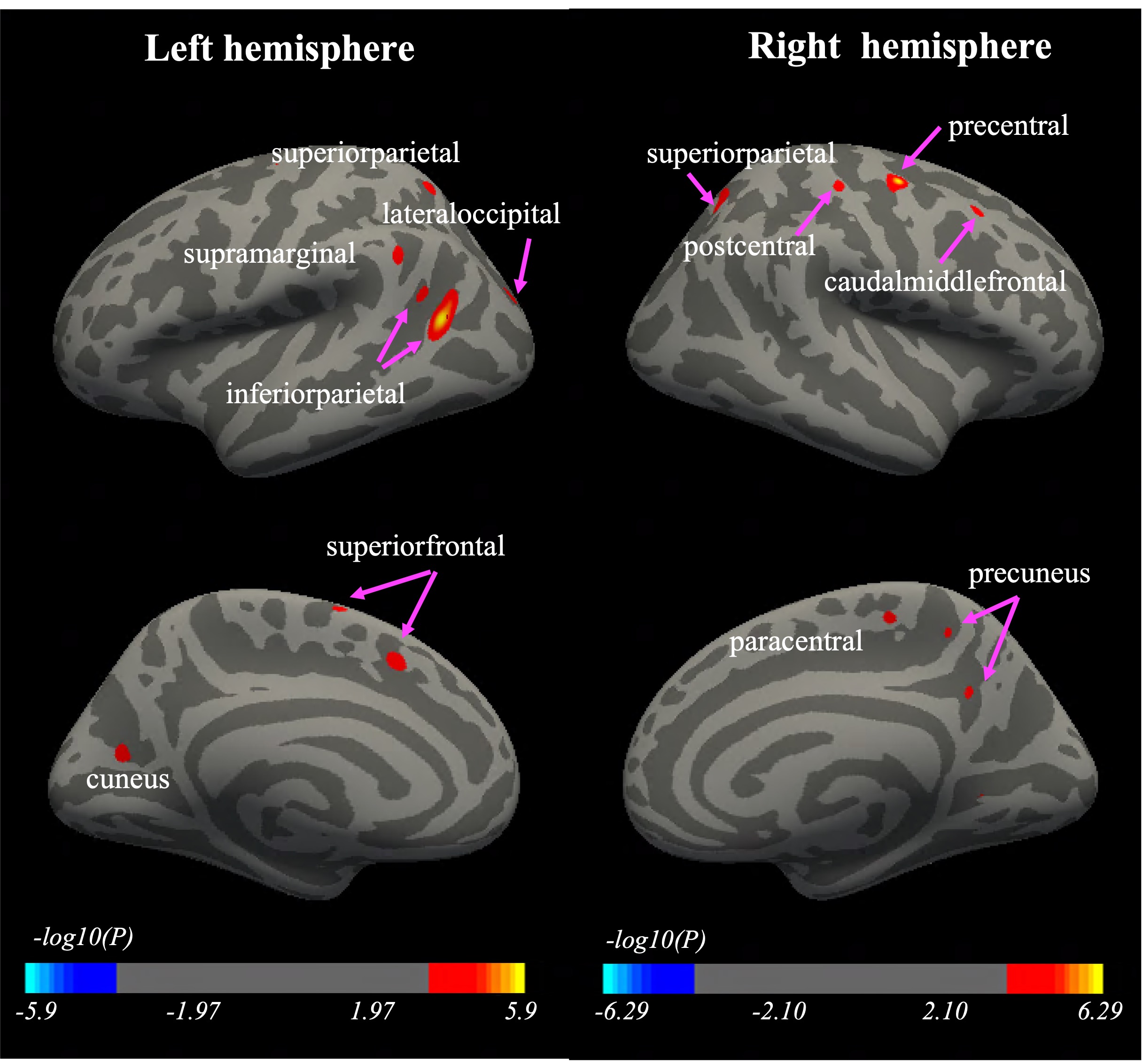
Group comparisons revealed statistically significant cortical thickening in the frontal, parietal, and occipital regions of the bilateral hemisphere youth boxers with SRC compared to the HCs group (Fig. 1). There was no cortical thinning in the SRC group.
Structural covariance network analyses
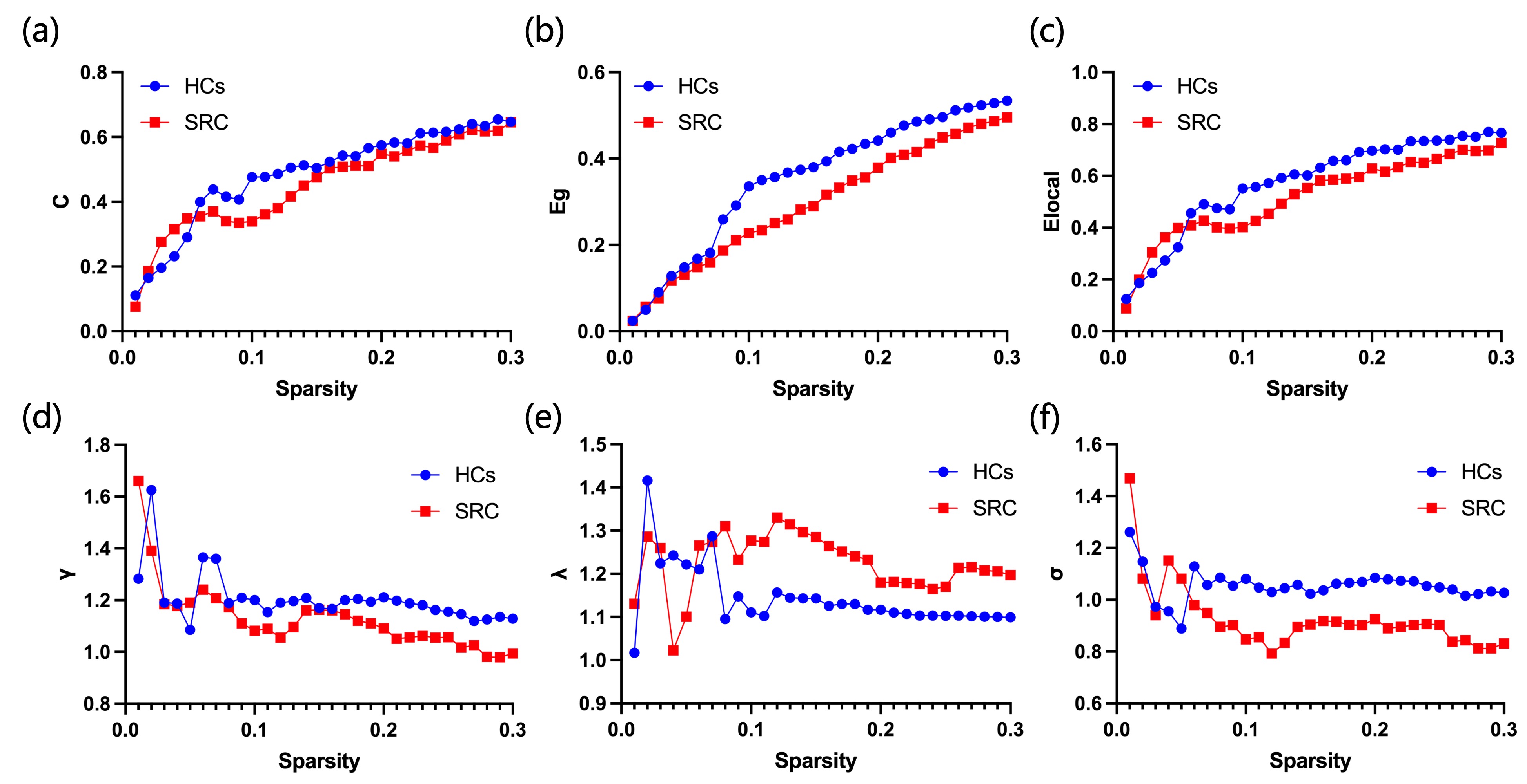
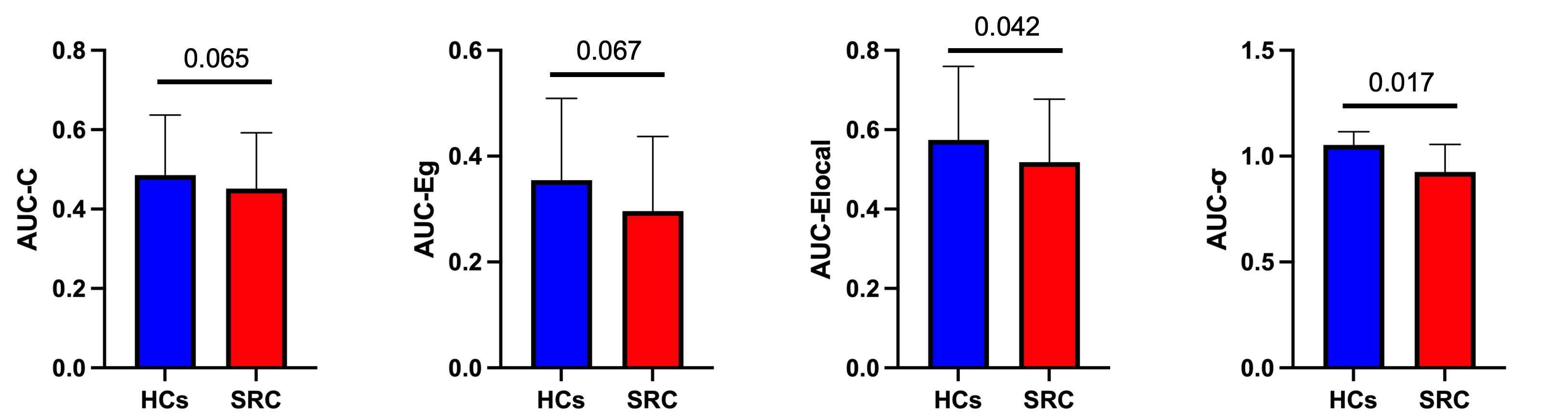
Clustering coefficients and global and local efficiency increased with network density (Fig. 2a–c). Only HCs demonstrated small-world properties between the two groups (σ > 1), and small-world properties were decreased in the SRC group (σ < 1), with a significant difference between groups (P = 0.017) (Fig. 3d). Compared with the HCs group, the SRC group had a reduced local efficiency (Elocal) (P = 0.042) and a trend toward reduced clustering coefficients (C) and global efficiency (Eg) (P = 0.065, P = 0.067) (Fig. 3a–c). There was no significant difference in local topology metrics.
Discussion
In this study, the thickness of the boxer's cortex was increased by quantitative morphological measurement. Previous studies have found evidence of increased cortical thickness in the subacute phase of traumatic brain injury2. Animal studies have found that repeated concussions lead to thicker and denser bones in the skull at the site of impact, highlighting the potential of the skull to adapt and respond to external stimuli3. The results of morphological measurements on the cortex of young boxers in this study suggest that the cerebral cortex of boxers may also undergo unique pathological changes in response to biomechanical shocks after repeated concussions. Significant differences in small-world properties characterize the structural covariance networks of the SRC group and the HCs group. The reduction of small-world characteristics indicates that the boxer's network information processing efficiency is reduced. Previous studies of traumatic brain injury also found that they are characterized by the loss of small-world characteristics4, 5. Compared with the HCs group, the local efficiency of the SRC group is lower, and the clustering coefficient and global efficiency have a downward trend. Given the inverse relationship between path length and local efficiency, the reduced local efficiency in SRC may reflect a decline in within-network communication.Conclusion
Compared with the HCs group, the young boxers had areas of increased cortical thickness in both cerebral hemispheres, which may be pathological changes in the brain in response to repeated concussions. In addition, the small-world properties of SRC groups are destroyed and the local efficiency is reduced. These results indicate that the topology of the boxer covariance network has changed.Acknowledgements
This work was supported by the National Natural Science Foundation of China (No. 81960309), the Gansu Province Clinical Research Center for Functional and Molecular Imaging (No. 21JR7RA438), and the Science and Technology Project of Gansu Province (No. 21JR7RA403).References
1. Sussman D, da Costa L, Chakrayarty MM, Pang EW, Taylor MJ, Dunkley BT. Concussion induces focal and widespread neuromorphological changes. Neuroscience Letters 2017;650:52-59.
2. Ware AL, Goodrich-Hunsaker NJ, Lebel C, et al. Post-Acute Cortical Thickness in Children with Mild Traumatic Brain Injury versus Orthopedic Injury. J Neurotrauma 2020;37:1892-1901.
3. Dill LK, Sims NA, Shad A, et al. Localized, time-dependent responses of rat cranial bone to repeated mild traumatic brain injuries. Sci Rep 2022;12:14175.
4. Pandit AS, Expert P, Lambiotte R, et al. Traumatic brain injury impairs small-world topology. Neurology 2013;80:1826-1833.
5. Imms P, Clemente A, Cook M, et al. The structural connectome in traumatic brain injury: A meta-analysis of graph metrics. Neuroscience and Biobehavioral Reviews 2019;99:128-137.
Figures
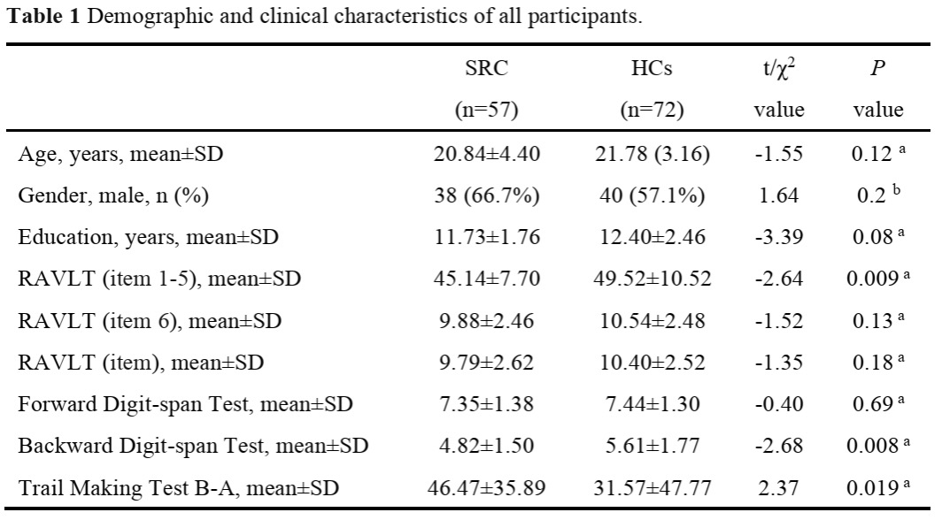
Table 1 Demographic and clinical characteristics of all participants.
Note: RAVLT = Rey Auditory Verbal Learning Test; NA = not available; SD = standard deviations; a two-sample t-test; b chi-square test.