1617
Quantitative UTE MRI of Demyelination in mTBI Mice Subject to Open-field Low-intensity Blast Injury1Radiology, UC San Diego, La Jolla, CA, United States, 2VA San Diego Healthcare System, La Jolla, CA, United States, 3Department of Explosive Engineering, Missouri University of Science and Technology, Rolla, MO, United States, 4Pathology and Anatomical Sciences, University of Missouri, Columbia, MO, United States, 5Bioengineering, UC San Diego, La Jolla, CA, United States
Synopsis
Keywords: Traumatic Brain Injury, Traumatic brain injury
Motivation: mTBI can lead to myelin damage, culminating in substantial cognitive function impairment. Yet, conventional neuroimaging methodologies often fall short in identifying abnormalities in a significant proportion of mTBI instances.
Goal(s): To assess the potential of UTE sequence in detecting demyelination in mTBI mice subject to an open-field LIB injury.
Approach: A new STAIR-UTE sequence was employed to study demyelination in mTBI mice subject to an open-field LIB injury at 3T.
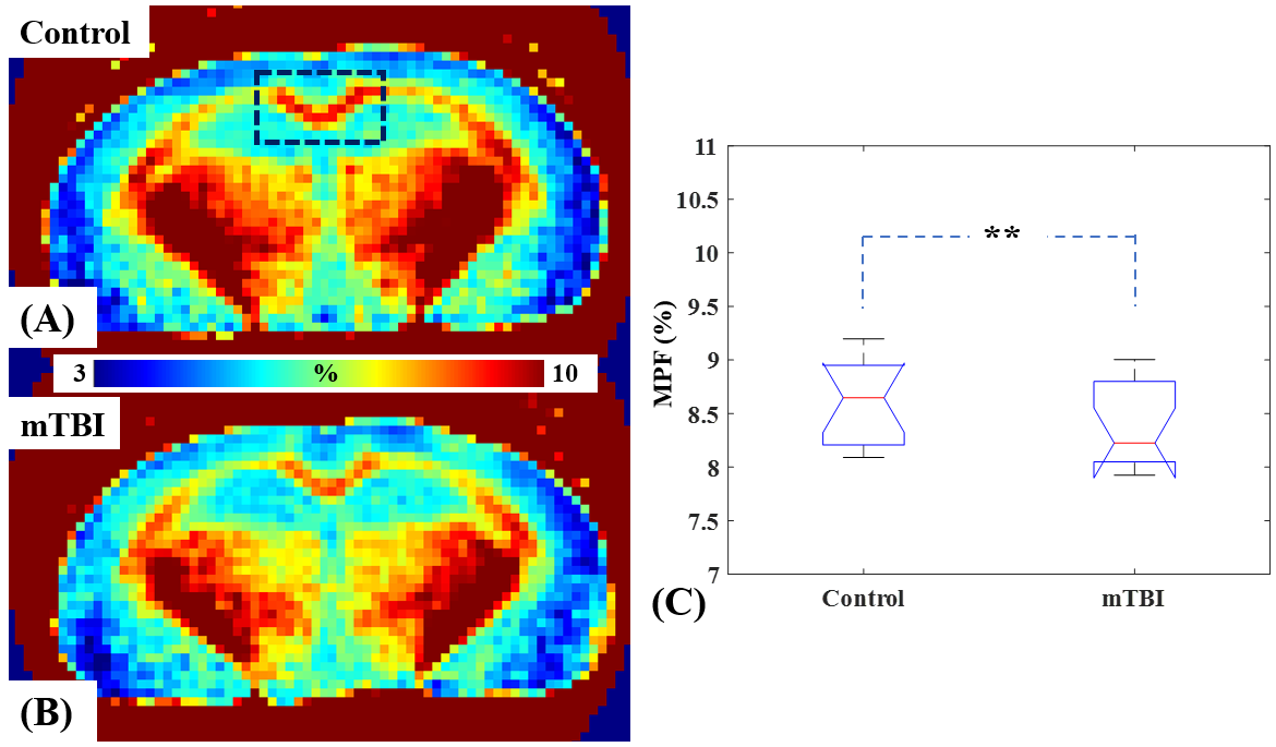
Results: The STAIR-UTE measured MPFs in the corpus callosum region for the mTBI mice are significantly lower (8.5±0.4% vs. 8.8±0.4%; p-value = 0.0055) than those for the control mice.
Impact: STAIR-UTE sequence enables quantitative myelin imaging at 3T MRI, facilitating the detection of demyelination within the mouse brain's white matter following open-field LIB exposure. This innovative STAIR-UTE technique holds significant promise for in vivo mTBI diagnosis and treatment monitoring.
Introduction
Mild traumatic brain injury (mTBI) is a leading cause of long-term disability. The mechanical forces induced by both linear and rotational brain acceleration can result in severe damage in both axon and myelin sheath (1-3). Impairment of myelin can disrupt axonal transport, integrity, and plasticity, leading to a substantial speed reduction in signal transmission (4-6). Given its pivotal role in the development and maintenance of complex cognitive functions, demyelination may play a crucial role in mTBI pathogenesis. However, conventional neuroimaging techniques cannot accurately assess myelin and may not detect abnormalities in most mTBI cases (7-9). Ultrashort echo time (UTE) sequences enable the direct detection of myelin signals (10-15). Recent investigations have revealed myelin sheath abnormalities within corpus callosum (CC) of mice exposed to a low-intensity blast (LIB) in a well-established murine model of mTBI (16). These myelin irregularities were characterized by widespread instances of split layers, pronounced degeneration, myelin ballooning, myelin disruption, or myelin detachment at 7 days post-blast injury (DPI). In addition, these myelin disruptions demonstrated a return to their normal appearance by 30 days DPI, aligning with findings from related studies (16). In this study, we aim to assess the potential of a new 3D short TR adiabatic inversion recovery UTE (STAIR-UTE) sequence in detecting demyelination in mTBI mice subject to an open-field LIB injury (14-16).Methods and Materials
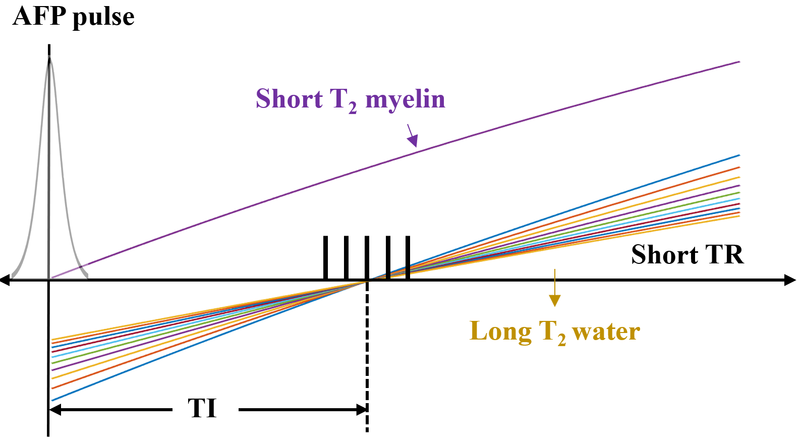
Figure 1 shows the major features of the 3D STAIR-UTE sequence (14,15). In this study, the STAIR-UTE sequence was implemented on a 3T Bruker MRI system equipped with a gradient strength of 450 mT/m strength and a slew rate of 4200 T/m/s. Ensuring robust suppression of long T2 signals necessitates the utilization of a short TR in the STAIR-UTE sequence. Employing a short TR (e.g.,150 ms) guarantees effective suppression of the extensive spectrum of long T2 components with varying T1 values. The optimal TI is determined by numerical simulation (14).The sequence parameters for 3D STAIR-UTE were set as follows: TR/TI=150/64.8ms, TE=0.015/1.6ms, field of view (FOV)=12×12×36mm³, resolution = 167×167×500µm³, flip angle (FA)=40º, number-of-spokes per TR=7, interspoke TR=4.4ms, bandwidth=50kHz, NEX=59, resulting in a total scan time of 5h42min. Additionally, a proton-density weighted UTE (PD-UTE) sequence was acquired in conjunction with STAIR-UTE to facilitate myelin proton fraction (MPF) quantification. The parameters for the proton-density weighted UTE sequence were TR=12ms, TE=0.015/1.6ms, FOV=12×12×36mm³, resolution=167×167×500µm³, FA=2º, bandwidth=50kHz, NEX=6, with a total scan time of 19min.
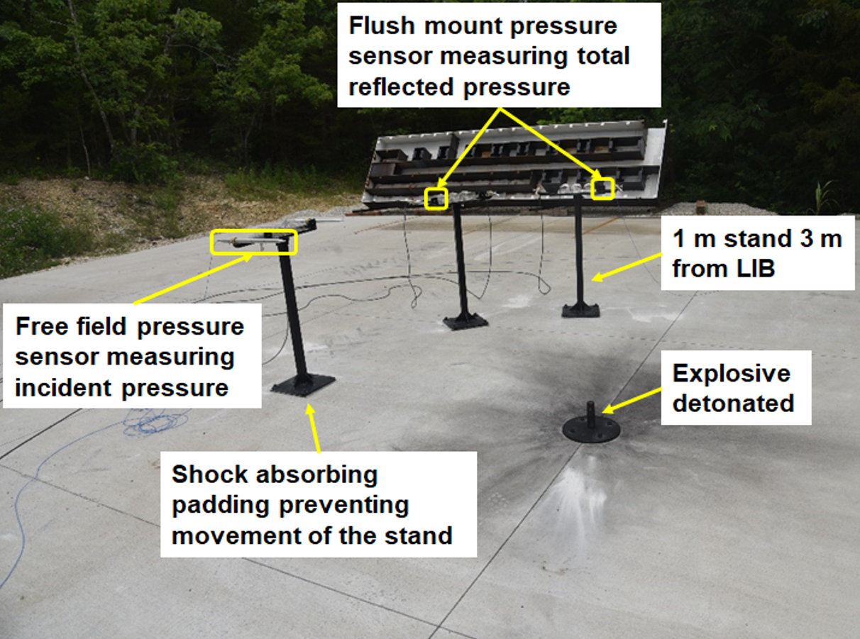
A total of 30 male C57BL/6 mice obtained from Jackson Laboratories, Bar Harbor, ME, and approximately 8 weeks of age, were included in this study in compliance with institutional guidelines. The mice were categorized into two groups, namely, the mTBI group (n=15) and the sham group (n=15). Two mice from each group were excluded from data analysis due to scanner malfunction. The mTBI group was subjected to a highly reproducible open-field LIB murine model as previously described (16). In this model, anesthetized mice were positioned prone, located at a distance of three meters from a 350g high-energy explosive C4 detonation (both 1 meter above ground) (Figure 2). One week following the blast procedures, the animals underwent MRI scanning.
Results and Discussion
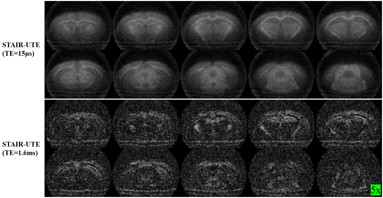
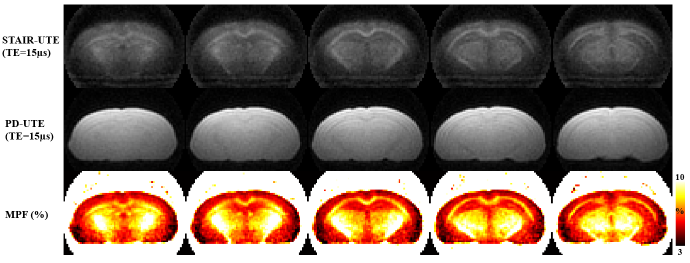
Figure 3 shows the representative STAIR-UTE brain images at TE=0.015ms and TE=1.6ms from an adult control C57BL/6 mouse. The myelin signals in white matter regions are selectively imaged at the first echo but decay to near zero at the second echo, consistent with their short T2* relaxation times.Figure 4 shows the representative STAIR-UTE and PD-UTE images as well as corresponding quantitative MPF maps from an adult control C57BL/6 mouse. The MPF values are much higher in white matter regions (e.g., CC) than those in grey matter regions.
Figure 5 shows representative brain MPF maps from a control mouse and an mTBI mouse as well as the summarized MPF measurements for CC in 13 controls and 13 mTBI. The measured MPF values in the body CC region for the mTBI mice are significantly lower (8.5±0.4% vs. 8.8±0.4%; p-value=0.0055) than those for the control mice.
Our results demonstrated the capability of the 3D STAIR-UTE technique for quantifying demyelination in mice subjected to open-field LIB injury-induced mTBI. Myelin staining for histological validation and behavioral assessments remain to be conducted or summarized to systematically compare the two groups of mice (normal vs. mTBI).
Conclusion
The 3D STAIR-UTE sequence enables quantitative myelin imaging in the murine brain using 3T MRI, facilitating the detection of demyelination within the mouse brain's white matter following open-field LIB exposure. This innovative STAIR-UTE technique holds significant promise for in vivo mTBI diagnosis and treatment monitoring.Acknowledgements
The authors acknowledge grant support from National Institutes of Health (RF1AG075717), VA Research and Development Services (Merit Awards I01CX001388).References
1. Armstrong RC, Mierzwa AJ, Marion CM, Sullivan GM. White matter involvement after TBI: clues to axon and myelin repair capacity. Experimental Neurology 2016; 275:328-333.
2. Jurick SM, Bangen KJ, Evangelista ND, Sanderson-Cimino M, Delano-Wood L, Jak AJ. Advanced neuroimaging to quantify myelin in vivo: application to mild TBI. Brain Inj. 2016; 30:1452-1457.
3. Weber AM, et al. Imaging the role of myelin in concussion. Neuroimaging Clin N Am 2018; 28:83-90.
4. Sinha K, Karimi-Abdolrezaee S, Velumian AA, Fehlings MG. Functional changes in genetically demyelinated spinal cord axons of shiverer mice role of juxtaparanodal Kv1 family K+ channels. Journal of Neurophysiology 2006; 95:1683-1695.
5. Lee Y, Morrison BM, Li Y., et al. Oligodendroglia metabolically support axons and contribute to neurodegeneration. Nature 2012; 487:443-448.
6. Chabas D, Baranzini SE, Mitchell D, Bernard CC, Rittling SR, Denhardt DT, Sobel RA, Lock C, Karpuj M, Pedotti R, Heller R, Oksenberg JR, Steinman L. The influence of the proinflammatory cytokine, osteopontin, on autoimmune demyelinating disease. Science 2001; 294:1731-1735.
7. Bigler ED, Orrison WW. Neuroimaging in sports-related brain injury. Eds. Lovell MR, Echemendia RJ, Barth JT, Collins MW. Pp. 71-94. Lisse, Netherlands: Swets and Zeitlinger 2004.
8. Kirkwood WM, Yeates KO, Wilson PE. Pediatric sport-related concussion: a review of the clinical management of an oft-neglected population. Pediatrics 2006; 117:1359-1371.
9. Johnston KM, Ptito A, Chankowsky J, et al. New frontiers in diagnostic imaging in concussive head injury. Clin J Sport Med 2001; 11:166-175.
10. Horch RA, Gore JC, Does MD. Origins of the ultrashort T2 1H NMR signals in myelinated nerve: a direct measure of myelin content? Magn Reson Med 2011; 66:24-31.
11. Wilhelm MJ, Ong HH, Wehrli SL, Li C, Tsai PH, Hackney DB, Wehrli FW. Direct magnetic resonance detection of myelin and prospects for quantitative imaging of myelin density. Proc Natl Acad Sci USA 2012; 109:9605-9610.
12. Du J, Ma G, Li S, Carl M, Szeverenyi N, VandenBerg S, Corey-Bloom J, Bydder GM. Ultrashort TE echo time (UTE) magnetic resonance imaging of the short T2 components in white matter of the brain using a clinical 3T scanner. NeuroImage 2013; 87C:32-41. PMID: 24188809.
13. Weiger M, Froidevaux R, Baadsvik EL, Brunner DO, Rosler MB, Pruessmann KP. Advances in MRI of the myelin bilayer. Neuroimage 2020; 217:116888.
14. Ma Y, Jang H, Wei Z, Cai Z, Xue Y, Chang EY, Bydder GM, Corey-Bloom J, Du J. Myelin Imaging in Human Brain Using a Short Repetition Time Adiabatic Inversion Recovery Prepared Ultrashort Echo Time (STAIR-UTE) MRI Sequence in Multiple Sclerosis. Radiology 2020; 297:392-404.
15. Ma YJ, Jang H, Wei Z, Wu M, Chang EY, Corey-Bloom J, Bydder GM, Du J. Brain ultrashort T2 component imaging using a short TR adiabatic inversion recovery prepared dual-echo ultrashort TE sequence with complex echo subtraction (STAIR-dUTE-ES). JMR 2021;323:106898.
16. Song H, Konan LM, Cui J, Johnson CE, et al. Ultrastructural brain abnormalities and associated behavioral changes in mice after low-intensity blast exposure. Behavioural Brain Research 2018; 347:148-157.
Figures