1616
In vivo Detection of Ball Heading-Associated Microstructure Changes at the Sulcal Fundi and Gyral Crowns in Amateur Soccer Players1Albert Einstein College of Medicine, Bronx, NY, United States, 2Columbia University, New York, NY, United States
Synopsis
Keywords: Traumatic Brain Injury, Traumatic brain injury, repetitive head impacts
Motivation: Neurodegenerative changes in the cortical sulci and gyri have been associated with repetitive head impacts (RHI) but have only been observed post-mortem.
Goal(s): To detect sulcal and gyral extrema white matter microstructure changes in vivo.
Approach: Automated image-analysis algorithms were used to average the fractional anisotropy (FA) of sulcal fundi and gyral crowns in a cohort of amateur adult soccer players and age- and sex- matched non-collision amateur athletes.
Results: We found soccer players’ FA was significantly lower within the fundi and crowns of the frontal, orbitofrontal, temporal regions, as well as the cingulate and parietal regions for fundi only.
Impact: Here we report the first in vivo imaging study demonstrating white matter microstructure changes within sulci and gyri due to RHI. We anticipate our approach to be a starting point for more sophisticated use of DTI to study RHI effects.
INTRODUCTION
Repetitive head impacts (RHI) are associated with cognitive decline1, and changes within the deep white matter microstructure2. Finite element modelling of RHI found that the depths of the cortical sulci received a high-strain rate deformation response3. Postmortem evidence of hyperphosphorylated tau deposition localized to the cortical sulci are hallmarks of Chronic Traumatic Encephalopathy (CTE)4 and have been associated with a history of RHI4. CTE pathology within the sulcal fundi has been observed post-mortem in young athletes exposed to repetitive head impacts5. A highly significant relationship of sulcal FA from DTI with pathologic evidence of axon disruption was demonstrated with ex vivo post-mortem MRI in American Football players6. Here we report the first in vivo magnetic resonance imaging evidence of RHI-related white matter microstructure changes at the sulcal fundi and gyral crowns of adult amateur soccer players.METHODS
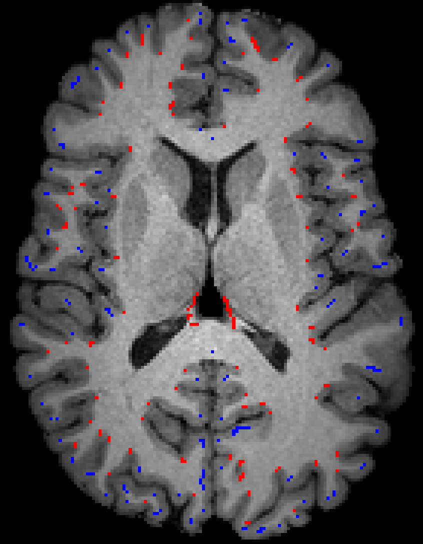
Healthy amateur adult soccer players and healthy non-collision athlete controls with no history of RHI were included in this analysis. Study procedures have been reported in detail2,7-10. In brief, amateur soccer players were recruited from the New York City metropolitan area. A validated, structured, computer-administered questionnaire (HeadCount11) was used to estimate soccer heading exposure over the prior year. Imaging was performed using a 3.0T Philips Achieva TX scanner and a 32-channel head coil. T1-weighted 3D magnetization-prepared rapid acquisition of gradient echo imaging was performed with TR/TE/TI = 9.9/4.6/900 ms, flip angle 8°, 1 mm3 isotropic resolution, 240 × 188 × 220 matrix. DTI was performed using 2D single-shot EPI with 32 diffusion encoding directions, b-value = 800 s/mm2, TR = 10 s, TE = 65 ms, 2 mm3 isotropic resolution, 128 × 120 matrix, 70 slices. Automated image analysis algorithms12 were used to extract fractional anisotropy (FA), separately for each player’s sulcal fundi and gyral crowns, averaged by cerebral lobe (Fig. 1). We tested for group differences (soccer vs. non-collision athlete) and for the association of heading with longitudinal change of FA within the fundi and crowns over 2 years within the soccer player group.RESULTS
65 soccer players and 75 age- and sex-matched control non-collision athletes were included in the analyses. Mean FA within sulcal fundi was lower amongst soccer players than non-collision athletes in five brain regions: frontal (P=2.04×10⁻⁵), orbitofrontal (P=5.26×10⁻¹¹) (Fig. 2), cingulate (P=2.83×10⁻³), temporal (P=1.96×10⁻⁴), and parietal (P=7.39×10⁻⁶). FA within the occipital region did not significantly differ (P=0.92). Similarly, FA was significantly lower within the gyral crowns of soccer players for the frontal (P=1.76×10⁻⁴), orbitofrontal (P=5.15×10⁻⁶), and temporal (P=5.03×10⁻⁶) regions. The parietal (P=0.11) and cingulate (P=0.87) regions did show group differences. Change in FA over two years within soccer players (n=363) showed heading was significantly associated with a decrease in FA in the sulcal fundi of the frontal (P=4.5×10⁻⁴), orbitofrontal (P=1.81×10⁻³), occipital (P=3.34×10⁻³), parietal (P=1.3×10⁻⁵), temporal (P=2.1×10⁻⁵), and cingulate (P=0.025) regions. A similar significant negative relationship was observed for heading and FA within the gyral crowns of the frontal (P=9.8×10⁻⁵), orbitofrontal (P=1.7×10⁻³), occipital (P=1.2×10⁻⁴), parietal (P=2.5×10⁻⁴), and temporal (P=5.3×10⁻⁴) regions. Cingulate was not significant (P=0.772).DISCUSSION
We identify an exposure-dependent relationship of soccer heading with lower FA within the sulcal fundi and gyral crowns. DTI may therefore identify regions of white matter microstructure changes that could co-locate with areas affected by phosphorylated tau pathology in RHI. The greater preponderance of lower FA in fundi and crowns within anterior regions of the brain is consistent with biomechanics of heading. The more widespread FA decreases within sulcal fundi compared to gyral crowns provides a preliminary clue that DTI may reveal the predilection of the depths of sulci to injury suggested by finite element modelling3 and post-mortem neuropathology4.CONCLUSION
Compared to matched non-collision athletes, adult amateur soccer players with high soccer heading exposure show lower FA within the sulcal fundi and gyral crowns, and also show decline of FA at these locations over two years. These findings in you otherwise healthy players suggest an in vivo MRI signature of pathologic findings reported due to RHI post mortem, could represent an early stage of the evolution of RHI-associated pathology and warrant further investigation.Acknowledgements
The Dana Foundation David Mahoney Neuroimaging Program, and the National Institute of Neurological Disorders and Stroke (R01NS082432 and R01NS123374).References
1. Strauss, S. B. et al. Framing potential for adverse effects of repetitive subconcussive impacts in soccer in the context of athlete and non-athlete controls. Brain imaging and behavior 15, 882-895 (2021).2. Lipton, M. L. et al. Soccer Heading Is Associated with White Matter Microstructural and Cognitive Abnormalities. Radiology 268, 850-857 (2013).
3. Braun, N. J., Yao, K. R., Alford, P. W. & Liao, D. Mechanical injuries of neurons induce tau mislocalization to dendritic spines and tau-dependent synaptic dysfunction. Proc Natl Acad Sci U S A 117, 29069-29079 (2020).
4. McKee, A. C. et al. Chronic traumatic encephalopathy (CTE): criteria for neuropathological diagnosis and relationship to repetitive head impacts. Acta Neuropathol 145, 371-394 (2023).
5. McKee, A. C. et al. Neuropathologic and Clinical Findings in Young Contact Sport Athletes Exposed to Repetitive Head Impacts. JAMA Neurology 80, 1037-1050 (2023).
6. Holleran, L. et al. Axonal disruption in white matter underlying cortical sulcus tau pathology in chronic traumatic encephalopathy. Acta Neuropathol 133, 367-380 (2017).
7. Hunter, L. E. et al. BDNF Val66Met positive players demonstrate diffusion tensor imaging consistent with impaired myelination associated with high levels of soccer heading: indication of a potential gene-environment interaction mechanism. Frontiers in neurology 10, 1297 (2019).
8. Hunter, L. E. et al. Associations of apolipoprotein E ε4 genotype and ball heading with verbal memory in amateur soccer players. JAMA Neurology 77, 419-426 (2020).
9. Hunter, L. E. et al. Heading and unintentional head impacts have opposing associations with Patient Reported Outcomes in amateur soccer players. Research in Sports Medicine 26, 390-400 (2018).
10. Levitch, C. F. et al. Recent and Long-Term Soccer Heading Exposure Is Differentially Associated With Neuropsychological Function in Amateur Players. Journal of the International Neuropsychological Society 24, 147-155 (2018).
11. Catenaccio, E. et al. Validation and calibration of HeadCount, a self-report measure for quantifying heading exposure in soccer players. Research in Sports Medicine 24, 416-425 (2016).
12. Lyu, I., Kim, S. H., Woodward, N. D., Styner, M. A. & Landman, B. A. TRACE: A Topological Graph Representation for Automatic Sulcal Curve Extraction. IEEE Trans Med Imaging 37, 1653-1663 (2018).
Figures
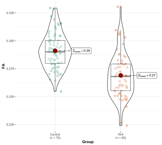
FA within the orbitofrontal region sulcal fundi is significantly lower (P=5.26 × 10⁻¹¹) in soccer RHI group compared to age- and sex- matched non-collision athletes.