1615
Multimodal MRI Investigation of Brain Structural and Functional Changes in Acute Mild Traumatic Brain Injury1Second Affiliated Hospital of Xi’an Jiaotong University, Xi’an, Shaanxi, China, 2Wang Jing Hospital of CACMS, Beijing, China, 3GE HealthCare MR Research, Beijing, China
Synopsis
Keywords: Traumatic Brain Injury, fMRI (resting state)
Motivation: Incidence of traumatic brain injury (TBI) are rising over years.
Goal(s): Accurately and objectively evaluate the degree of brain injury and judge the prognosis of patients with mTBI.
Approach: Neuropsychological assessment results, brain volume from morphological MR data, and brain functional connectivity from fMRI data were compared between 48 acute mTBI patients and 30 healthy controls.
Results: Acute mTBI patients exhibit reduced volume in the right frontal lobe compared to the healthy control group, which is associated with cognitive decline.
Impact: The morphological changes in the right frontal lobe observed in this study may serve as a potential biomarker for acute mTBI, offering a more objective measure for the diagnosis and treatment of brain injury in acute mTBI patients.
Introduction
Traumatic brain injury (TBI) represents a prominent central nervous system disorder characterized by a substantial incidence of mortality and disability. Notably, mild traumatic brain injury (mTBI) constitutes a significant proportion, estimated to encompass approximately 60-95% of all TBI cases1. Presently, the diagnostic approach for mTBI predominantly relies upon the assessment of clinical symptoms and signs exhibited by patients. Consequently, there exists a compelling imperative to identify an objective and non-invasive method for the detection of structural brain damage and functional abnormalities post-mTBI. The purpose of this study is to provide objective evidence for the diagnosis and treatment of mTBI by analyzing the changes in brain structure and resting-state fMRI in mTBI patients, as well as their relationship with neuropsychology.Methods
PatientsThe study was approved by the Medical Ethics Committee and informed consent forms were received from all subjects. Patients with acute mTBI admitted to the Emergency Department from December 2021 to December 2022 were enrolled. According to criteria, 43 patients with acute mTBI were finally included, along with 30 healthy gender-, age- and education level- matched controls.
Data acquisition
All subjects underwent neuropsychological questionnaire assessment, including Rivermead Post-Concussion Symptoms Questionnaire (RPQ), minimum mental state examination (MMSE), Hamilton Depression Scale (HAMD), and Fatigue Severity Scale (FSS). MR examinations were conducted on a 3T scanner (Architect, GE Healthcare, USA) equipped with a 48-channel head coil. Sequences scanned include routine brain MRI (T1WI, T2WI, sagittal fat-suppressed T2FLAIR), susceptibility weighted imaging, 3D T1WI with 1mm isotropic resolution and resting-state functional MRI.
Data processing and Statistics
VBM analysis was performed with FMRIB Software Library (FSL6.0.5.2). SPM12-based tool CONN20b was used for group-wise comparison of function connection. All subjects' rs-fMRI data were processed with both one- and two-level analysis based on GRETNA, with preprocessing step include noise reduction. Statistical analysis was performed with SPSS (version 18) and bilateral P < 0.05 was considered to be statistically significant in all tests.
Results
Neuropsychological assessmentThe average RPQ score for the mTBI group was 20.77±15.11, indicating that patients with acute phase mTBI commonly experience symptoms such as headache, dizziness, and nausea, with some patients also reporting symptoms of tinnitus and vomiting. The MMSE score in the mTBI group was significantly lower than that in the healthy control group (P=0.008). HAMD and FSS score of the mTBI group was significantly higher than that of the control group (P=0.018, P=0.016).
Brain volume and its correlation with neuropsychological scale scores
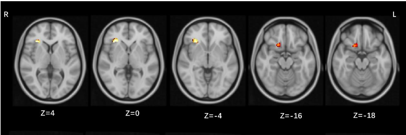
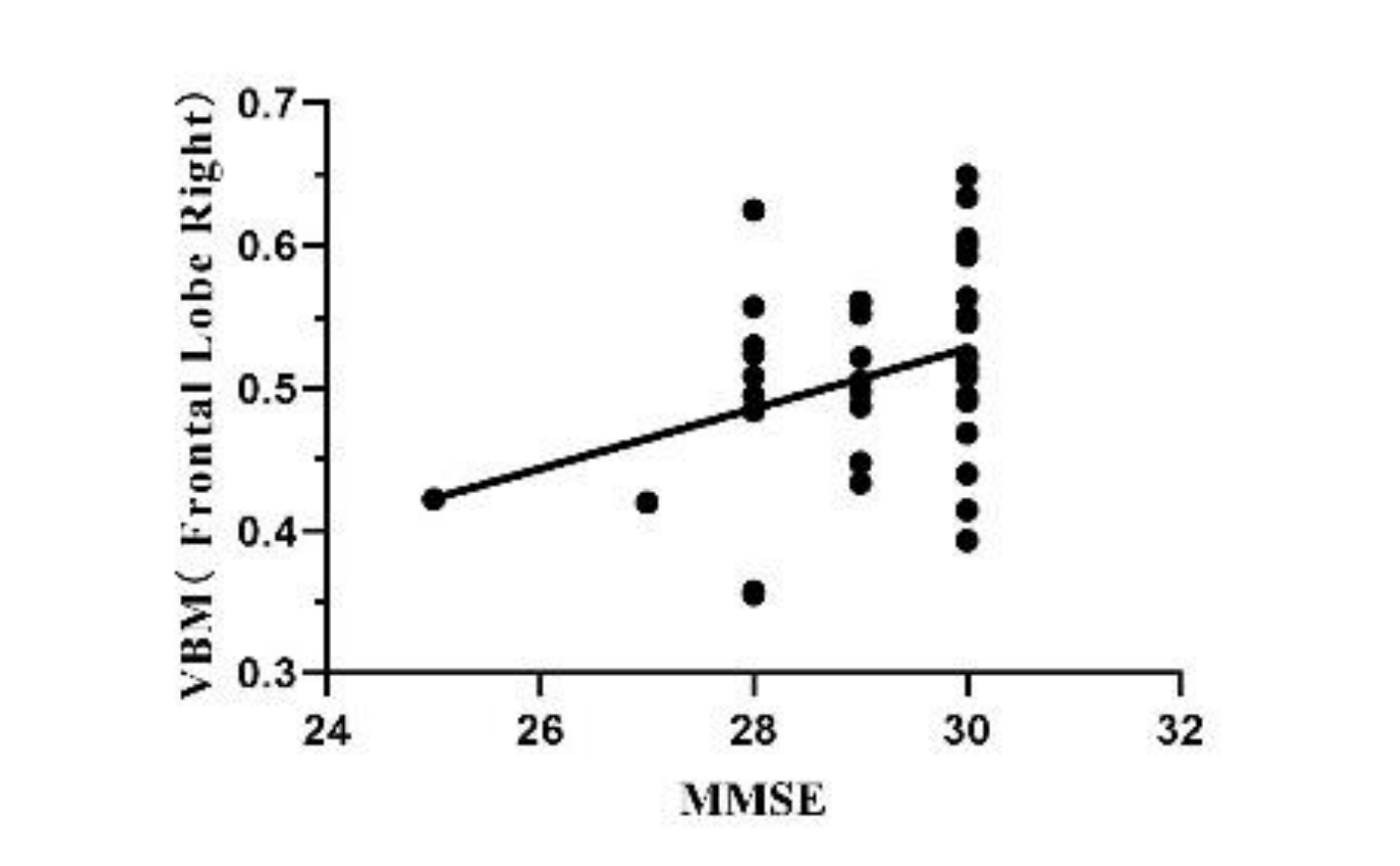
Compared with the healthy control group, region with reduced brain for mTBI group was located in the right frontal lobe (P < 0.001, Figure1), and the reduction of GMV in this region was positively correlated with MMSE score (P=0.026, Figure 2).
Functional connectivity (FC) analysis based on substantia nigra and amygdala
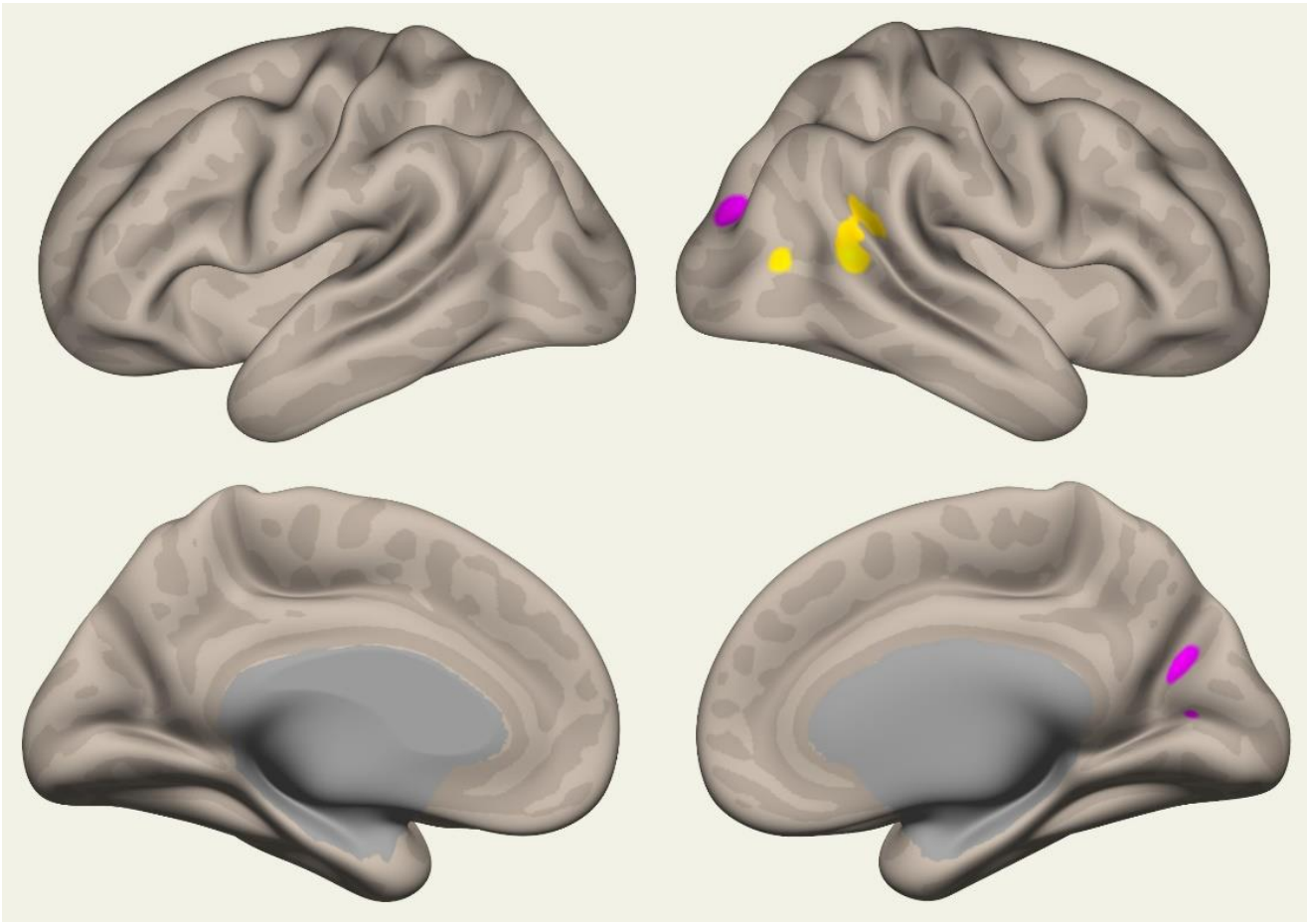
Compared with the HC group, the right substantia nigra pars compa in the mTBI group had increased FC with the right cuneus cortex (P=0.03) and decreased FC with the right angular gyrus (P=0.004, Figure 3). The FC between the right amygdala and the left inferior temporal gyrus was increased (P=0.042).
Complex brain network analysis based on graph theory
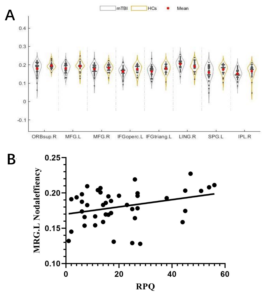
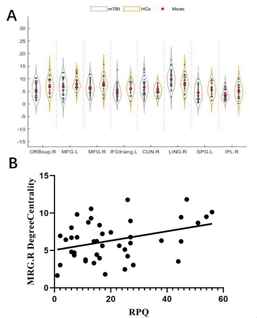
The node efficiency of the left middle frontal gyrus and the degree centrality of the right middle frontal gyrus are related to the physical symptoms after concussion. The RPQ score was positively correlated with the left middle frontal gyrus (P=0.041, Figure 4) and the right middle frontal gyrus (P=0.019, Figure 5).
Discussion
A recent MRI study of post-traumatic PTSD patients has unveiled notable functional alterations within the prefrontal-marginal and striatal regions, as well as reduced connectivity between marginal and striatal networks 2. This finding might shed light on the observed positive correlation between morphological changes and MMSE scores in current study. Previous studies have shown that the level of dopamine in striatum is decreased in patients with TBI, and dopamine predominantly resides in the substantia nigra pars compacta3. This insight may offer an explanation for the observed abnormal functional connectivity in the substantia nigra pars compacta, while concurrently no conspicuous irregularities were observed in the reticular part within the context of the present study.Conclusions
Our study suggests that morphological changes in the right frontal lobe may serve as potential image-marker for mild traumatic brain injury. Additionally, the efficiency of nodes in the left middle frontal gyrus and the centrality of nodes in the right middle frontal gyrus may contribute to additional and unique mechanisms of post-traumatic symptoms in patients with acute mild traumatic brain injury. This provides insights for further exploration of the mechanisms underlying concussion syndrome.Acknowledgements
Funding for this project was provided by: Shaanxi Provincial Department of Science and Technology; Project category: General project - Social development area; Project No. : 2021SF147.
References
1. Maas A I R, Menon D K, Adelson P D, et al. Traumatic brain injury: integrated approaches to improve prevention, clinical care, and research[J]. Lancet Neurol, 2017, 16(12):987-1048.
2. Meng L, Jiang J, Jin C, et al. Trauma-specific Grey Matter Alterations in PTSD[J]. Sci Rep, 2016, 6:33748.
3. Jenkins P O, De Simoni S, Bourke N J, et al. Dopaminergic abnormalities following traumatic brain injury[J]. Brain, 2018, 141(3):797-810.
Figures