1611
High-Resolution Diffusion-MRI Detects Accelerated White Matter Degeneration in Alzheimer's Mice after Neurotrauma
Ning Hua1, Olga Minaeva1, Douglas Parsons1, Juliet Moncaster1, Hernan Jara1, and Lee Goldstein 1
1Boston University, Boston, MA, United States
1Boston University, Boston, MA, United States
Synopsis
Keywords: Traumatic Brain Injury, Traumatic brain injury
Motivation: It is important to understand how neurotrauma accelerates the progression of Alzheimer's disease.
Goal(s): The project's goal is to use ultra-high isotropic resolution (60μm) diffusion-MRI for exploring changes in white matter integrity after neurotrauma.
Approach: Ex vivo diffusion-MRI was acquired using a 9.4T Bruker scanner in a transgenic mouse model of Alzheimer's disease with and without neurotrauma.
Results: We observed decreased quantitative anisotropy in mice 8 months post the brain injury.
Impact: Our preliminary finding demonstrated that neurotrauma can induce accelerated white matter degeneration in Alzheimer's disease.
Introduction
Traumatic brain Injury (TBI) is a risk factor for earlier onset of Alzheimer’s disease (AD)1, and the more severe the injury, the greater the risk of developing AD2. Given the prevalence of AD in modern society, the possibility that TBI may predispose individuals to develop AD has significant social and economic implications. Therefore, it is important to understand how TBI triggers accelerated AD progression. In this study, we explored how neurotrauma accelerated white matter degeneration in a transgenic mouse model of AD using high-resolution ex vivo diffusion MRI.Methods
3xTg-AD mice (Jackson Laboratory, ME) were bred and maintained at Boston University. Unanesthetized mice were pretreated with a non-sedating dose of the analgesic buprenorphine (0.2mg/kg, i.p.) prior to TBI. 3xTg-AD mice were subjected to left-lateral closed-head impact injury (Figure 1) at 10-12 weeks of age as described in our previous work3. At six months post-TBI, mice were sacrificed using transcardial perfusion. The harvested brains were submerged in 10% formalin for 24 hours and then stored in Gadavist-doped PBS (1:400 dilution) until MRI. MRI data were acquired using a 9.4T Bruker scanner (Bruker BioSpec 94/20) and a 2-element transmit/receive MRI cryoprobe. Key parameters are TR=300ms, TE=27.7ms, b=3000 s/mm2 (64 directions) and 5000 s/mm2 (96 directions), FOV=13.92x10.20x7.20mm3, Matrix=232x170x120, resolution=60 μm3. Diffusion MRI was analyzed in DSI Studio (http://dsistudio.labsolver.org). Age, gender-matched 3xTg-AD mice without TBI were used as controls.Results
Figure 2 shows the representative quantitative anisotropy (QA) maps obtained from a mouse with TBI. At 6 months post TBI, decreased QA values were observed in the brain regions ipsilateral to the injury (arrows) compared to the corresponding contralateral regions. Figure 3 shows the representative QA maps of the regions around the hippocampus in mice with and without TBI. Compared to the control mouse, the mouse with neurotrauma showed decreased QA values in the corpus callosum and bilateral hippocampus, especially in the CA1 subregion.Discussion
Our results demonstrated that ultra-high resolution diffusion MRI can capture subtle alterations of white matter integrity in different brain sub-regions. Using this technique, we were able to detect trauma-accelerated white matter degeneration in a mouse model of Alzheimer’s disease.Conclusion
Neurotrauma accelerates white matter deterioration, which may contribute to the worsening of Alzheimer’s disease.Acknowledgements
No acknowledgement found.References
1. Graves AB, White E, Koepsell TD, Reifler BV, van Belle G, Larson EB, Raskind M. The association between head trauma and Alzheimer's disease. Am J Epidemiol. 1990;131(3):491-501. doi: 10.1093/oxfordjournals.aje.a115523. PubMed PMID: 24056482. Johnson VE, Stewart W, Smith DH. Traumatic brain injury and amyloid-beta pathology: a link to Alzheimer's disease? Nat Rev Neurosci. 2010;11(5):361-70. doi: 10.1038/nrn2808. PubMed PMID: 20216546; PMCID: PMC3979339.
3. Tagge CA, Fisher AM, Minaeva OV, et al. Concussion, microvascular injury, and early tauopathy in young athletes after impact head injury and an impact concussion mouse model. Brain. 2018;141:422-58. doi: 10.1093/brain/awx350. PubMed PMID: WOS:000424217900019.
Figures
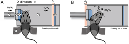
Figure 1. Illustration of our left-lateral closed-head impact (CHI) system before (A) and after (B) impact.
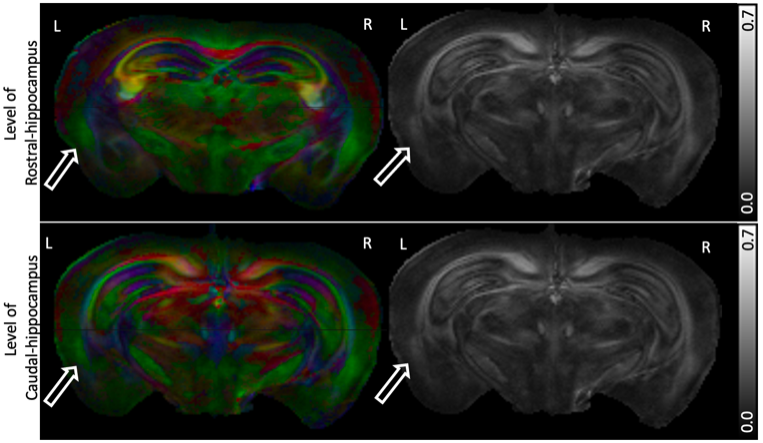
Figure 2. Representative quantitative anisotropy (QA) maps from a 3xTg-AD mouse at 6 months post left-side neurotrauma exposure. Reduced QA values were observed at the ipsilateral side (arrows) of the brain injury. Left column, color showing the axonal diffusion directions (red: right-left; green: posterior-anterior; blue: superior-inferior). Right column, QA maps.
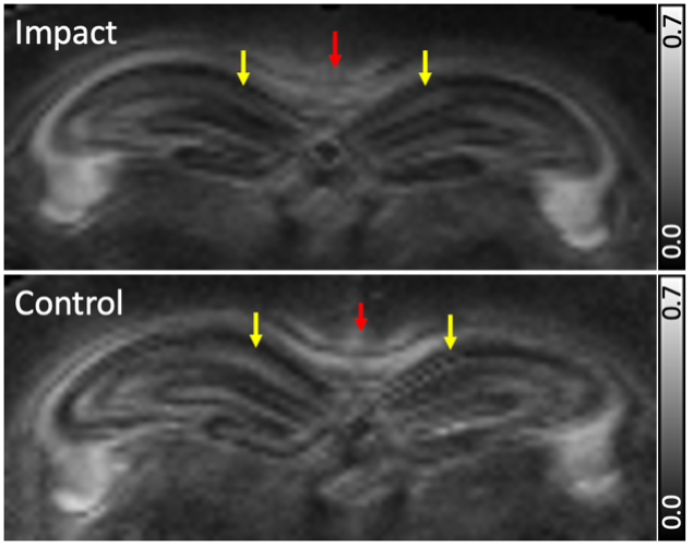
Figure 3. Representative quantitative anisotropy (QA) maps from 3xTg-AD mice with (Impact) and without (Control) neurotrauma around the area of hippocampus. Decreased QA values were observed in the corpus callosum (red arrows) and hippocampi, especially in the CA1 subregions (yellow arrows)
DOI: https://doi.org/10.58530/2024/1611