1609
Decreased water exchange rate across blood-brain barrier within 14 days after cardiac arrest1Department of Radiology, Beijing Chaoyang Hospital, Capital Medical University, Beijing, China, 2Department of Emergency Medicine, Beijing Chaoyang Hospital, Capital Medical University, Beijing, China, 3Laboratory of FMRI Technology (LOFT), Mark & Mary Stevens Neuroimaging and Informatics Institute, Keck School of Medicine, University of Southern California, Los Angeles, CA, United States, 4MR Research Collaboration, Siemens Healthineers, Beijing, China
Synopsis
Keywords: Traumatic Brain Injury, Brain
Motivation: The alteration of the blood-brain barrier (BBB) and cerebral perfusion in the short period of time after cardiac arrest remains unclear.
Goal(s): To determine the changes of BBB and cerebral blood flow (CBF) in patients with cardiac arrest.
Approach: Diffusion prepared pseudo-continuous arterial spin labeling was used to estimate the water exchange rate across the BBB (kw) and CBF in 21 cardiac arrest patients and 21 matched healthy controls.
Results: The kw was significantly decreased in cardiac arrest patients compared to healthy controls, whereas CBF was not significantly different. Reduced kw was associated with neurologic dysfunction in surviving patients after cardiac arrest.
Impact: This study demonstrated that the kw decreased within 14 days after cardiac arrest, suggesting that BBB dysfunction occurred in the short term after cardiac arrest, which will help clarify the pathophysiological changes in the brain after cardiac arrest.
Introduction
Hypoxic-ischemic brain injury is the leading cause of death and disability in patients with cardiac arrest1. Structural destruction of the BBB is one of the secondary injuries resulting from ischemia and hypoxia2. However, it is unclear whether there is functional impairment of the BBB after cardiac arrest. Diffusion prepared pseudo-continuous arterial spin labeling (DP-pCASL) can quantify water exchange rate across BBB (kw) to reflect structural and functional damage of the BBB3. Moreover, DP-pCASL allows for computation of cerebral perfusion metrics such as cerebral blood flow (CBF). Therefore, this study applied DP-pCASL to assess the alterations of kw and CBF in cardiac arrest patients.Methods
In this study, 21 patients (57.43 ± 14.07 years, 14 males) with cardiac arrest within 14 days and 21 matched healthy controls (50.57 ± 7.42 years, 9 males) were enrolled. All participants underwent brain MRI on a 3T scanner (MAGNETOM Prisma, Siemens Healthcare, Erlangen, Germany). DP-pCASL data were obtained using a 3D gradient-and-spin-echo diffusion-prepared pCASL sequence (resolution = 3.5 ×3.5 × 8 mm3, TE = 36.5ms, TR = 4000ms, flip angle = 120°, FOV = 224 mm × 224 mm, matrix size = 64 × 64, 12 slices (10% oversampling), label/control duration = 1500ms)4. The cerebral performance categories (CPC) score at discharge and the modified Rankin scale (mRs) score after 6 months was applied to evaluate the neurological prognosis of patients with cardiac arrest, CPC > 2 or mRs > 3 indicated a poor outcome5. Spearman rank correlation analysis was used to assess the relationship between the kw, CBF and the neurological prognosis in surviving patients after cardiac arrest at discharge.Results
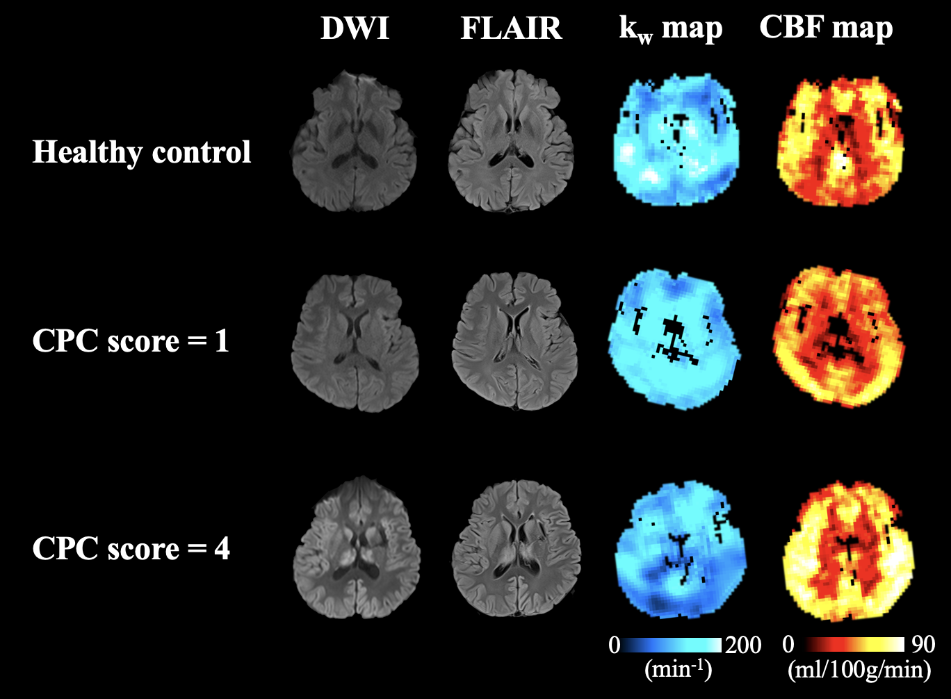
The kw was significantly lower in cardiac arrest patients compared to healthy controls (p = 0.002), whereas CBF was not significantly different (p = 0.818) (Fig.1, Table 1A). A total of 13 patients survived after cardiac arrest at discharge, of which 5 were in the group of good neurological outcome and 8 in the group of poor neurological outcome. Compared with the patients with good neurological prognosis, patients with poor neurological prognosis had significantly lower kw (Fig.1, Table 1B). Reduced kw was significantly associated with neurologic prognosis at discharge and after 6 months in surviving patients after cardiac arrest (r = -0.635, p = 0.020; r = -0.704, p = 0.007).Discussion and Conclusion
This study focused on investigating the damage to the BBB and the alterations of cerebral perfusion in patients with cardiac arrest within 14 days. The results of the study showed that there was decreased kw in the whole brain after cardiac arrest, and there was no significant change of perfusion in the whole brain. These findings suggest that the cerebral perfusion of patients with cardiac arrest is recovered after successful cardiopulmonary resuscitation, and the BBB dysfunction is the second injury caused by ischemia and hypoxia. Furthermore, the present study also demonstrated that the kw has a good performance in predicting neurologic dysfunction in surviving patients after cardiac arrest.Acknowledgements
This work was supported by grants from the National Natural Science Foundation of China (81961128030, 82025018).References
1. Snider SB, Fischer D, McKeown ME, et al. Regional Distribution of Brain Injury After Cardiac Arrest: Clinical and Electrographic Correlates. Neurology. 2022;98(12):e1238-e1247.
2. Park JS, You Y, Min JH, et al. Study on the timing of severe blood-brain barrier disruption using cerebrospinal fluid-serum albumin quotient in post cardiac arrest patients treated with targeted temperature management. Resuscitation. 2019;135:118-123.
3. Li Y, Ying Y, Yao T, et al. Decreased water exchange rate across blood-brain barrier in hereditary cerebral small vessel disease. Brain. Published online January 10, 2023:awac500.
4. Shao X, Ma SJ, Casey M, D’Orazio L, Ringman JM, Wang DJJ. Mapping water exchange across the blood-brain barrier using 3D diffusion-prepared arterial spin labeled perfusion MRI. Magn Reson Med. 2019;81(5):3065-3079.
5. Sandroni C, Cronberg T, Sekhon M. Brain injury after cardiac arrest: pathophysiology, treatment, and prognosis. Intensive Care Med. 2021;47(12):1393-1414.
Figures