1604
A Wireless Tx/Rx Litzcage coil for 1.5 T Knee MRI1Research center, Sino Canada Health Institute Inc., Winnipeg, Manitoba, Canada, Winnipeg, MB, Canada, 2Sino Canada Health Engineering Research Institute (Hefei) Ltd, Hefei, China, 3Hubei Key Laboratory of Intelligent Conveying Technology and Device, Hubei Polytechnic University, Huangshi, China, 4Department of Physics, The University of Winnipeg, Winnipeg, MB, Canada, 5Department of Radiology and Radiological Sciences, Vanderbilt University Medical Center, Nashville, TN, United States, 6Vanderbilt University Institute of Imaging Science, Vanderbilt University Medical Center, Nashville, TN, United States, 7Department of Electrical and Computer Engineering, Vanderbilt University, Nashville, TN, United States
Synopsis
Keywords: RF Arrays & Systems, RF Arrays & Systems, Knee, MRI, wireless coil, SNR, Tx/Rx, RF coil.
Motivation: The conventional Tx/Rx knee coil optimizes transmission and reception using a complex dual-layer structure with multiple components, requiring local transmit power and a limited diameter.
Goal(s): Developing a knee coil for that offers superior performance, patient-comfort, and affordable, without the need for a local transmit connection.
Approach: Design a wireless Tx/Rx knee coil incorporating system body coil in transmit phase and a flex phased array during receive phase, evaluating its performance against the flex phased array alone using phantom and in vivo imaging.
Results: Enhanced SNR is achieved when compared to a 13-channel flexible phased array, effectively eliminating wrap-around artifacts in knee image.
Impact: The advancement of inductive RF resonator technology with a wireless birdcage resonator incorporating a Figure-of-Eight (Fo8) conductor design is aiming to achieve cost-effective and improved performance for knee MRI which improves the way of designing MRI coils and their applications
INTRODUCTION
Standard commercial knee coil optimizes RF transmission as well as reception, effectively preventing the wrap-around artifact from another knee and meanwhile maximizing the SNR. For instance, the widely used commercial knee coil employs a local birdcage coil for RF transmission and 15 local receive coils for RF reception1. However, this configuration has a complex two-layer structure and intricate electronic components such as preamplifiers, detune circuits, baluns, cables, and connectors, leading to high costs. It also requires the local transmission capability of the scanner and has a relatively small inner diameter of ~15.4cm. In this work, we introduce a low-cost and simple wireless Litzcage resonator to enhance both transmission and reception performance for knee MRI.METHODS
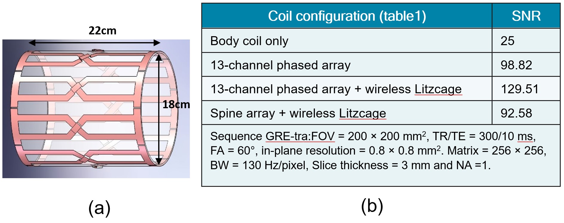
A wireless high-pass quadrature Litzcage coil with 9 pairs of rungs was constructed, as shown in Figure 1. The wireless Litzcage has a length of 22 cm and an inner diameter of up to 18 cm, allowing it to accommodate the knees of a large population.During the transmit phase, the body coil couples with the Litzcage resonator, producing a strong and uniform B1+ field within the Litzcage, enabling local transmission without the need for local RF transmission. This addresses a limitation in cost-effective MRI scanners where conventional Tx/Rx knee coils are not applicable.
During the receive phase, the local receive array, rather than the body coil, serves as the primary coil to maximize receive SNR. Together, the proposed wireless coil and setup enhance both the transmit and receive performance with a low-cost and simple solution. The phantom and volunteer experiments were performed on a 1.5T whole-body scanner (Siemens MAGNETOM Sempra).
RESULTS
Receive SNRTable 1 summarizes the average SNR over the central axial slice on a 5.3L bottle phantom (diameter 16 cm and height 45 cm) using different coil setups. Using the wireless Ltizcage insert and the 13-channel flexible receive array, it can achieve an SNR of 129.51, which is 5.2x improvement compared to the body coil alone and 1.31x improvement compared to the local receive array alone. We noticed that the built-in spin array could be used as the primary coil for RF reception, but its SNR is lower than the 13-ch array alone. The imaging sequence for SNR evaluation is in table1 The reference amplitude for a 180-degree flip angle with 1ms rectangular pulse length reduced from 280V without a coupled resonator to about 42V with the coupled resonator for phantom and 36V for human knee.
Phase-wrapping artifact
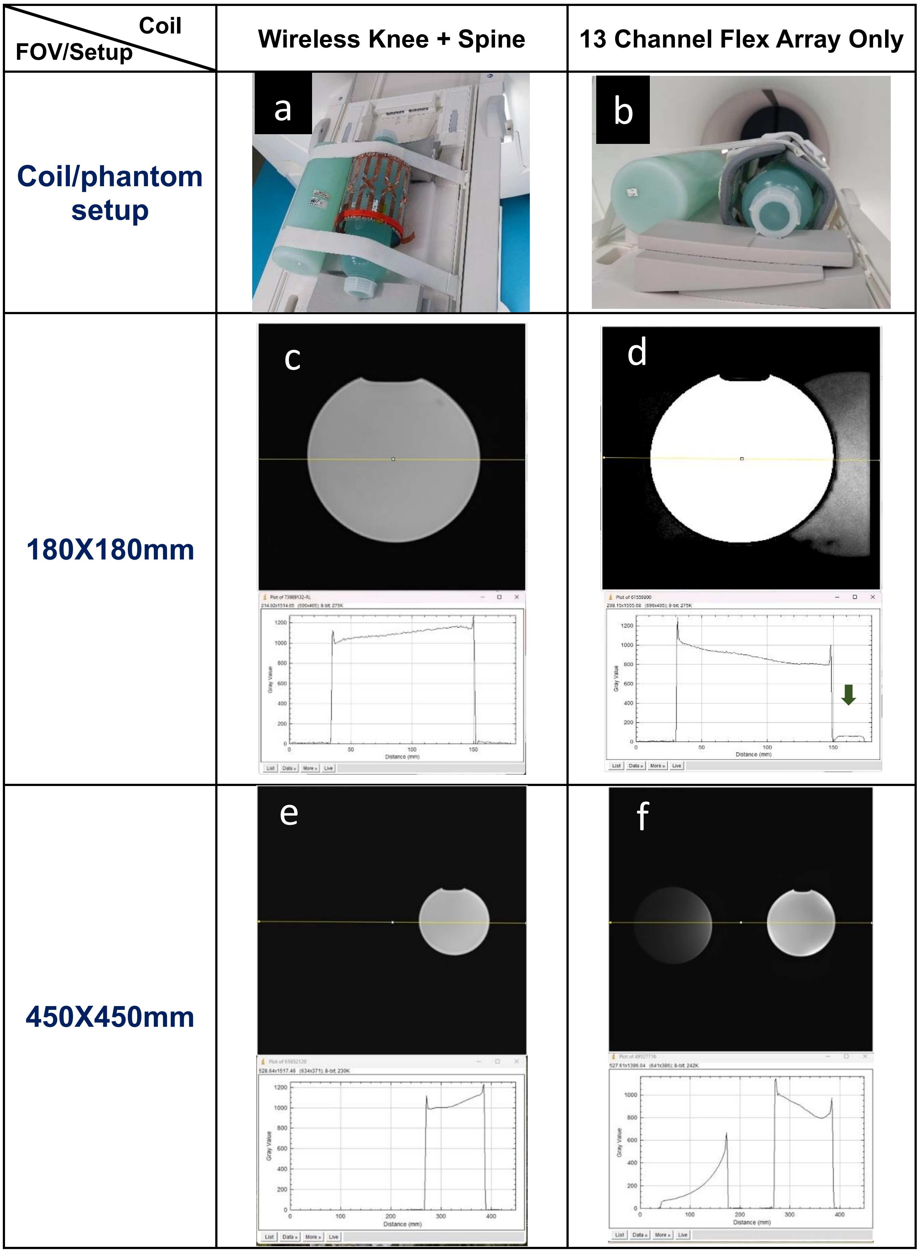
Figures 2a and 2b depict the MRI setups for assessing phase-wrapping artifacts. In addition to two 1900mL imaging phantoms placed inside the testing coil, one 5.3L phantom was positioned outside the coil to replicate the conditions of the other knee/leg during the MRI. For images with a field of view measuring 18x18cm and 45x45cm, the wireless Litzcage resonator in Figure 2c eliminates phase-wrapping artifacts, while they remain imperceptible in Figure 2e. In contrast, the 13-channel flex body array exhibits wrap-around artifacts, as illustrated in Figure 2d and clearly visible in Figure 2f
Human knee imaging
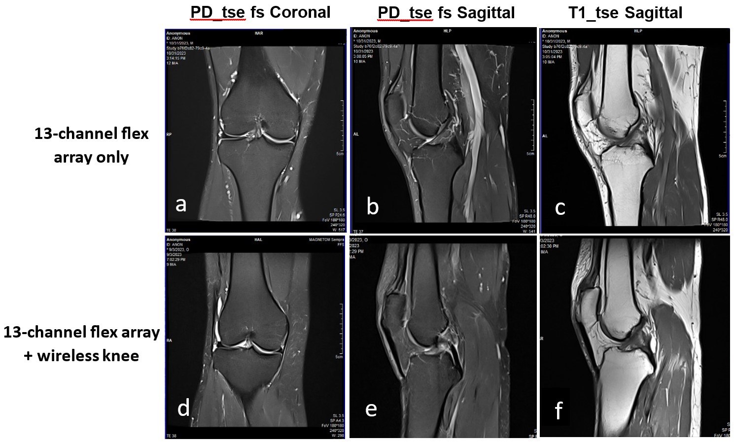
Figure 3 shows measured knee images on a healthy volunteer, using the wireless Litzcage TR/Rx coil and 13-channel flex Rx array.
DISCUSSIONS
The wireless Litzcage knee coil demonstrated a significantly reduced RF reference amplitude of 36V, in stark contrast to the 280V of a traditional phased array coil and even lower than the local Tx/Rx knee coil (~70V). This indicates that the wireless Litzcage offers the following advantages:- Enhances RF transmission efficiency by increasing the B1+ field by 7.7 times with the same RF power.
- Increases the B1- field within the wireless Litzcage, improving SNR.
- Effectively eliminates wrap-around artifacts from another knee.
- Reduces SAR, particularly in the shoulder region, enhancing overall safety for the body.
Acknowledgements
No acknowledgement found.References
- https://www.siemens-healthineers.com/magnetic-resonance-imaging/options-and-upgrades/coils/tx-rx-15-channel-knee-coil
- W. Wang et al., “Inductive Coupled Local TX Coil Design,” in ISMRM, 2010, p. 1510
- Y. Yi et al., “In vivo MRI of knee using a metasurface-inspired wireless coil,” Magnetic Resonance in Medicine, vol. n/a, no. n/a, doi: 10.1002/mrm.29870.
- H. Amat et al., “A flexible metasurface to improve knee MRI,” in Medical Imaging 2023: Physics of Medical Imaging, SPIE, Apr. 2023, pp. 448–453. doi: 10.1117/12.2651679.
- A. Shchelokova, R. Schmidt, A. Slobozhanyuk, T. Kallos, A. Webb and P. A. Belov, "Enhancement of magnetic resonance imaging with metasurfaces: From concept to human trials," 2017 11th International Congress on Engineered Materials Platforms for Novel Wave Phenomena (Metamaterials), Marseille, France, 2017, pp. 31-33, doi: 10.1109/MetaMaterials.2017.8107800.
- A. V. Shchelokova et al., "In vivo magnetic resonance imaging of human knee with metasurface," 2017 Progress In Electromagnetics Research Symposium - Spring (PIERS), St. Petersburg, Russia, 2017, pp. 3657-3660, doi: 10.1109/PIERS.2017.8262393.
Figures