1602
A Simulation Framework for Comparison of Volumetric B1+ Mapping Methods1Wellcome Centre for Integrative Neuroimaging, FMRIB, Nuffield Department of Clinical Neurosciences, University of Oxford, Oxford, United Kingdom, 2Oxford Centre for Clinical Magnetic Resonance Research, John Radcliffe Hospital, University of Oxford, Oxford, United Kingdom, 3Department of Imaging Methods, Institute of Measurement Science, Slovak Academy of Sciences, Bratislava, Slovakia, 4Research & Collaborations GB&I, Siemens Healthcare Ltd, Frimley, United Kingdom
Synopsis
Keywords: High-Field MRI, High-Field MRI, B1+ Mapping, SatTFL, AFI, DREAM, Sandwich, SA2RAGE
Motivation: To benefit from all the advantages that ultra-high field MRI offers, knowledge of the RF transmit field (B1+) is required. Many B1+ mapping methods have been developed, but no single method has become the ‘gold-standard’, with many sites opting for their own implementations.
Goal(s): To evaluate five promising sequences and understand their sensitivity to B0, T1, flow and SNR on the accuracy and dynamic range of the measured B1+.
Approach: SatTFL, Sandwich, SA2RAGE, AFI, and 3DREAM were investigated in MATLAB using open-source EPG and Bloch simulations.
Results: A simulation framework to compare B1+ mapping sequences was developed.
Impact: This simulation framework is beneficial for understanding the impact of various factors on the accuracy of B1+ mapping sequences and can help to inform better pulse sequence design and parameter optimisation.
Introduction
Ultra-high field MRI offers significant advantages over clinically available field strengths, but challenges remain in overcoming the increased B1+ inhomogeneity. Parallel transmission (pTx) is the solution but pTx often requires fast and accurate B1+ maps in order to realise its full potential. To date, many methods of B1+ mapping have been proposed in the literature, but no ‘gold standard’ has emerged. This work aims to evaluate the most promising volumetric B1+ mapping methods by simulating the signal evolution and quantifying the effects of B0, T1, flow, diffusion and SNR on the accuracy, point spread function and dynamic range of the measured B1+ map.Methods
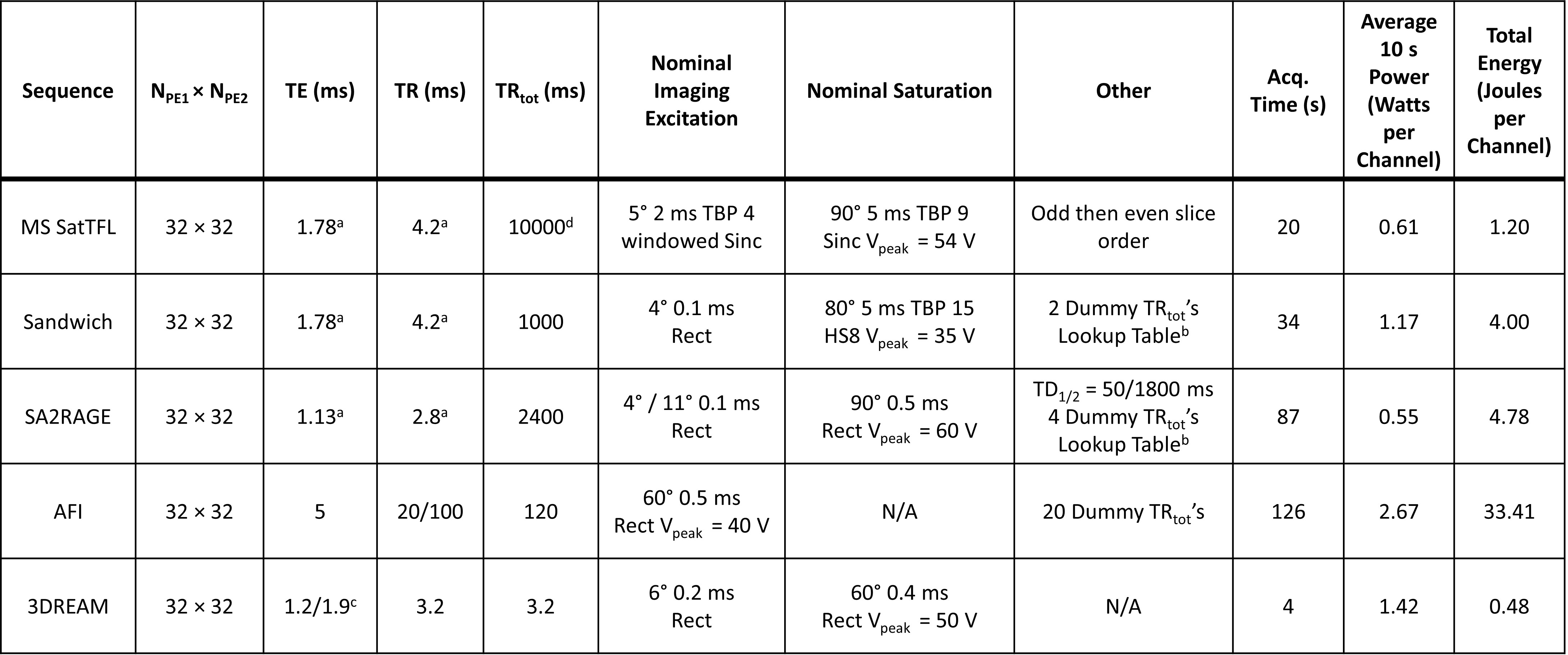
Using both EPG and Bloch simulations, both 2D multi-slice and 3D B1+ mapping sequences were simulated in MATLAB (MathWorks, USA). These were a) 2D Multislice SatTFL [1] b) 3D Sandwich [2] c) 3D SA2RAGE [3] d) 3D AFI [4] e) single-shot 3DREAM [5,6]. Simulations used parameters taken from the literature as shown in Table 1 including RF spoiling and slice profile effects. A dynamic range of B1+ was simulated from 0 to 3 times the nominal flip angle, B0 off-resonances from 0 to 1.5 kHz and T1 times of 0.5 to 3 seconds. The T2 for all simulations was kept fixed at 25 ms. The sensitivity of each method to flow and diffusion was evaluated by simulating a range of coherent flows with a velocity in the range of 0 to 0.2 ms-1 and an isotropic free water diffusion coefficient of 3×10⁻⁹ m2s-1. The relative effects of noise across the different schemes were evaluated using a Monte Carlo method with 1000 repetitions, with a fixed level (peak SNR 60 dB) of zero-mean complex Gaussian noise added independently to each image. The flip angle was calculated as reported in the literature, except for the Sandwich and SA2RAGE sequences where flip angles were calculated using a lookup table with a global T1 assumed to be 1.5 s. Results were normalized by their dynamic range for plotting. The dynamic range of 1 was defined as the lowest B1+ measured, on resonance, where the difference in the mean flip angle and standard deviation was less than 10% of the simulated value. The simulation code is openly available at https://github.com/jameslewiskent/B1-mapping-simulations.Results
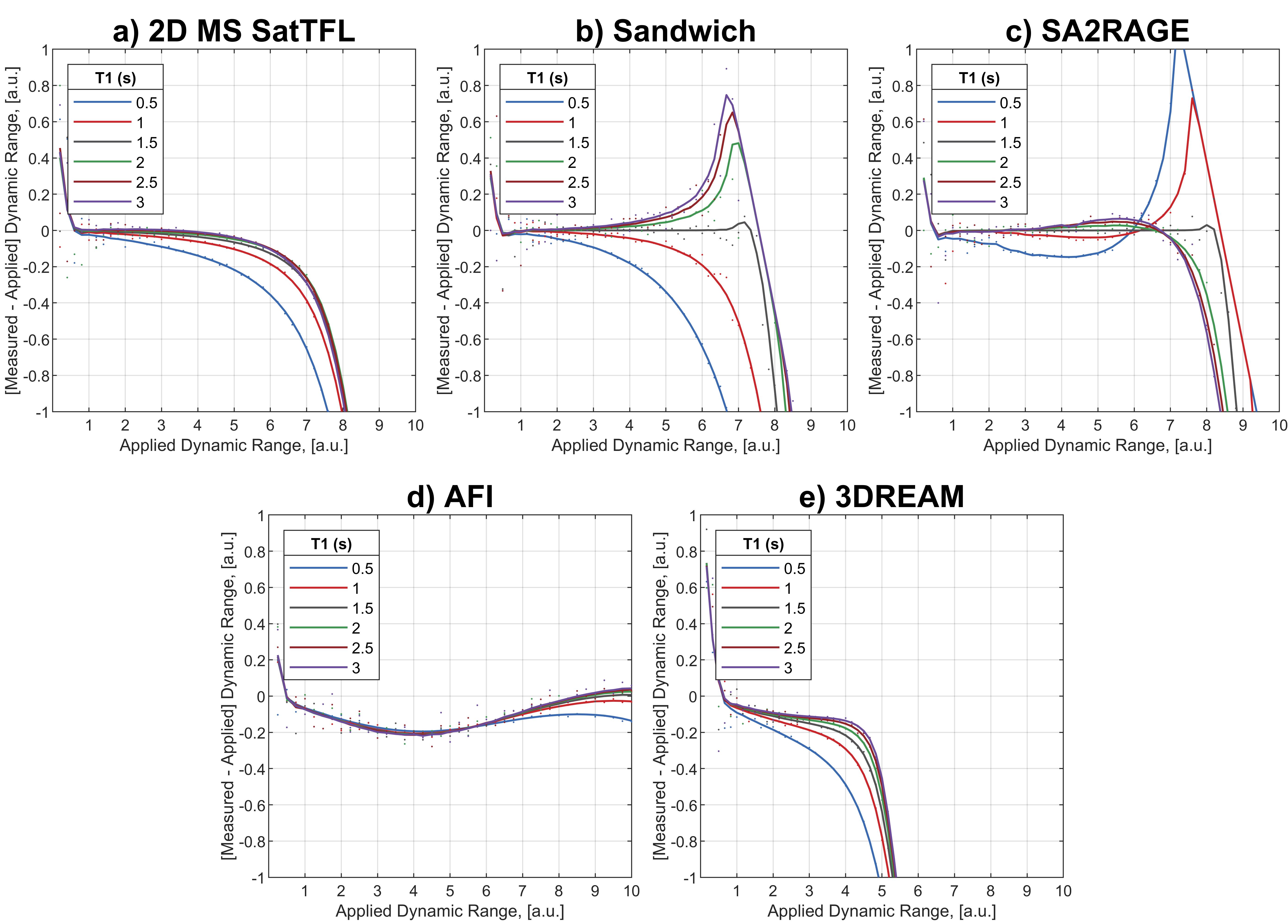
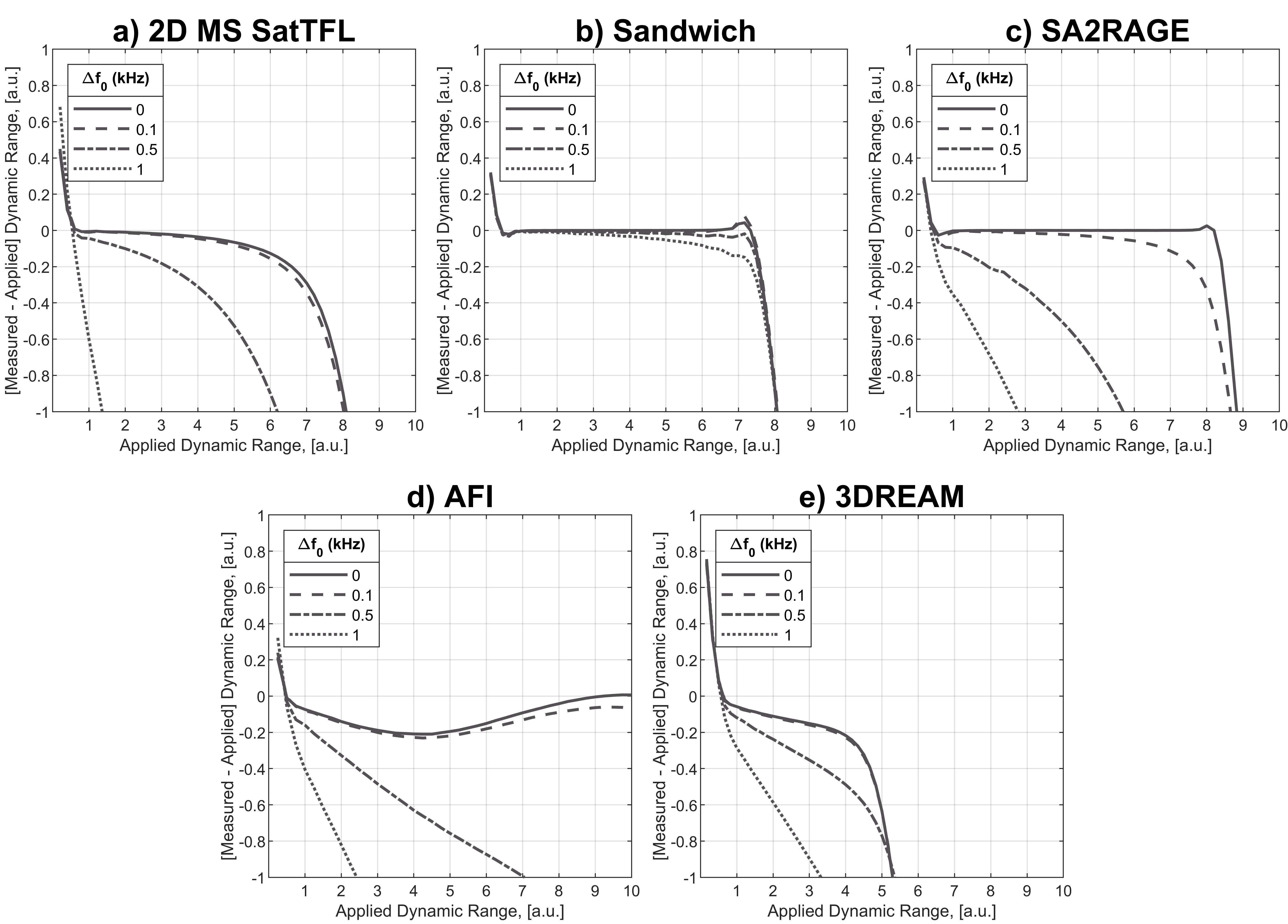
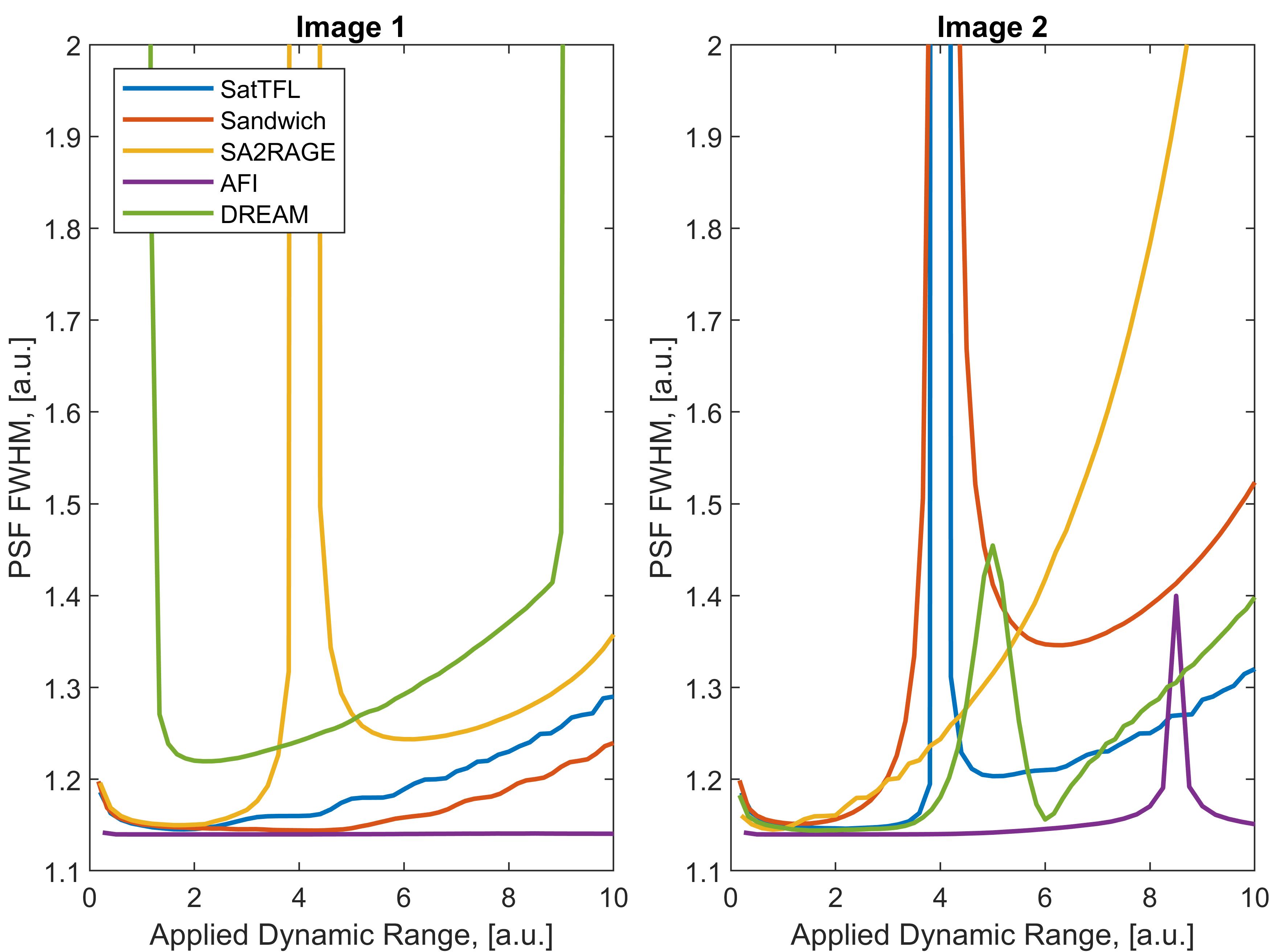
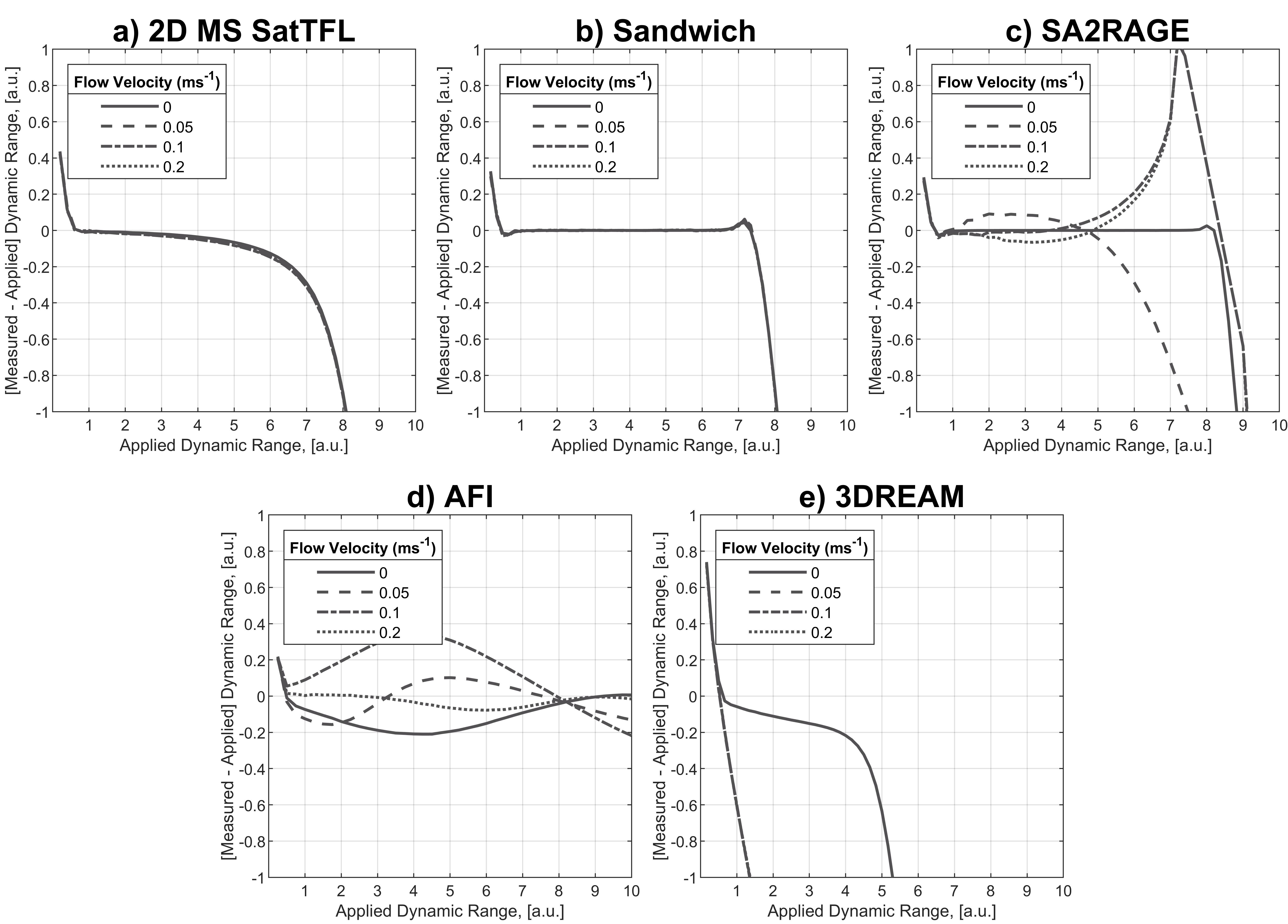
Figure 1 shows the response of the measured B1+ to different T1 values. All methods using long readout trains (a,b,c,d) suffer from large T1 dependence, which can be reduced by shortening the readout train, minimising the image train TR or using variable flip angles. AFI shows the least T1 dependence. SatTFL, Sandwich and SA2RAGE also show similar dynamic ranges of approximately 5 to 6. AFI displays the largest dynamic range but these results indicate a slight underestimation (2.5° underestimation at 50°) of the measured flip angle which varies with the applied B1+. 3DREAM shows the lowest dynamic range with a similar amount of flip angle underestimation within the usable dynamic range but has the lowest acquisition time and total energy of any method. Figure 2 shows the response of the measured flip angle to different B0 off-resonances. The Sandwich sequence shows the best robustness to B0 thus requiring no additional B0 correction. In addition, Sandwich has the lowest peak voltage, as seen in Table 1, which is beneficial when mapping regions of low B1+. Analysis of the full width at half maximum of the point spread function in Figure 3 suggests that intravoxel blurring is low for all schemes. Figure 4 shows 3DREAM and AFI display the most significant sensitivity to flow.Discussion
In our experience, the majority of B0 off-resonances in the cerebrum are expected to be below 100 Hz where all compared sequences work well in this range. However, off-resonances can be as much as 1200 Hz when including the brain stem and neck [7]. Across the heart, off-resonances can be on the order of a few hundred Hz and B1+ mapping must be robust to these off-resonances [8]. Thus, the sandwich sequence appears to be the ideal candidate for gated volumetric cardiac B1+ mapping due to its insensitivity to flow and B0 as well as its ability to have a non-fixed TRtot time. SA2RAGE could also benefit from using an HS8 pulse for better B0 insensitivity. Future work will corroborate these results in phantom and in vivo scans on a 7T MRI system.Conclusion
The choice of a single B1+ mapping technique, as originally implemented, seems to be impossible. However, our simulation framework provides better understanding of the trade-offs between the different B1+ mapping sequences, potentially useful for improved sequence design.Acknowledgements
JK acknowledges the support of EPSRC through an iCASE award in collaboration with Siemens Healthcare Ltd. The Wellcome Centre for Integrative Neuroimaging is supported by core funding from the Wellcome Trust (203139/Z/16/Z and 203139/A/16/Z). LV is funded by a Sir Henry Dale Fellowship awarded jointly by the Wellcome Trust and the Royal Society (221805/Z/20/Z) and also acknowledges the support of the Slovak Grant Agencies VEGA (2/0004/23) and APVV (#21–0299).References
[1] Chung S, Kim D, Breton E, Axel L. Rapid B1+ mapping using a preconditioning RF pulse with TurboFLASH readout. Magn Reson Med 2010;64:439–446.
[2] Kent JL, Dragonu I, Valkovič L, Hess AT. Rapid 3D absolute B1+ mapping using a sandwiched train presaturated TurboFLASH sequence at 7 T for the brain and heart. Magn Reson Med 2023;89:964–976.
[3] Eggenschwiler F, Kober T, Magill AW, Gruetter R, Marques JP. SA2RAGE: A new sequence for fast B1+ mapping. Magn Reson Med 2012;67:1609–1619.
[4] Yarnykh VL. Actual flip-angle imaging in the pulsed steady state: A method for rapid three-dimensional mapping of the transmitted radiofrequency field. Magn Reson Med 2007;57:192–200.
[5] Ehses P, Brenner D, Stirnberg R, Pracht ED, Stöcker T. Whole-brain B1-mapping using three-dimensional DREAM. Magn Reson Med 2019;82:924–934.
[6] Nehrke K, Versluis MJ, Webb A, Börnert P. Volumetric B1+ Mapping of the Brain at 7T using DREAM. Magn Reson Med 2014;71:246–256.
[7] de Buck MHS, Kent JL, Jezzard P, Hess AT. Head‐and‐neck multichannel B1+ mapping and RF shimming of the carotid arteries using a 7T parallel‐transmit head coil. Magn Reson Med 2023:1–15.
[8] Tao Y, Hess AT, Graeme KA, Rodgers CT, Robson MD. B0-Independent quantitative measurement of low B1 field for human cardiac MRI at 7T. Proc Intl Soc Mag Reson Med 2013;21:2596.Figures