1599
A Novel Transceive 2-channel Orthogonally Arranged Dual Loop Solenoid RF Coil Design for Improving Low Field MRI SNR1The First People’s Hospital of Lianyungang, Jiangsu Province, Lianyungang, China, 2Jiangsu LiCi Medical Device Co. Ltd, Lianyungang, China, 3Singapore University of Technology and Design, Singapore, Singapore
Synopsis
Keywords: Low-Field MRI, Low-Field MRI
Motivation: There are growing interest in low field specialised MRI R&D but they are currently burdened by their low SNR images.
Goal(s): A novel 2-channel orthogonally arranged dual loop solenoid RF coil design has been proposed to improve low field MR image SNR.
Approach: Two transceive 2-Channel RF coil prototypes using the novel design and using conventional design were constructed and tested in a 0.35T MRI system.
Results: In comparing the acquired MR images, it is shown that the proposed novel design can improve low field MR image SNR by ≥20% as compared to the conventional design.
Impact: A novel 2-channel orthogonally arranged dual loop solenoid transceive RF coil has been constructed and tested in a 0.35T MRI system. Using this new RF coil design it is shown that the acquired image SNR can be increased by ≥20%.
Introduction
Low field open permanent magnet for designing specialised MRI systems, such as dedicated MSK MRI [1], dedicated baby head MRI [2] and portable MRI [3], has received growing interest in the field of MRI R&D. These specialised MRI systems primary used ≤0.35T open permanent magnet where it has the advantages that the magnet can be freely arranged or designed for adaptation to a specific clinical application and the weight of the permanent magnet system is light enough to allow these specialised MRI systems to be installed as an in-suite MRI system. Although there are specific advantages with these specialised low field MRI systems, they are however burdened by their low SNR images. Hence, most of the effort in low field MRI R&D has been concentrated in improving the SNR. The conventional RF coil design for low field MRI system is mainly based on a 2-channels design, which consisted of a solenoid and saddle/helmholtz coils having their vector B1 fields perpendicular to each other and to the B0 field. However, using such conventional design, the acquired image SNR obtained from individual RF coil differ greatly from each other (unbalanced SNR). The SNR of the saddle/helmholtz coil is often 10~20% lower than the solenoid coil thus, after SOS combining the images, the final combined image SNR still remains low. In this work, a novel 2-channel orthogonally arranged dual loop solenoid coils design has been proposed to mitigate this unbalanced SNR issue. Transceive 2-Channel RF coil prototypes using the proposed design and the conventional design were constructed and tested in a 0.35T MRI system. From the MR images acquired with the two prototypes, it is shown that the SNR of the proposed RF coil design can be improved by ≥20% as compared to the conventional design.Method
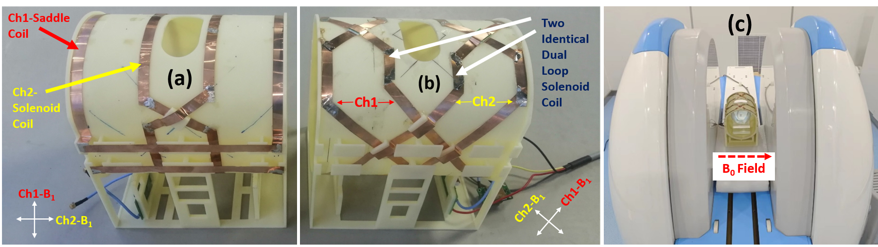
Depicted in Figs 1(a) and (b) are the constructed transceive 2-Channel RF coil prototypes with 145mm inner diameter and 200mm in length. Fig 1(a) is constructed using the conventional design, which consisted of a dual loop solenoid coil and a saddle coil arranged orthogonally. As indicated the transmit B1 fields are in the vertical and horizontal directions (“Cross” configuration). Fig 1(b) is the prototype constructed using the proposed design, which consisted of 2 identical dual loop solenoid coils arranged orthogonal to each other. Differ from the conventional design, the transmit B1 fields of the proposed design are rotated 45⁰ clockwise (“X” configuration). Additional capacitive mutual decoupling circuits are used to improve the isolation between channels. As depicted in Fig 1(c), the prototypes are tested in a 0.35T open MRI system. Homogenous ball phantom (130mm diameter), pineapple and volunteer wrist images are acquired and compared.Results
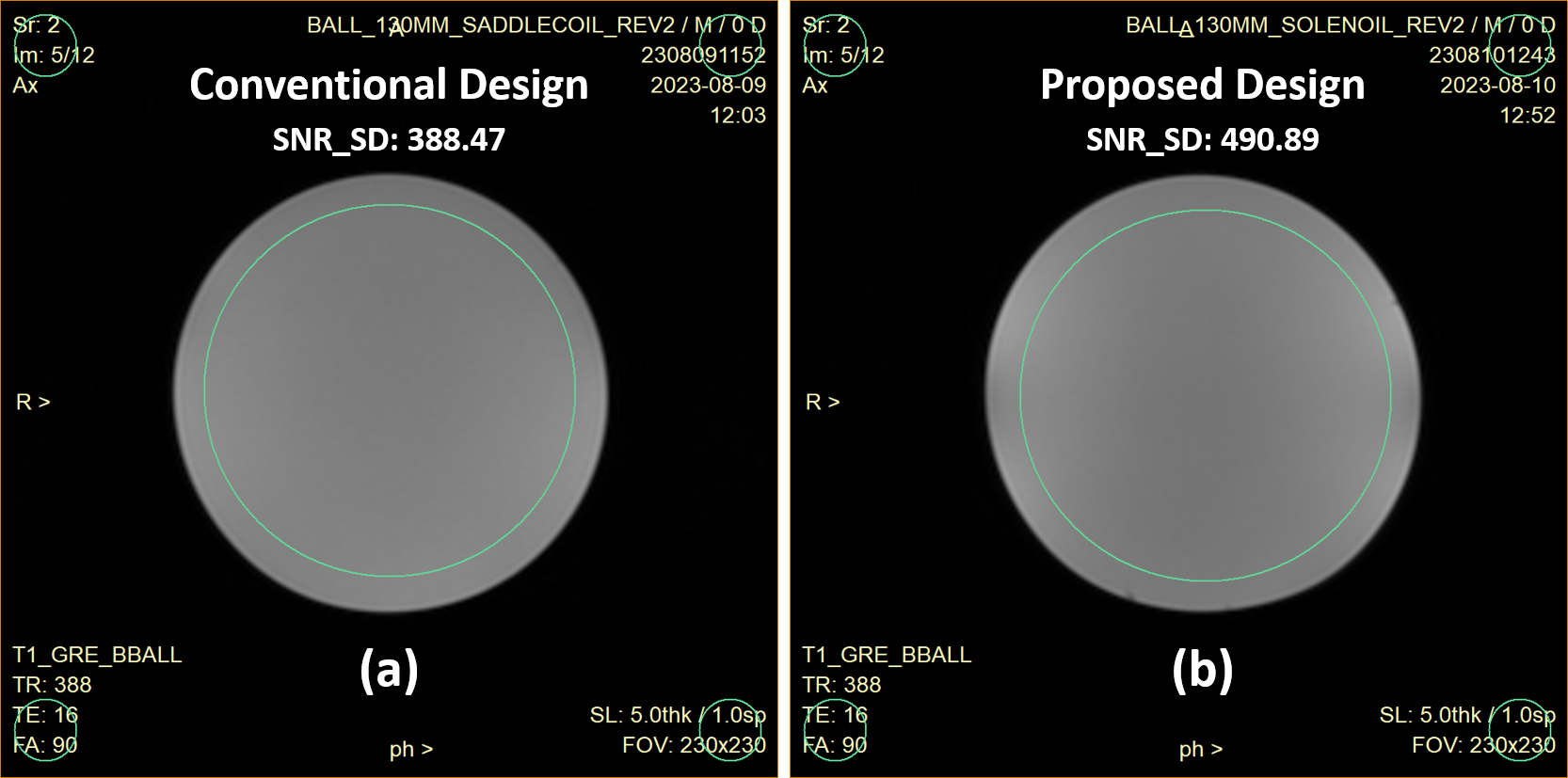
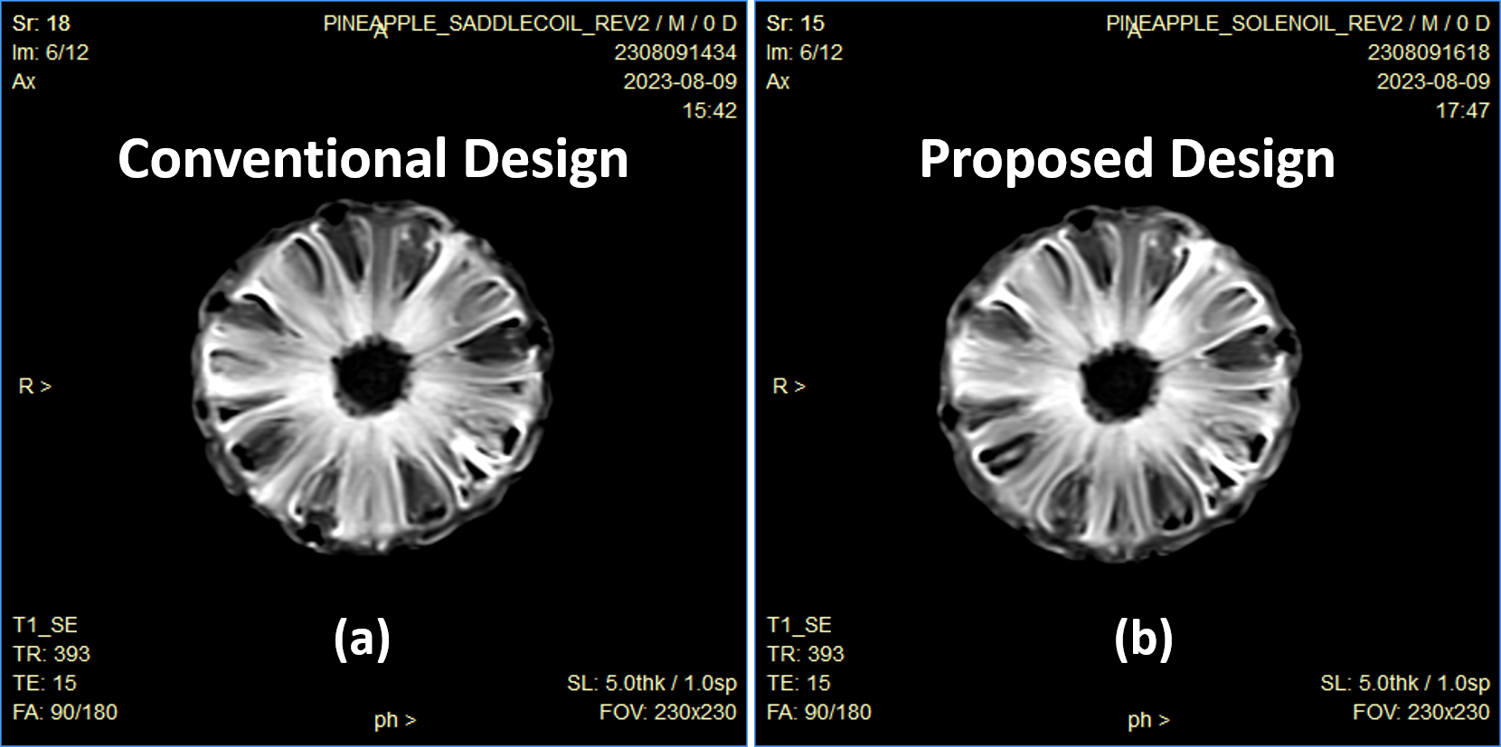
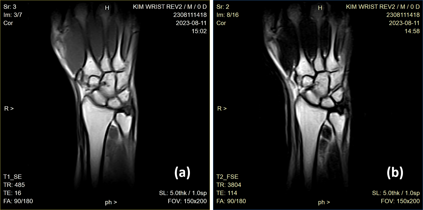
Shown in Fig 2 is the acquired T1 GRE homogenous ball phatom images while Fig 3 is the T1 SE pineapple images using the conventional and the proposed designed prototypes (identical imaging parameters are used when acquiring these images). According to IEC 62464-1:2018 SNR measurement standards, the standard deviation (SD) SNR of the homogenous ball phatom images were calculated and as indicated in Figs 2(a) and (b), with the conventional design the SD SNR is 388.47 while the SD SNR of the proposed design is 490.89. Using the proposed design, the image SNR can be improved by ~26%. In comparing the T1 SE pineapple images, there is apparent improvement in the contrast and higher image detail can be obtained with the proposed design. In additional, as demostated from the acquired coronal wrist images of Fig 4, the “X” configured transmit B1 fields of the proposed design does not display any deteriorate effect to the transmit B1 homogeneity. Homogenous wrist images can be acquired, an indication that the transmit B1 fields need not be designed according to the MRI standard XYZ Cartesian coordinates system.Conclusion
In this work, we have shown that the proposed 2-channel orthogonally arranged dual loop solenoid RF coil design can be used to ameliorate the unbalanced SNR issue with conventional designed RF coil. The images acquired using two identical RF have approximately the same MR signal strength and background noise. Hence, after a SOS image combination the final image SNR can be greatly improved.Acknowledgements
No acknowledgement found.References
[1] YF Zou et al, MRI of the knee during weight-bearing standing postures involving positions of joint extension and flexion, ISMRM, pp. 2815, 2020. [2] M Sheng et al, A Novel Dedicated 0.35T Open Neonatal-Infant Brain MRI system, ISMRM, pp. 4029, 2021. [3] E Kuoy et al, Point-of-Care Brain MRI: Preliminary Results from a Single-Center Retrospective Study. Radiology. 2022; 305(3):666-671.
Figures