1595
Magnetically Coupled Resonant Wireless RF Coil for MRI1Chinese Academy of Sciences, Shenzhen Institute of Advanced Technology, Shenzhen, China, 2Key Laboratory for Magnetic Resonance and Multimodality Imaging of Guangdong Province, Shenzhen, China, 3Shanghai United Imaging Healthcare Co., Ltd, Shanghai, China, 4Department of Biomedical Engineering, State University of New York, Buffalo, Armenia
Synopsis
Keywords: New Devices, Brain, wireless coil
Motivation: The bulky cables of the MRI RF coil not only complicate the coil replacement procedure but also lead to a waste of examination time.
Goal(s): In order to achieve a lightweight and user-friendly wireless RF coil with high-resolution imaging capability.
Approach: In this study, we made a wireless coil and compared its SNR and high-resolution imaging performance with the rat coil RAC and the knee coil alone, using the knee coil as the pickup coil.
Results: The wireless coil achieved high-resolution imaging of up to 0.13 mm × 0.13 mm × 1mm on the 3T system.
Impact: The experiment demonstrated the potential of wireless RF coils for high-resolution imaging.
INTRODUCTION
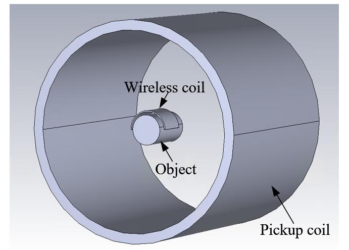
The bulky cables of the MRI RF coil not only complicate the coil replacement procedure but also lead to a waste of examination time, as the usage time of the MRI machine is expensive. To address this, some researchers have designed wireless coils based on analog signal transmission systems1 and digital signal transmission systems2,3. However, even though RF transmitters and receivers can replace the role of cables, these coils require the integration of additional wireless transmission modules, and a wireless method is required to provide power to the active components on the coil and transmit control signals. This undoubtedly increases the cost and complicates the system. In recent years, resonant structures with homogeneous field enhancement and adaptive resonant modes have been reported4,5. Inspired by this, we envisioned a scenario for the operation of a wireless coil (Fig. 1): the wireless coil only includes the essential components for resonance and detuning, and the acquired MRI signal is transmitted through magnetic coupling to a universal pickup coil. When performing MRI imaging of different body parts, it’s only necessary to replace the wireless coil, eliminating the need to change the pickup coil. In this study, we made a wireless coil and compared its SNR and high-resolution imaging performance with the rat coil RAC and the knee coil alone, using the knee coil as the pickup coil. The imaging subject in this work was the brain of a freshly deceased rat.METHOD
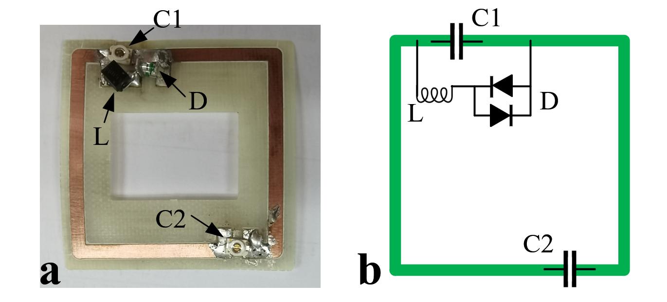
The wireless coil, etched from a 0.2 mm thick copper-clad board, features a 3 mm wide trace and has a length of 30 mm on each side. As shown in Fig. 2, D is a bilateral diode that conducts during the transmission phase and cuts off during the reception phase. When D is conducting, inductor L resonates in parallel with capacitor C1, causing detuning of the wireless coil. C2 is a variable capacitor that can be adjusted to make the wireless coil resonate at the operating frequency.At the beginning of the experiment, the wireless coil was wrapped around the rat's brain and placed inside the knee coil. In subsequent experiments, the rats remained in a prone position. A fast- spin-echo (FSE) sequence with TR = 4500 ms, TE = 100.64 ms, FOV = 60 × 60 mm2, matrix = 128 × 128, slice thickness = 2 mm, and NEX =1 was performed for SNR comparison on 3T MRI scanner. (uMR 790, Shanghai United Imaging Healthcare, Shanghai, China). When performed high-resolution imaging, the parameters were: TR = 4500 ms, TE = 93 ms, FOV = 60 × 60 mm2, matrix = 320 × 320, slice thickness = 2 mm, NEX =2. Additionally, TR = 4500 ms, TE = 92.7 ms, FOV = 60× 60 mm2, matrix = 448 × 448, slice thickness = 1 mm, NEX =2. High-resolution imaging took 2:56 minutes and 4:08 minutes, respectively. For comparison, imaging of the rat brain was conducted using both the rat coil and the knee coil alone.RESULTS AND DISCUSSION
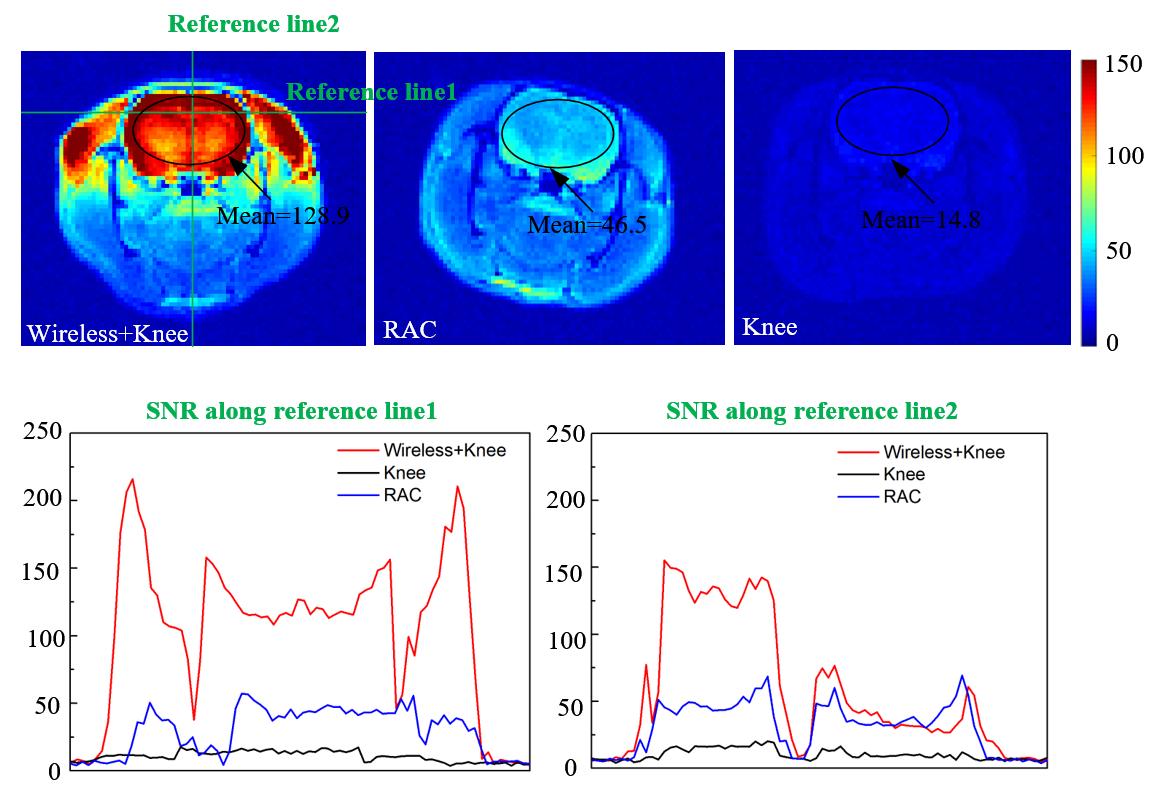
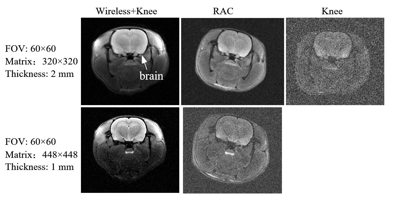
In the SNR comparison experiment, as shown in Fig. 3, the combination of the wireless and knee coils exhibited the best SNR performance. It demonstrated a 2.8-fold improvement in SNR compared to using the RAC and an 8.8-fold improvement compared to using the knee coil alone. In high-resolution imaging experiments (Fig. 4), under similar resolution conditions, the wireless coil exhibited the best image quality. Moreover, the wireless coil achieved high-resolution imaging of up to 0.13 mm × 0.13 mm × 1mm on the 3T system.CONCLUSION
In this article, we manufactured a wireless coil and performed experiments to evaluate the SNR and high-resolution imaging of the rat brain, utilizing a knee coil as the receiving coil. The experimental results indicate that the magnetically coupled wireless coil, in conjunction with the pickup coil (knee coil), outperforms the dedicated rat coil in terms of high-resolution imaging capabilities.Acknowledgements
This work was supported in part by the National Key Scientific Instrument Development Project (Grant No. 81927807); the Project on Global Common Challenges of Chinese Academy of Sciences(No. 321GJHZ2022081GC); the NSFC grant (81627901); the Key Laboratory for Magnetic Resonance and Multimodality Imaging of Guangdong Province (2023B1212060052); the Funding Program of Shenzhen, China (RCYX20200714114735123); the Chinese Academy of Sciences Youth Innovation Promotion Association funded project (Y2021098).References
1. Riffe M J, Yutzy S R, Jiang Y, et al. Device localization and dynamic scan plane selection using a wireless magnetic resonance imaging detector array[J]. Magnetic resonance in medicine, 2014, 71(6): 2243-2249.
2. Jutras J D, Fallone B G, De Zanche N. Efficient multichannel coil data compression: A prospective study for distributed detection in wireless high‐density arrays[J]. Concepts in Magnetic Resonance Part B: Magnetic Resonance Engineering, 2011, 39(2): 64-77.
3. Aggarwal, K., Joshi, K. R., Rajavi, Y., Taghivand, M., Pauly, J. M., Poon, A. S., & Scott, G. (2016). A millimeter-wave digital link for wireless MRI. IEEE transactions on medical imaging, 36(2), 574-583.
4. Chao Luo, Xiaoqing Hu, Xiaoliang Zhang, Xin Liu, and Ye Li. “Preliminary Metamaterial Design and Fabrication for MRI at 3T”, Proc. 25th Annual Meeting of ISMRM, Honolulu, USA, 2017, p.2679.
5. Chi Z, Yi Y, Wang Y, et al. Adaptive cylindrical wireless metasurfaces in clinical magnetic resonance imaging[J]. Advanced Materials, 2021, 33(40): 2102469.
Figures