1591
Improving Long-distance Surface Imaging Performance with a Cryogenic Coil1Shenzhen Institutes of Advanced Technology, Chinese Academy of Sciences, Shenzhen, China, 2Key Laboratory for Magnetic Resonance and Multimodality Imaging of Guangdong Province, Shenzhen, China
Synopsis
Keywords: Non-Array RF Coils, Antennas & Waveguides, RF Arrays & Systems, signal-to-noise ratio, high-resolution, cryogenic, RF coils.
Motivation: The signal-to-noise ratio of high-field RF surface coils decays with distance, and we propose cryogenic coils to compensate for the SNR of MR Imaging.
Goal(s): The cryogenic coil in our work aims to significantly improve the SNR of the images at long distances while guaranteeing a large imaging FOV.
Approach: We design a large size RF coil and cool it down for rat imaging.
Results: The experimental results show that the cryogenic coil obtained a SNR of 1.8-fold higher than a room-temperature coil and 1.1-fold higher than that of a commercial multi-channel rat coil.
Impact: The designed coil can help to improve the quality of MR imaging in some scenarios where the object to be measured is far away from the RF receive coil.
Synopsis
Many methods exist to enhance MRI imaging performance under a constant main magnetic field. This study proposes that reducing the coil temperature can significantly enhance the signal-to-noise ratio (SNR) of the images at a greater imaging distance. The cryogenic coil can provide a 1.8-fold increase in the SNR gain compared to a room-temperature coil. It is noteworthy that our homemade cryogenic coil has a 1.1-fold higher SNR gain times than the commercially available rat coils. As cooling down the coil without a complex cryogenic system is easy to use and cost-effective, it can work as an additional option for acquiring high-resolution images for scientific research and clinical application.Introduction
Cryogenic coils have been reported that they can enhance the SNR of MR images for general small-animal imaging. The general SNR gain of a receive coil is determined by sample noise, coil noise,etc1,2, where the distance between the sample and the coil significantly affects the signal noise. Previous studies have shown that cryogenic coils (including superconducting coils) can improve the overall SNR by reducing the coil noise, but usually require the cryogenic coil to be very close to the sample, which puts high demands on the cooling system. In this work, we demonstrate that cryogenic coils can still improve the SNR of images at a longer distance for MR imaging.Methods
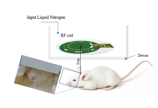
Fig.1 shows our homemade single-channel RF receive-only coil. The diameter of the typical cryogenic copper coil is less than 2 cm3,4, but considering the larger size of the rat compared to smaller animals, we choose a diameter of 4 cm, which guarantees a better radiofrequency field and FOV. When the coil works at 77K, we add liquid nitrogen to the Dewar. In addition, another room-temperature coil with a same circuit is used for comparison. The coil is located approximately 3 cm above the head surface of a rat as shown in Fig.1, which is almost an order of magnitude larger than the previously reported typical imaging distance of 5 mm of cryogenic surface coils3,5.We then evaluate the Q value of the two coils in both the unloaded and loaded cases based on a commercial network analyzer. In this work, we investigate three different RF receive coils: room-temperature coil (RC), cryogenic coil (CC), and commercial 12-channel rat coil (Com) with an inner diameter of 6.8 cm. For SNR calculation and high-resolution images at a test distance of 3cm, we scan the transverse (TRA) plane and the sagittal (SAG) plane of the rat on a 3T MRI scanner (uMR 790, Shanghai United Imaging Healthcare) using a T2 FSE sequence ( For high-resolution scanning: TR/TE=4500ms/85.1ms, FOV= 50mm× 50mm for TRA, thickness = 2.0mm, flip angle= 90°, average number of repetitions = 2, scan time = 02:56min).
Results
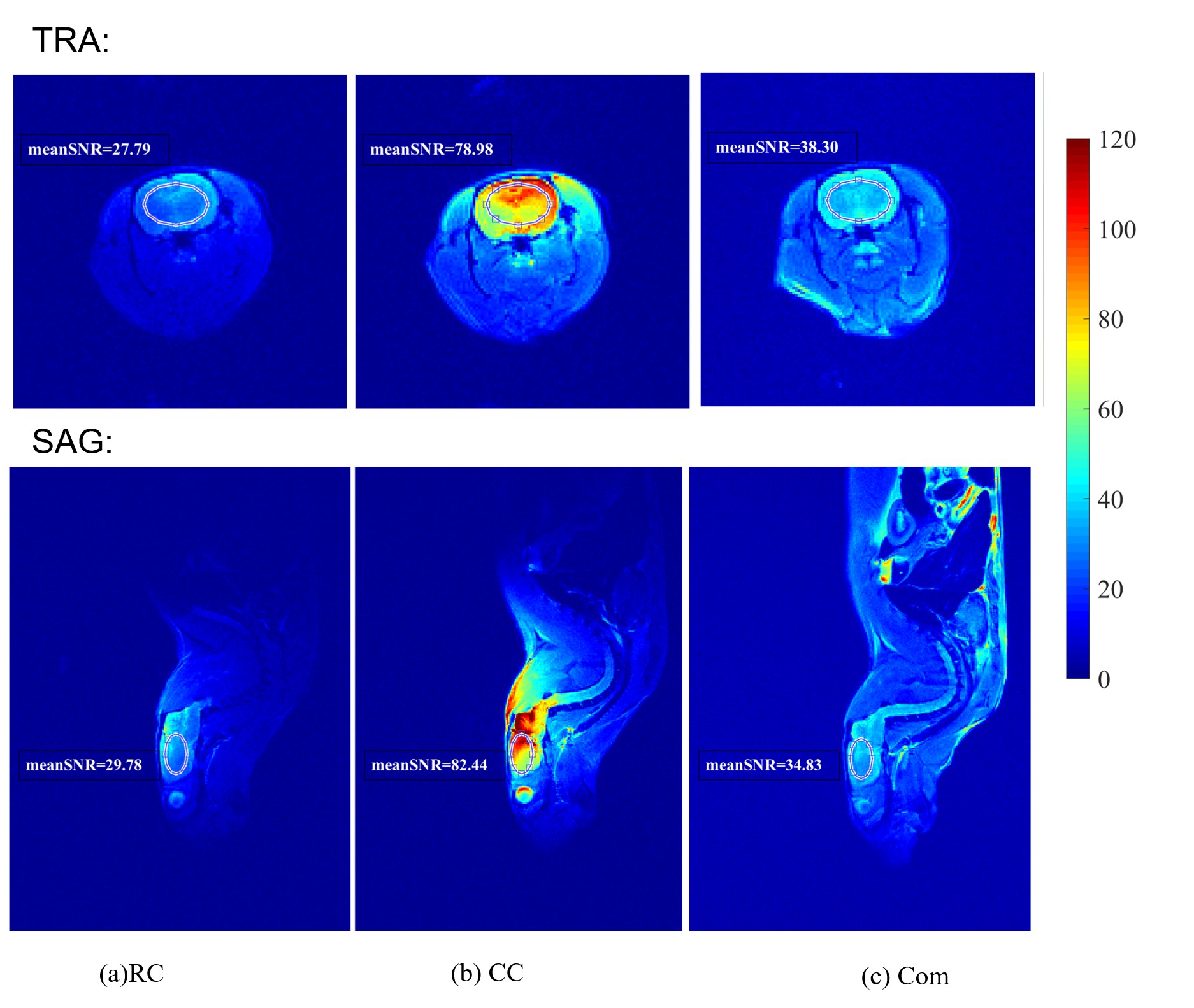
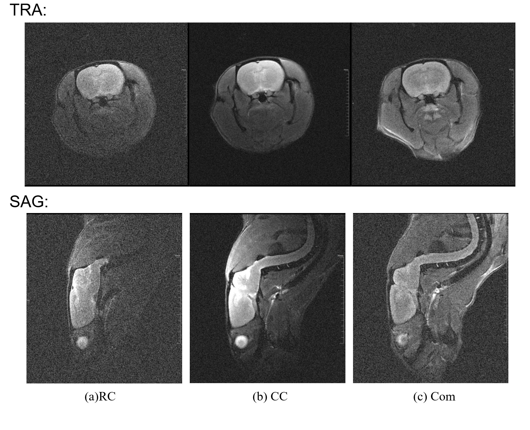
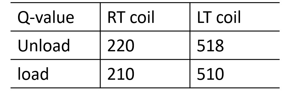
According to Table I, the loaded Q values of both coils vary slightly compared to the unloaded Q. This suggests that the coil noise dominates the whole noise of this experiment at a distance of 3 cm. Then we can see the loaded Q of CC is enhanced by a factor of 1.43 compared to that of RC which is approximately consistent with the comparison of the SNR map of CC and RC according to the classical calculation model between Q and SNR6,7. Therefore, we can demonstrate that decreasing the coil noise can result in a considerable enhancement of its intrinsic performance, even at an imaging distance of 3 cm.Furthermore, it also offers significantly clearer images than the commercial 12-channel coil in the upper half of the field of vision (FOV). The SNR map shown in Fig.2 (a) and Fig.2 (b) demonstrates that the cryogenic coil achieves a 1.8-fold SNR gain and it is significantly better than the commercial rat coil at a 3 cm distance. This strongly validates the performance advantage of the cryogenic coil. The imaging results also show that we can obtain a high SNR gain under the premise of expanding the coil diameter and imaging FOV. Fig. 3 presents high-resolution images of the same slice of the rat brain region under three conditions. The results indicate that cooling down the coil enhan over all three cases.
Discussions and Conclusions
The cryogenic coil in our work can significantly improve the SNR of the images at long distances while guaranteeing a large imaging FOV, and demonstrates a better performance than a type of commercial rat coil under the same conditions. The designed coil can help to improve the quality of MR imaging in some scenarios where the object to be measured is far away from the RF receive coil.Acknowledgements
This work was supported in part by the Project on Global Common Challenges of Chinese Academy of Sciences (No. 321GJHZ2022081GC), the NSFC grant (U22A20344), the Key Laboratory for Magnetic Resonance and Multimodality Imaging of Guangdong Province (2023B1212060052), the Funding Program of Shenzhen, China (RCYX20200714114735123), the Chinese Academy of Sciences Youth Innovation Promotion Association funded project (Y2021098).References
1 Labbé, A. et al. Recent advances and challenges in the development of radiofrequency HTS coil for MRI. Frontiers in Physics 9, 705438 (2021).
2 Wosik, J., Nesteruk, K., Tan, I.-C., Qin, K. & Bankson, J. A. in ISMRM. 1791.
3 Junge, S. Cryogenic and Superconducting Coils for MRI. (eMagRes, 2012).
4 Hu, B. et al. A Novel Receive-Only Liquid Nitrogen ($\hbox {LN} _ {2} $)-Cooled RF Coil for High-Resolution In Vivo Imaging on a 3-Tesla Whole-Body Scanner. IEEE Transactions on Instrumentation Measurement Science and Technology 61, 129-139 (2011).
5 Niendorf, T. et al. Advancing cardiovascular, neurovascular, and renal magnetic resonance imaging in small rodents using cryogenic radiofrequency coil technology. Frontiers in pharmacology 6, 255 (2015).
6 Sack, M., Wetterling, F., Sartorius, A., Ende, G. & Weber‐Fahr, W. Signal‐to‐noise ratio of a mouse brain 13C CryoProbe™ system in comparison with room temperature coils: spectroscopic phantom and in vivo results. NMR in Biomedicine 27, 709-715 (2014).
7 Darrasse, L. & Ginefri, J.-C. J. B. Perspectives with cryogenic RF probes in biomedical MRI. 85, 915-937 (2003).
Figures