1588
Development of a double resonant fluorine-19 and proton coil for low-field MRI experiments1Electrical Engineering and Information Technology, South Westphalia University of Applied Sciences, Luedenscheid, Germany
Synopsis
Keywords: Low-Field MRI, Low-Field MRI, X-nuclei
Motivation: Our project aims to refine MRI technology for a specific clinical purpose, focusing on a double-resonant coil design. This innovation bridges knowledge gaps and enhances diagnostic precision.
Goal(s): Our goal during the current semester is to create a double-resonant coil for MRI experiments on fluorine and protons. Achieving this involves circuit design and hardware construction.
Approach: Our approach includes capacitor and inductor integration, two separate output channels, and the development of essential hardware for signal acquisition using a didactic low field MR system.
Results: The design allows us to measure T1 and T2 relaxation times and enables us to perform imaging as well.
Impact: With the coil we performed proof-of-principle fluorine and proton measurements in a didactic low MR-system. It can be used to investigate samples with 1H and 19F without moving the sample. The non-proton channel can be tuned to other x-nuclei frequencies.
Author contributions
Ibrahim Alami Merrouni, Amira Alouane and Mina Sakhi contributed equally to this work.Introduction
MRI is mainly based on hydrogen nuclei signals. Since hydrogen atoms are ubiquitous in the human body in the form of water molecules and other metabolic products, they are ideally suited for this type of imaging. MRI of hydrogen nuclei enables the investigation of many diseases and their effects on the human body. However, hydrogen MRI does not provide direct insights into biochemical processes or cellular metabolism.1In contrast to conventional MRI, in which hydrogen nuclei are aligned by a magnetic field, X-nuclei MRI aligns nuclei of other elements, such as fluorine. X-nuclei MRI was developed to provide additional information about metabolic processes, as hydrogen MRI alone is not sufficient to adequately capture these processes. The low natural abundance of these nuclei results in low signal intensity, which is why high-field MRI devices are often used. A more cost-effective alternative is low-field MRI.2
The selection of these two nuclides is based on their importance in biological and medical applications. Hydrogen-1 is the most commonly used in MRI because it is present in water and many biological molecules. On the other hand, fluorine-19 is of great interest due to its unique chemical properties and its use in contrast agents and labeling agents.1
Therefore we developed a double-resonant coil for low-field capabilities to detect fluorine and hydrogen using low-field MRI and to acquire the T1 and T2 relaxation times of 19F and 1H.
Methods
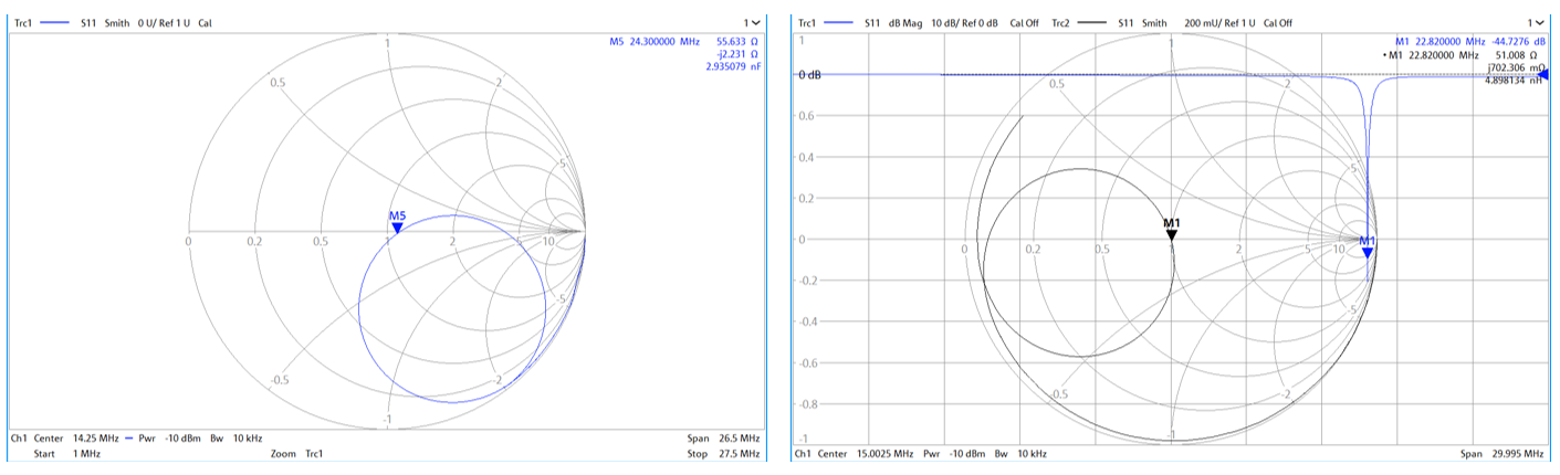
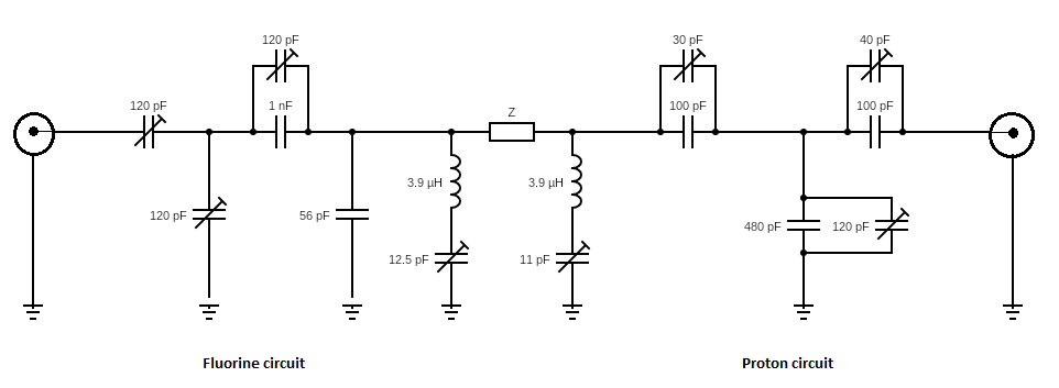
A circuit was developed for proton fluorine imaging that is used in conjunction with a cylindrical coil. The coil with seven windings was built from self-adhesive copper foil on a 3D-printed holder. Both channels were tuned using a vector network analyzer (ZNL3, Rohde & Schwarz GmbH & Co. KG, Munich, Germany) to the resonant frequencies of 19F (22.8 MHz) and 1H (24.3 MHz) and matched to 50 Ω. Figure 1 shows smith charts of the tuned and matched proton and fluorine double resonant coil. Printed circuit boards holding the gradient coils of the MR system were attached to the 3d printed coil probehead. A circuit diagram of the double-resonant coil is shown in Figure 2.The probehead was inserted into magnet (Magspec Benchtop Magnet, Pure Devices GmbH, Rimpar, Germany) with a flux density of B=0.57 T (Tesla). Two phantoms were used to maximize the signal in low-field MRI and because it is a double-resonant coil targeting two different nuclides (protons and fluorine). Phantoms with a perflouorotributylamine solution and a proton solution (oil) in 10 mm NMR tubes (Deutero GmbH, Kastellaun, Germany) were used during the experiments. The T1 relaxation time is determined using a saturation recovery sequence with a repetition time of TR = 10s for both measurements, while the T2 relaxation time is measured using an echo spin sequence with an echo time of TE = 1ms for protons and TE = 3ms for fluorine measurements. The MRI images of both phantoms were recorded using 2D spin echo sequence with sequence parameters of TR = 1s and TE = 6ms.Results
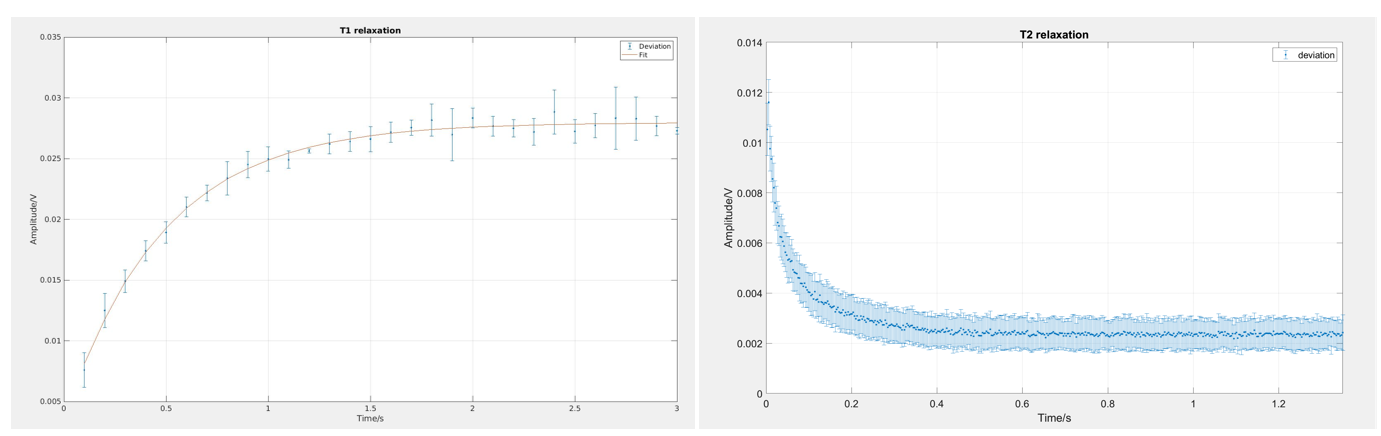
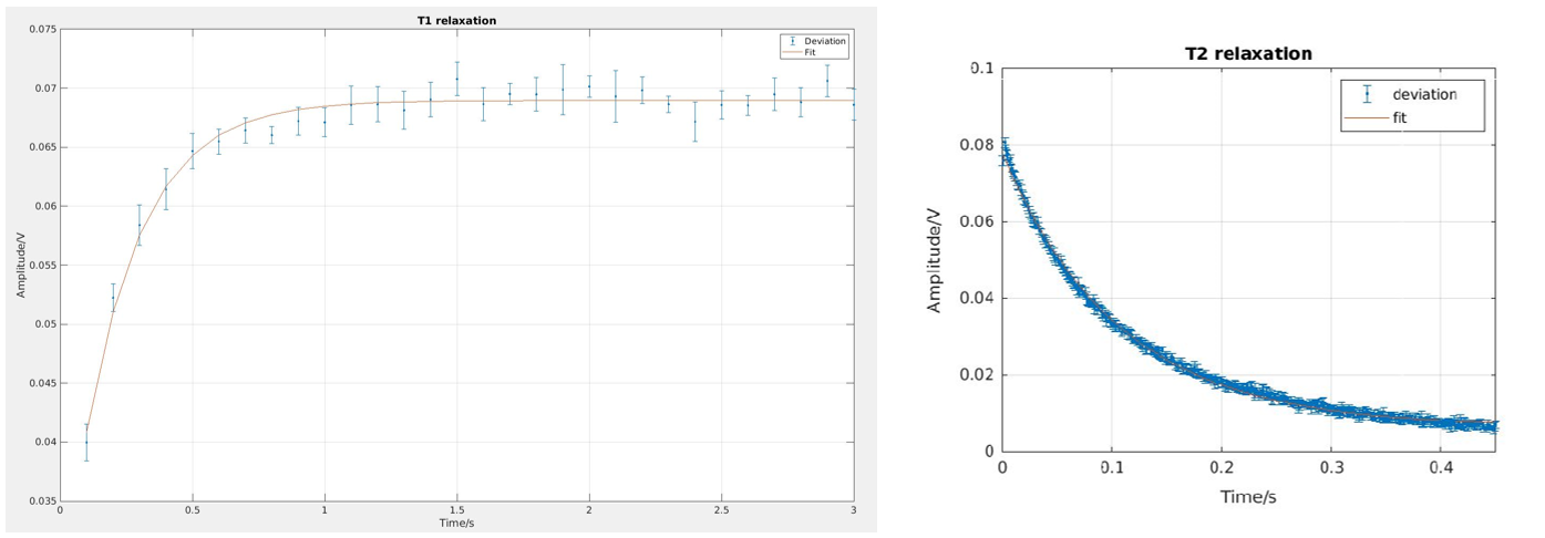
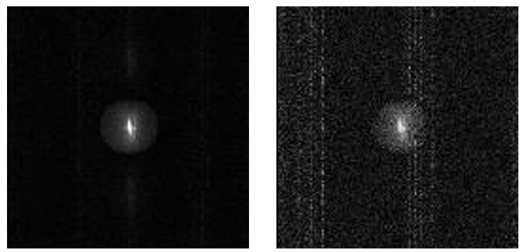
Regarding protons, the T1 relaxation time was calculated to be 0.2218 seconds and the T2 relaxation time was calculated to be 0.1065 seconds. Figure 3 shows the T1 and T2 fitted graph.For fluorine-19, the T1 relaxation time was determined to be 0.4823 seconds and the T2 relaxation time to be 0.0648 seconds. Figure 4 shows the T1 and T2 fitted data.The acquired fluorine and proton spin echo images are displayed in Figure 5. Both images exhibit RF-artefacts and a central brightening.Discussion
Small disturbances are caused by the unprotected circuit board, which is influenced by external RF sources. One way to prevent this interference would be to optimize the circuit board by shielding it appropriately or by miniaturizing the circuit board. In addition, the MR images have artifacts that occur due to external high-frequency interference. The central brightening occurs possibly due to insufficient shimming or gradient coil misplacement which will be adressed by redesigning the 3D printed probehead holder. Nevertheless, the coil delivers promising results in low-field MRI.Conclusion
In this project, a method for simultaneous MRI imaging of hydrogen-1 and fluorine-19 was developed. The results showed T1 and T2 relaxation times of 0.2218s and 0.1065s for the oil (proton channel) and 0.4823s and 0.0648s for fluorine. This technique opens up possibilities to capture biochemical and metabolic processes that are not accessible to conventional MRI techniques. In addition, the production of an inexpensive coil enables institutions that do not have a large budget available to carry out experiments and tests.Acknowledgements
No acknowledgement found.References
[1] Madelin, G. (2022). X-Nuclei Magnetic Resonance Imaging (1st ed.). Jenny Stanford Publishing. S.37-3
[2] Marques, J. P., Simonis, F.F.J., Webb, A. G. (2019). Low-Field MRI: An MR Physics Perspective. Journal of Magnetic Resonance Imaging 49(6):1528-1542
Figures