1587
A dedicated RF coil design for 31P MRS in the occipital lobe at 3T1Medical Imaging Center, University of Science and Technology of China, Hefei, Anhui, China, 2GE Healthcare, Shanghai, China, 3Anhui Fuqing Medical Equipment Co., Ltd., Hefei, Anhui, China
Synopsis
Keywords: Non-Array RF Coils, Antennas & Waveguides, RF Arrays & Systems
Motivation: The occipital lobe is the site of a variety of brain diseases. 31P MRS can noninvasively detect the metabolites to monitor and diagnose related diseases timely.
Goal(s): To collect 31P MRS signals in the occipital lobe at 3T, the dedicated RF coil setup is designed as a double-channel transceiver integrated surface coil.
Approach: The coil was designed as a "pillow” and verified by finite element simulation, physical production, bench tests and phantom experiments.
Results: The coil showed good uniformity and strong magnetic field intensity at the imaging region. Scanning with phantom, the spectral lines had a smooth baseline, high peaks, and excellent SNR.
Impact: we propose a transceiver and transmitter integrated surface coil for 31P magnetic resonance spectroscopy in the occipital lobe, which provides an excellent signal-to-noise ratio for excitation and acquisition of 31P spectral signals at 3T.
Keywords
Surface RF Coil, 31P MRS, MNS, Occipital Lobe.Introduction
The occipital lobe is the site of a variety of brain diseases, such as migraine, epilepsy, cerebral infarction and brain tumors, etc. Phosphorus magnetic resonance spectroscopy (31P-MRS) offers a very unique window to non-invasively look at tissue metabolism in vivo [1-2]. In vivo energy metabolism studied by phosphorus magnetic resonance spectroscopy (31P-MRS) can be of critical importance to diagnose different diseases [7]. The complexity of spectral patterns of 31P-MRS is reduced compared with 1H spectra, because fewer metabolites have a 31P nucleus present, and fewer resonances overlap [3-4]. Dedicated hardware is needed to detect MR signals from 31P nuclei. A clever design of this hardware can contribute to an improved signal-to-noise ratio (SNR) for detection of these signals.RF surface coils, regardless of their operating frequency, have been frequently used for their excellent, albeit spatially constrained SNR [8]. In this work, we propose a transceiver and transmitter integrated surface coil for 31P magnetic resonance spectroscopy in the occipital lobe, which provides a good signal-to-noise ratio for excitation and acquisition of 31P spectral signals at 3T.
Methods
31P MRS coil design: Considering that the phosphorus content in the human body is much lower than that of hydrogen protons and the magnetic resonance signal intensity is low, the RF coil of excitation and reception signal has higher sensitivity and signal-to-noise ratio requirements. Referring to the "pillow" used by humans during sleep, we designed the occipital lobe 31P coil as an arc structure to wrap the part of the human occipital lobe for magnetic resonance phosphorus spectrum signal acquisition (Fig.1). The surface coil was placed close to the occipital lobe of the brain to maximize the acquisition signal intensity.Electromagnetic field simulation: The coil EMF simulation was performed using FEKO software. The coil structure (Fig.2(a)) was established and the finite element simulation was performed to obtain the S-parameters (Fig.2(b)). The magnetic field distribution of the two coil channels at 51.71MHz is shown in Fig.2(c-d). The magnetic field in the space close to the coil is evenly distributed, with high intensity B1 field.
Bench tests: Frequency tuning and impedance matching of the surface coil were performed using a network analyzer (Keysight Technologies, model E5071C) [6]. The S-parameters obtained by bench debugging are shown in Figure 3. The test bandwidth is 5MHz and S11/S22/S21 are -26.23 dB /-28.92 dB /-15.63 dB, respectively.
Phantom experiments: NaH2PO4·2H2O and NaCl were used to prepare a cylindrical phantom with a concentration of about 8% phosphorus-containing solution. The phantom was placed on the coil, connected to the circuit through the MNS TR Switch, and tested on the GE 3T scanner (MR750, GE Healthcare, WI) for 31P MRS (Fig.4). During the phantom test, the scout image was scanned through the 1H transmitting coil, and then the 31P MRS signal was acquired. Calibration to a stable MNS center frequency was first performed by manual 2-3 pre-scans with the fidall bloch siegert sequence. Subsequently, the fidall csi 6×6×6 multi-voxel spectral acquisition sequence was scanned to obtain the phantom signal spectral lines shown in Figure 5.
Results and Discussion
Electromagnetic simulation showed that the magnetic field in the region of interest was evenly distributed, and the RF field signal strength was high. In the actual production, each channel coil is resonant and matched by adjustable non-magnetocapacitance. The bench test showed that the two channels were well isolated, and the quality factor was about 180 in no-load and 80 in load. Because of the good isolation of the two channels, one as transmitting and the other as receiving, there was no trap circuit built in the coil. As shown in Fig. 5(a), three curves were the sum of the collected signals, the maximum value and the root mean square, with a narrow and strong signal peak at the center frequency. Compared with the peak, the baseline was smooth and low in intensity, indicating the prominence of the phantom signal with a high SNR. Fig.5(b) corresponded to the layer with the strongest signal in the sampling voxel and the spatial location with the strongest signal.Conclusion
The dual-channel occipital lobe surface coil designed in this paper shows satisfactory signal transmission and reception performance in the 3T 31P MRS phantom signal acquisition. The spectral baseline with the multi-voxel MNS sequence acquisition is stable and the signal peak is sharp.Acknowledgements
Special thanks are extended to Professor Xiaoxiao Wang and Engineer Yong Zhang for technical guidance. We are indebted to the equipment support from “Anhui Fuqing Medical Equipment Co., Ltd.” The authors would like to thank Information Science Laboratory Center of University of Science and Technology of China for the measurement services.
References
[1] Alejandro Santos-Díaz a c, D M D N B C. Phosphorus magnetic resonance spectroscopy and imaging (31P-MRS/MRSI) as a window to brain and muscle metabolism: A review of the methods[J]. Biomedical Signal Processing and Control, 60[2023-10-31]. DOI: 10.1016/j.bspc.2020.101967.
[2] Schmitz B, Wang X, Barker P B, et al. Effects of Aging on the Human Brain: A Proton and Phosphorus MR Spectroscopy Study at 3T[J]. Journal of Neuroimaging, 2018. DOI: 10.1111/jon.12514.
[3] Bank B L V D, Orzada S, Smits F, et al. Optimized 31P MRS in the human brain at 7T with a dedicated RF coil setup[J]. NMR in Biomedicine, 2015, 28(11): 1570-1578.DOI:10.1002/nbm.3422.
[4] Ediv P, Kipfelsberger M, M. Dezortová, et al. Dynamic 31P MR spectroscopy of plantar flexion: influence of ergometer design, magnetic field strength (3 and 7 T), and RF-coil design[J]. Medical physics, 2015, 42(4): 1678-89.DOI:10.1118/1.4914448.
[5] Isaac G, Schnall M D, Lenkinski R E, et al. A design for a double-tuned birdcage coil for use in an integrated MRI/MRS examination[J]. Journal of Magnetic Resonance, 1990, 89(1): 41-50. DOI: 10.1016/0022-2364(90)90160-B.
[6] AOR, ATK, AP, et al. Design and test of a double-nuclear RF coil for 1H MRI and 13 C MRSI at 7T[J]. Journal of Magnetic Resonance, 2016, 267:15-21.
[7] Reyngoudt H, Paemeleire K, Descamps B, et al. 31P-MRS demonstrates a reduction in high-energy phosphates in the occipital lobe of migraine without aura patients[J]. Cephalalgia (Sage Publications Ltd, 2011(12). DOI:10.1177/0333102410394675.
[8] Wright SM, Wald LL. Theory and application of array coils in MR spectroscopy. NMR Biomed. 1997; 10(8): 394–410.
Figures
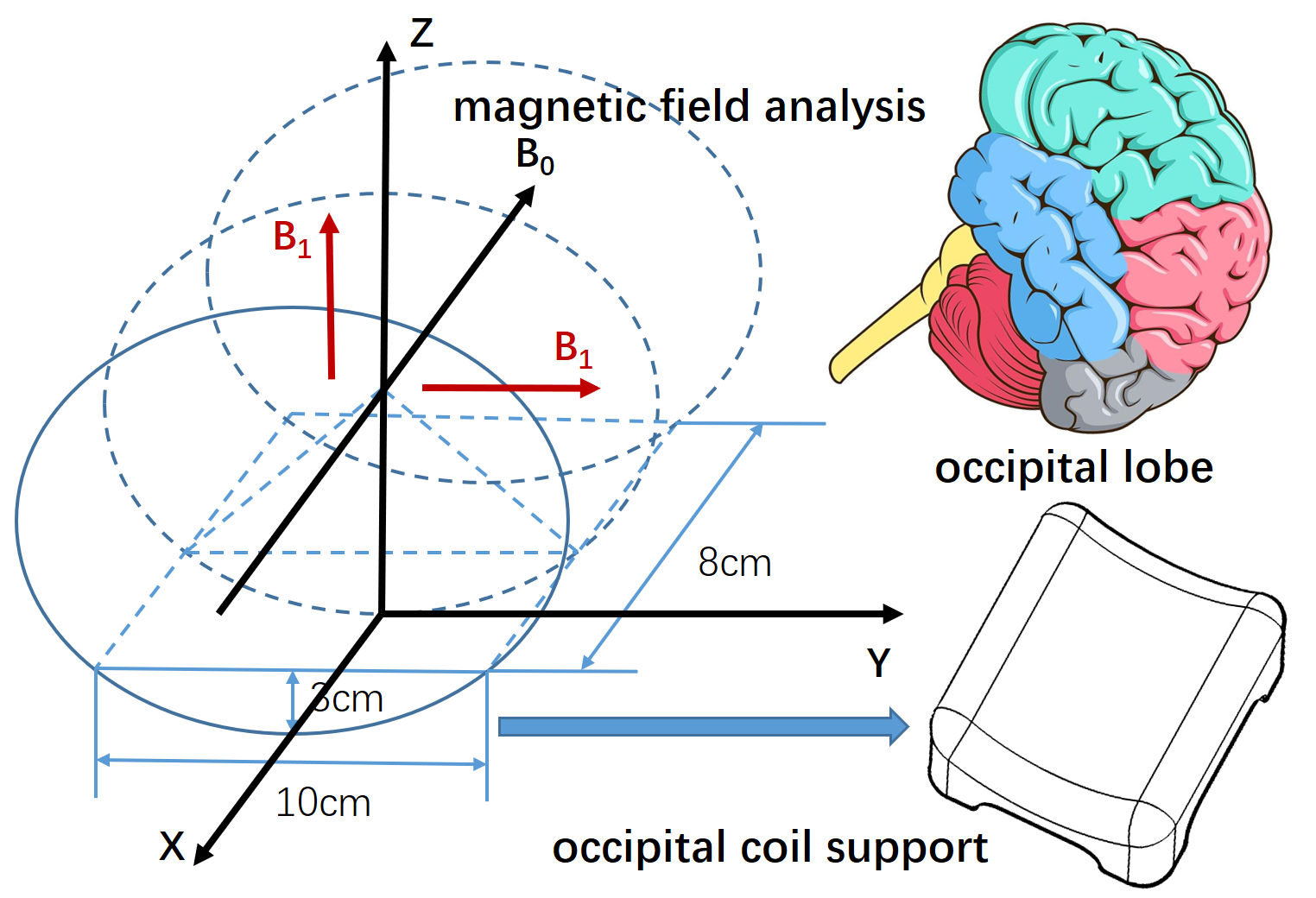
Fig.1 Design idea and size of brain occipital lobe 31P MRS coil. The coil scaffold is 100mm in axial width, 80mm in sagittal plane, and 30mm in depth, which wraps the occipital lobe in an arc.
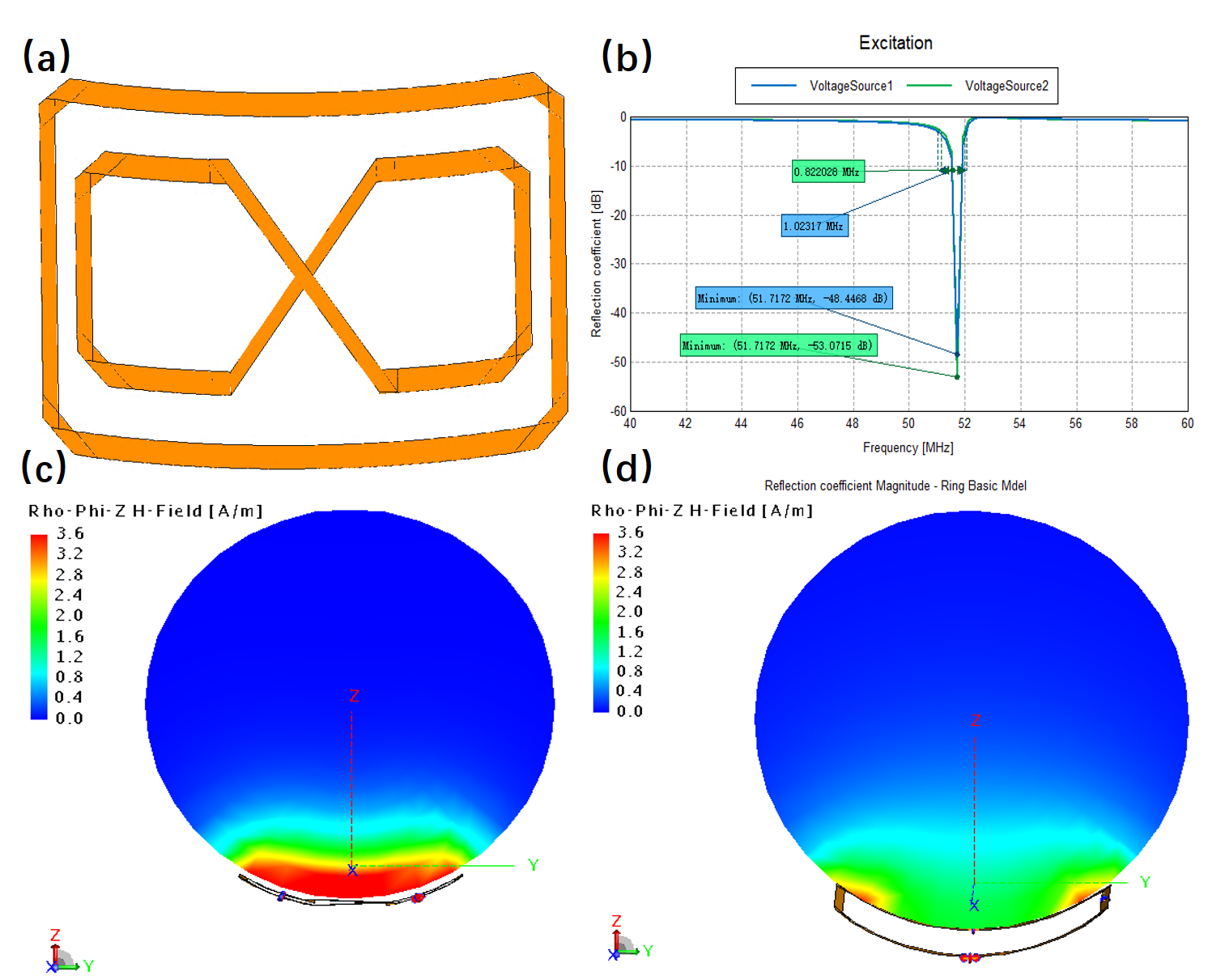
Fig.2 Coil structure modeling and electromagnetic field simulation results with the FEKO software. (a) Coil model; (b) S parameters obtained by adjusting capacitance parameters; (c) and (d) show the axial simulated magnetic fields of the single-channel butterfly and circular coils, respectively.
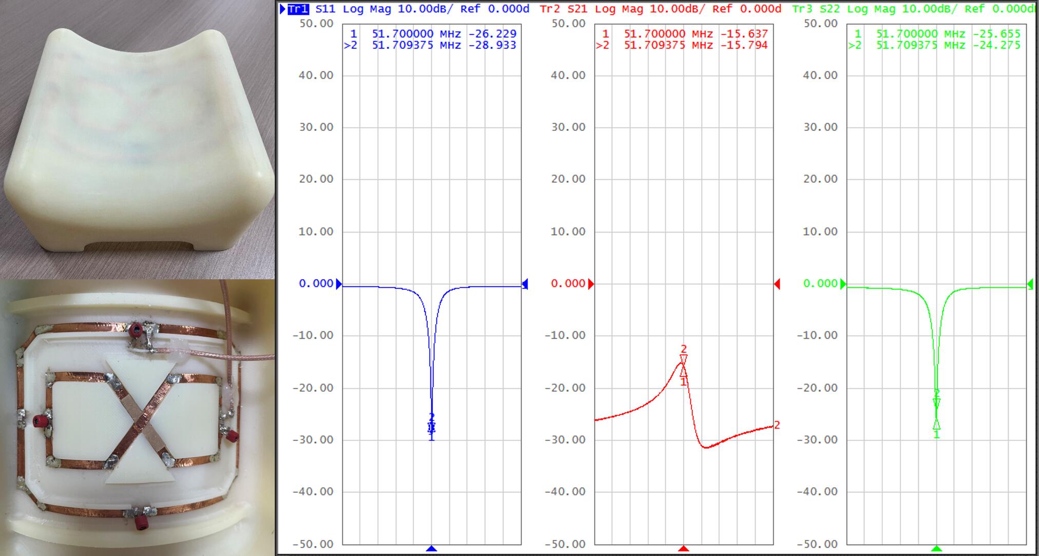
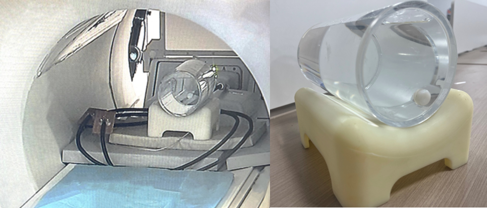
Fig.4 The cylindrical phantom containing phosphorus solution and the occipital lobe coil were scanned by phosphorus magnetic resonance spectroscopy.
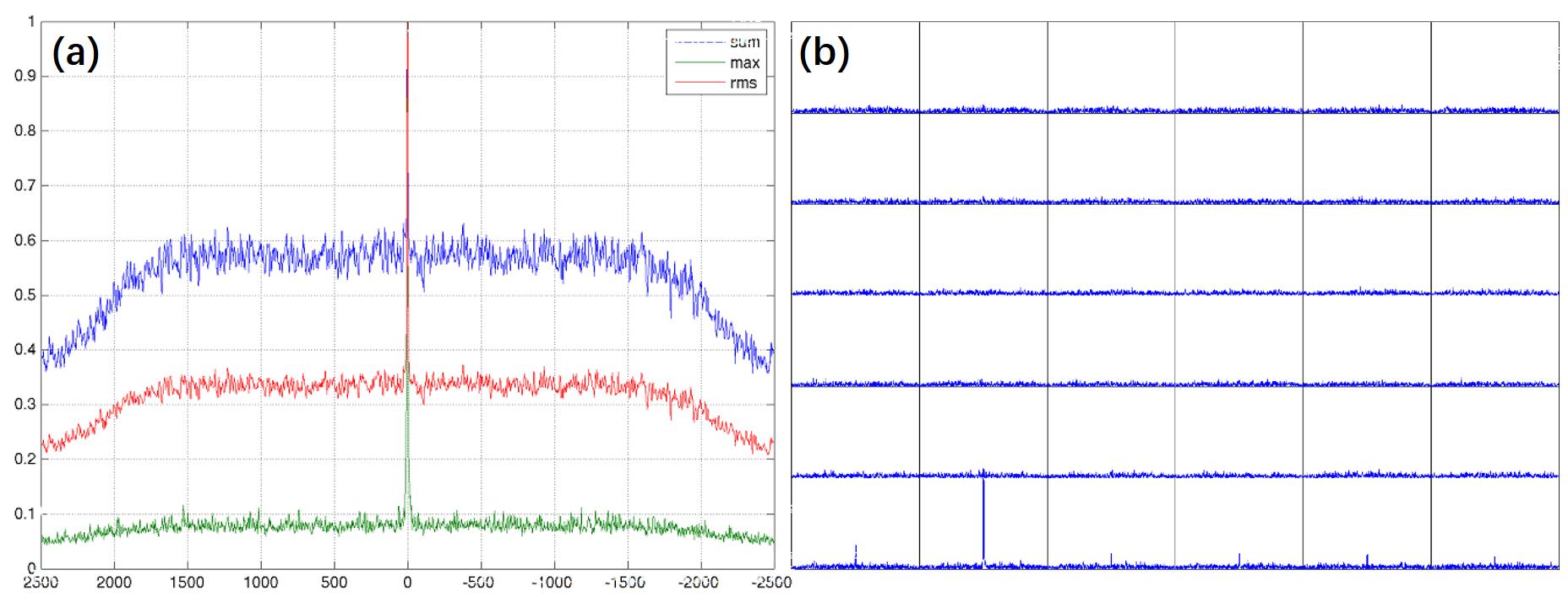
Fig.5 The center frequency and spectral lines of phosphor magnetic resonance spectra. The three curves in (a) are the sum of the collected signals, the maximum value and the root mean square, with a narrow and strong signal peak at the center frequency. (b) corresponded to the layer with the strongest signal in the sampling voxel and the spatial location with the strongest signal.