1586
Inductively induced triple-frequency tuned transceiver coil for multinuclear imaging at 7T1Center for Magnetic Resonance Research, University of Minnesota, Minneapolis, MN, United States, 2Department of Radiology, University of Minnesota, Minneapolis, MN, United States
Synopsis
Keywords: Non-Array RF Coils, Antennas & Waveguides, Non-Array RF Coils, Antennas & Waveguides, Novel RF coil
Motivation: Great potentials arise from multinuclear imaging modalities of 2H, 17O, 31P and 1H, which could depict the complexity of tissue and physiopathology related to the disease progression from multiple dimensions.
Goal(s): To develop a triple-tuned RF coil which can operate at three resonant frequencies of interest (2H, 17O, and 1H) or (2H, 31P and 1H).
Approach: A triple-frequency tuned RF coil was developed and simultaneously tuned and match to three resonant frequencies, and evaluated using a head-size phantom at 7T.
Results: We demonstrated a novel triple-frequency tuned coil design with robust imaging results for all three imaging frequencies.
Impact: As deuterium MRS imaging (DMRSI) is gaining more attention for brain tumor imaging, this triple-frequency tuned coil design can help add another X-nuclear frequency to the current DMRSI coil design, thus creating additional contrast for the brain tumor diagnosis.
Introduction
X-nuclei MRSI is a powerful imaging tool for non-invasively imaging various metabolites within the human body. It adds additional imaging contrasts to the 1H-based structure images, aiding the distinction between lesion tissue and normal tissue for diagnosing disease and guiding treatment. However, challenges arise when developing a robust multi-frequency tuned RF coil, which usually brings trade-offs between image performances across different Larmor frequencies. Traditional multi-nuclear coil design uses reverse bias PIN diodes to control the activation of additional capacitors or using LC trap circuits to introduce additional resonant frequencies 1-3. On the other hand, novel multi-tuned coil designs such as the nested coil 4 and common-mode differential-mode (CMDM) coil 5 only provide two resonating frequencies. To the best of our knowledge, there are no existing triple-tuned coil designs that do not rely on traditional concepts (i.e., PIN diode, LC trap circuit) that cause significant performance trade-offs between difference imaging frequencies.In this work, we present a novel triple-frequency coil based on a modified design from our previous double-tuned/matched coil concept6. This coil successfully generated high-quality 1H MRI images and two X-nuclear MRSIs in two different scenarios (2H, 17O, and 1H) and (2H, 31P and 1H).
Methods:
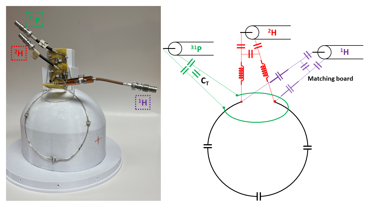
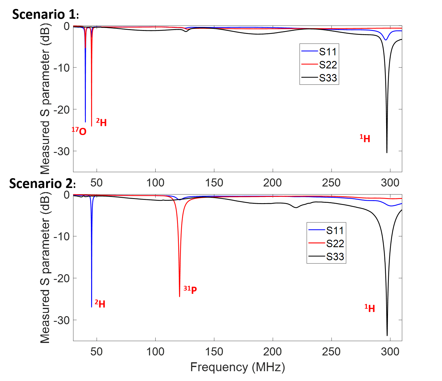
The triple-tuned coil is designed and constructed with a 16cm-diameter primary coil loop (black line in Fig. 1). The tuning to 2H and 1H resonant frequencies is achieved through the in-series inductors and capacitors, respectively 6. To generate the third resonant frequency, the self-resonant frequency of the secondary (small) loop (green line in Fig. 1) is adjusted by tuning the variable capacitor CT , which then inductively introduces the third frequency (31P or 17O) into the primary coil loop.This triple-tuned coil was loaded with an inorganic phosphate (Pi) head-size phantom and then tuned and matched to two scenarios: (2H, 17O, and 1H) and (2H, 31P and 1H), as shown in Fig. 2. In each scenario, the coil can be simultaneously matched and tuned to three resonant frequencies of interest.
The imaging of 1H MRI and X-nuclear MRSI was performed in the Siemens MAGNETOM 7T scanner. For all X-nuclear MRSI studies, the FSW-chemical shift imaging (CSI) was applied and acquired with: 18x18x18 cm field of view (FOV) and 9x9x7 matrix size under fully relaxed condition. The signal-to-noise ratio (SNR) of the X-nuclear MRSI was quantified as the spectrum peak divided by the noised level.
Results
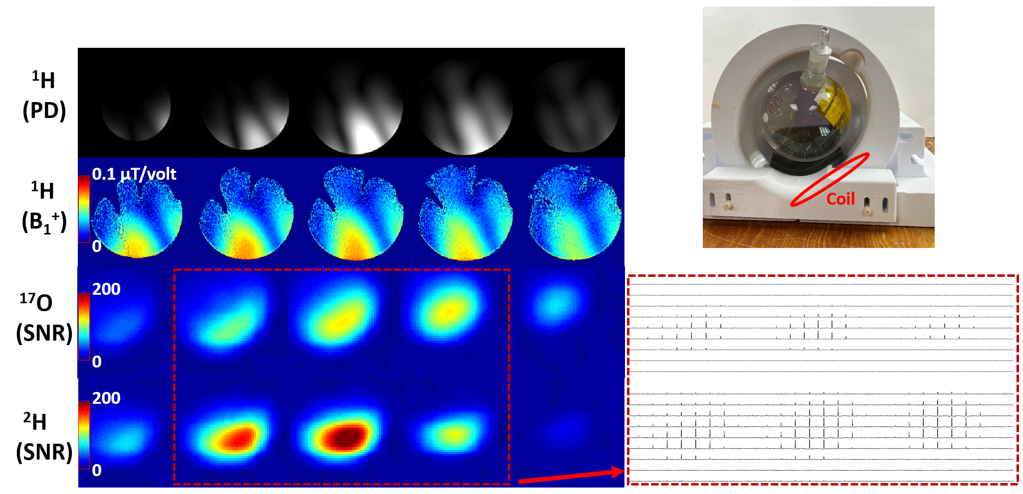
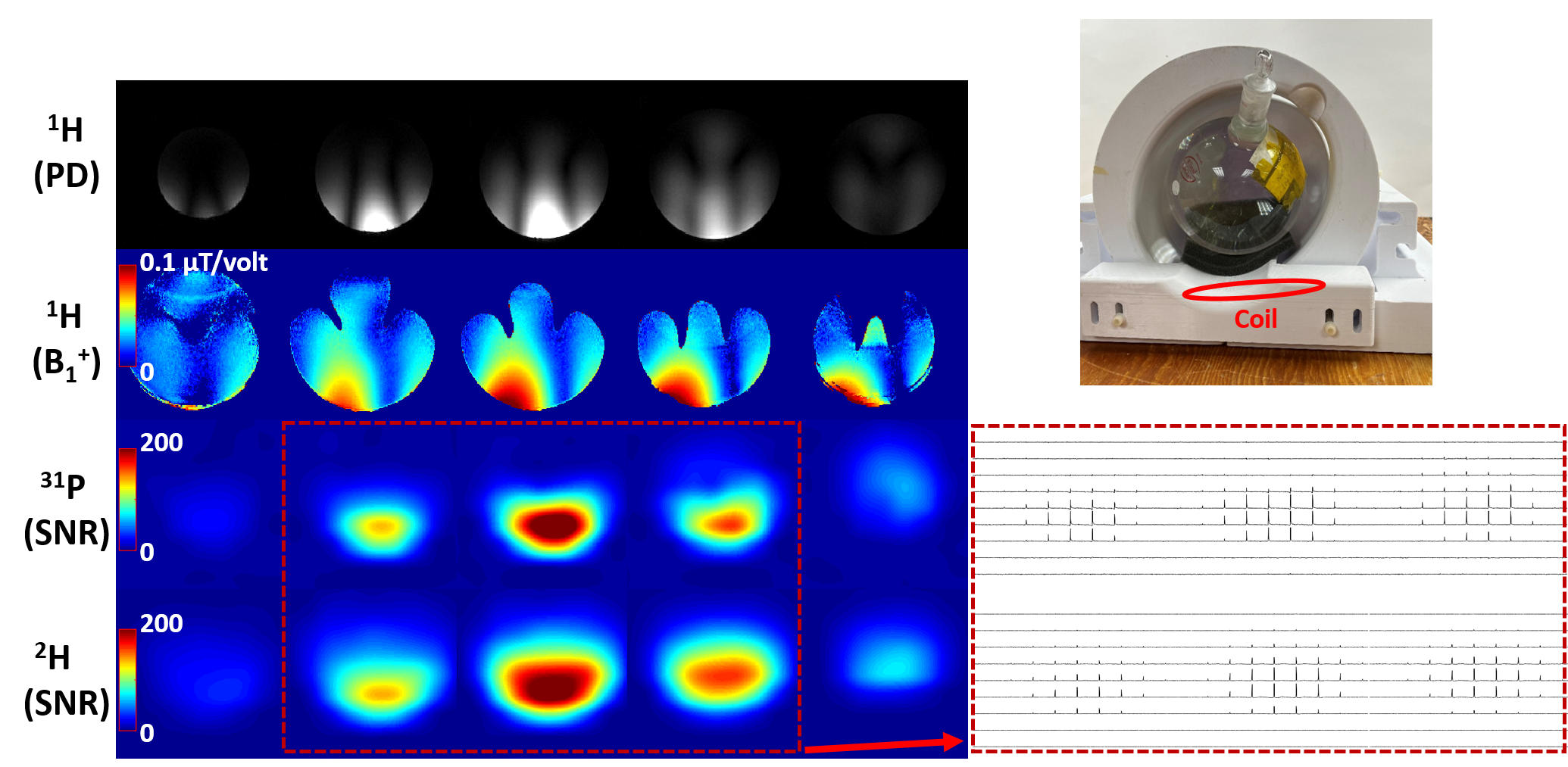
The triple-tuned coil under loaded condition can be tuned and matched to two scenarios: (2H, 17O, and 1H) & (2H, 31P and 1H), all with a reflection coefficient better than -20 dB as shown in Fig. 2. The imaging results of Scenario I and II are shown in Fig. 3 and Fig. 4, respectively. High-quality proton density images and proton RF transmit field (B1+) can be obtained for both scenarios, with consistent SNR values for all X-nuclei imaging measurements. It is worth noting that 17O MRSI has larger spectrum linewidth thus has a relatively lower SNR of natural abundance water signal as compared to 2H MRSI.Discussion:
We introduced a novel triple-frequency tuned coil design by using a secondary loop which inductively introduces an additional resonant frequency on the main coil loop. This additional frequency can be adjusted by changing the self-resonant frequency of the secondary loop (via tuning CT, see Fig. 1). The results demonstrate the third resonant frequency can be adjusted to between or close to 17O and 31P operating frequencies, and thus it is also possible to reach 23Na and 13C resonant frequencies at 7T. The proposed triple-frequency tuned coil design avoids the use of PIN diodes or LC traps, which are commonly used in traditional multi-frequency tuned coils to generate additionally resonant frequencies, and thus is easier to operate and can lead to more robust imaging results.Conclusion:
We have developed a novel triple-frequency tuned transceiver RF coil that can be tuned and matched to three resonant frequencies of interest simultaneously. This triple-tuned transceiver RF coil demonstrated robust imaging performance at multiple operating frequencies. Moreover, the coil design can be used to construct an array-coil for whole-brain triple-frequency MRI/MRS imaging applications.Acknowledgements
This work was supported, in part, by NIH grants: R01CA240953, U01 EB026978, R01NS133006 and P41EB027061.References
1. Choi, C.-H., et al., The state-of-the-art and emerging design approaches of double-tuned RF coils for X-nuclei, brain MR imaging and spectroscopy: A review. Magnetic Resonance Imaging, 2020. 72: p. 103-116.
2. Dai, J., et al., An RF coil design to enable quintuple nuclear whole-brain MRI. Magnetic Resonance in Medicine, 2023. 89(5): p. 2131-2141.
3. Avdievich, N.I., et al., Double‐tuned 31P/1H human head array with high performance at both frequencies for spectroscopic imaging at 9.4T. Magnetic Resonance in Medicine, 2020. 84(2): p. 1076-1089.
4. Brown, R., et al., A nested phosphorus and proton coil array for brain magnetic resonance imaging and spectroscopy. NeuroImage, 2016. 124: p. 602-611.
5. Pang, Y., et al., Common-mode differential-mode (CMDM) method for double-nuclear MR signal excitation and reception at ultrahigh fields. IEEE Trans Med Imaging, 2011. 30(11): p. 1965-73.
6. Li, X., et al. DOuble tuned and DOuble matched large-size loop coil (DODO) design and evaluation for 17O MRSI and 1H MRI application at 7T. in ISMRM & ISMRT 2023 Annual Meeting & Exhibition 03-09 June in Toronto, Canada, p. 5077.
Figures