1584
Potassium birdcage resonator design for multi-nuclear imaging at 7T1King's College London, London, United Kingdom, 2London Collaborative Ultra high field System (LoCUS), London, United Kingdom, 3MR Research Collaborations, Siemens Healthcare Limited, Camberley, United Kingdom, 4Stark Contrast, Erlangen, Germany, 5Department of Physics, The Royal Marsden NHS Foundation Trust, Sutton, United Kingdom, 6Division of Radiotherapy and Imaging, The Institute of Cancer Research, London, United Kingdom, 7Department of Medical Physics, GSTT, London, United Kingdom
Synopsis
Keywords: Non-Array RF Coils, Antennas & Waveguides, RF Arrays & Systems
Motivation: Potassium(39K) imaging provides an opportunity to probe cellular processes and has recently been demonstrated in human calf muscle at 7T
Goal(s): Developing an in-house birdcage resonator for potassium imaging, enabling simultaneous imaging with proton/sodium using previously developed dipole/loop array.
Approach: A low-pass eight-rung birdcage resonator was constructed using copper strip, chosen for its high Q factor. The resonator was optimized for maximum power efficiency and phantom images were acquired at 7T.
Results: The birdcage resonator demonstrated exceptional tuning and matching on the bench, with a homogeneous phantom image acquired in around 6 minutes. The findings suggest significant potential for in-vivo potassium quantification.
Impact: A birdcage resonator was designed and constructed in-house to create a capability for enhanced potassium imaging for neurological, musculoskeletal and oncology research. Potential integration with 1H/23Na array would allow simultaneous acquisition with three nuclei.
Introduction
Potassium(39K) plays a vital role in many cellular processes in neurological activities1 as well as skeletal muscle function2 and in oncology3. However, potassium imaging faces great challenges due to exceptionally low SNR arising from the low gyromagnetic ratio (~22 times lower than proton) and rapid T2 relaxation. Clinical feasibility of 39K quantification has recently been demonstrated with a commercial coil in skeletal muscle4. In this study, we designed and built a birdcage resonator for potassium integrating with a previously presented proton(1H)/Sodium(23Na) dipole/loop array5, with the eventual aim of imaging all three nuclei in one efficient examination. Here we present first results for the birdcage at 39K.Methods
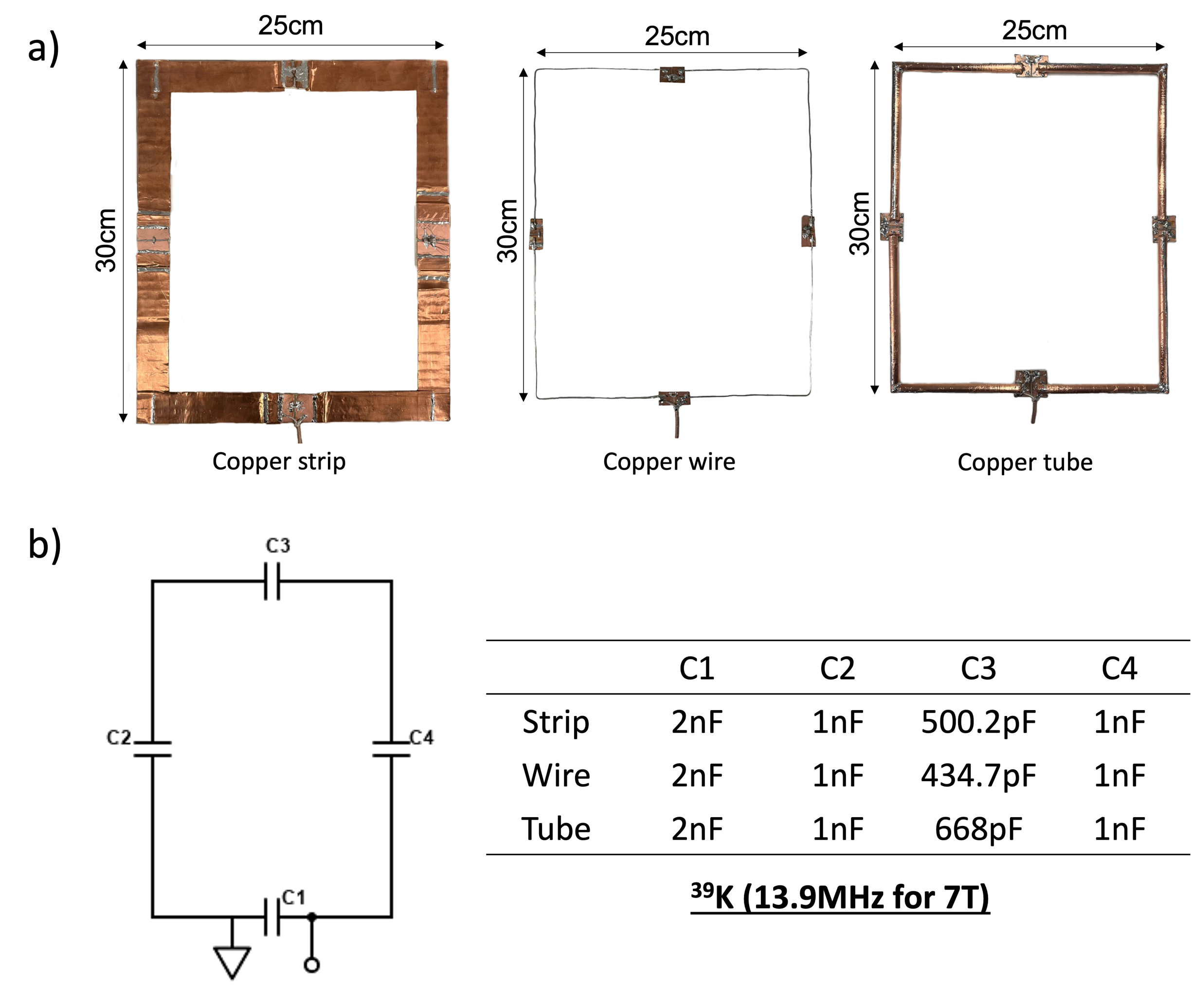
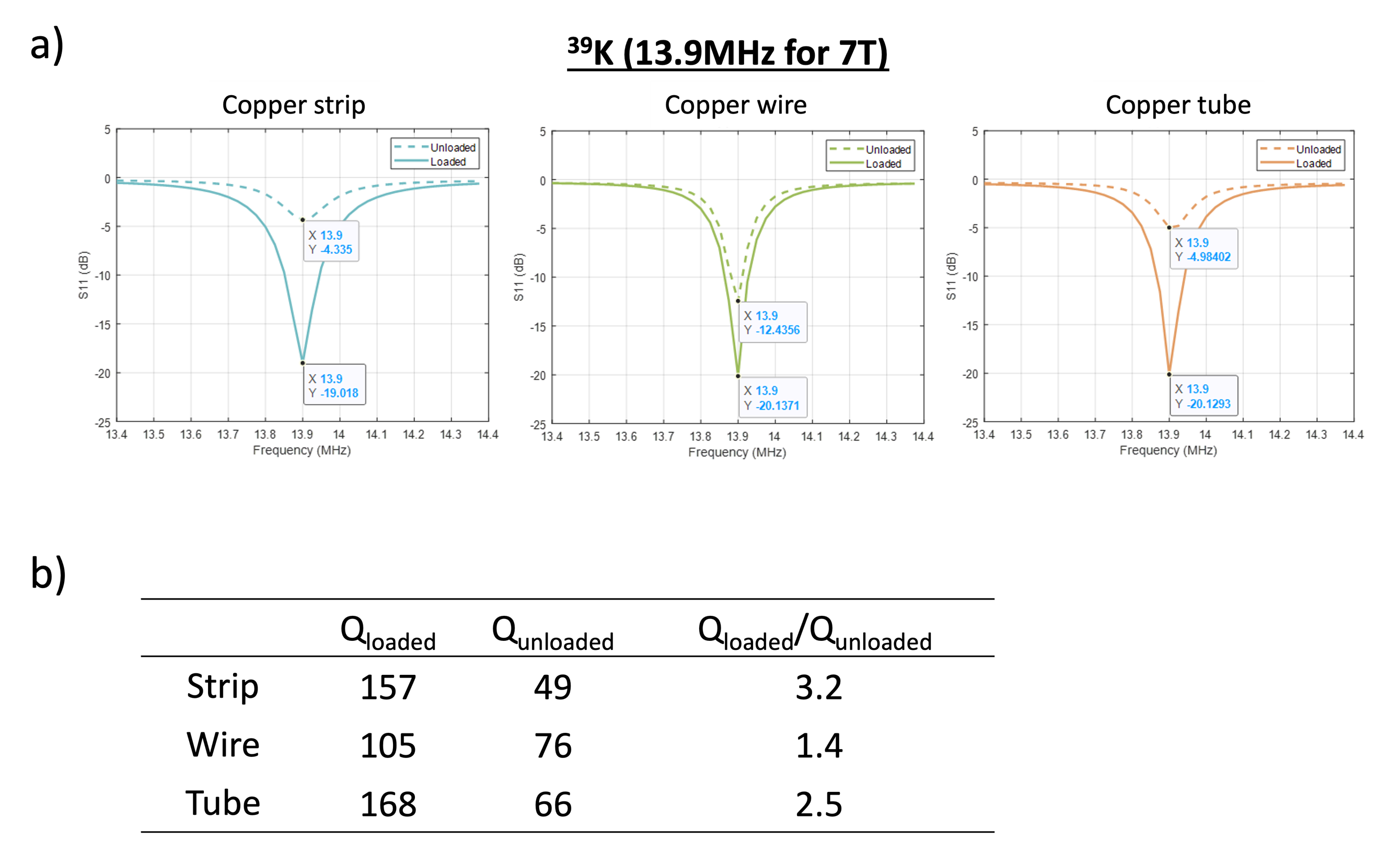
A 15 cm diameter spherical phantom containing 300mM KCl (3M KCl, Supelco mixed with tap water) was used to characterise the coil.Conductor comparison Prior to the birdcage construction, three loops of common conductors were built: copper strip (width=25mm, thickness=0.035mm,RS-PRO 542-5303), copper wire(diameter=1.22mm,RS-PRO 335-057), and copper tube (d=7.9mm) made by wrapping the copper tape around a hollow plastic tube. Four capacitors were distributed symmetrically along the loop coil to tune and match to 13.9MHz for 39K at 7T(Fig.1). Q factors were recorded on the bench from a vector network analyser (VNA) (E5063A ENA, Keysight) when the coil was either unloaded or loaded with the phantom (Fig.2).
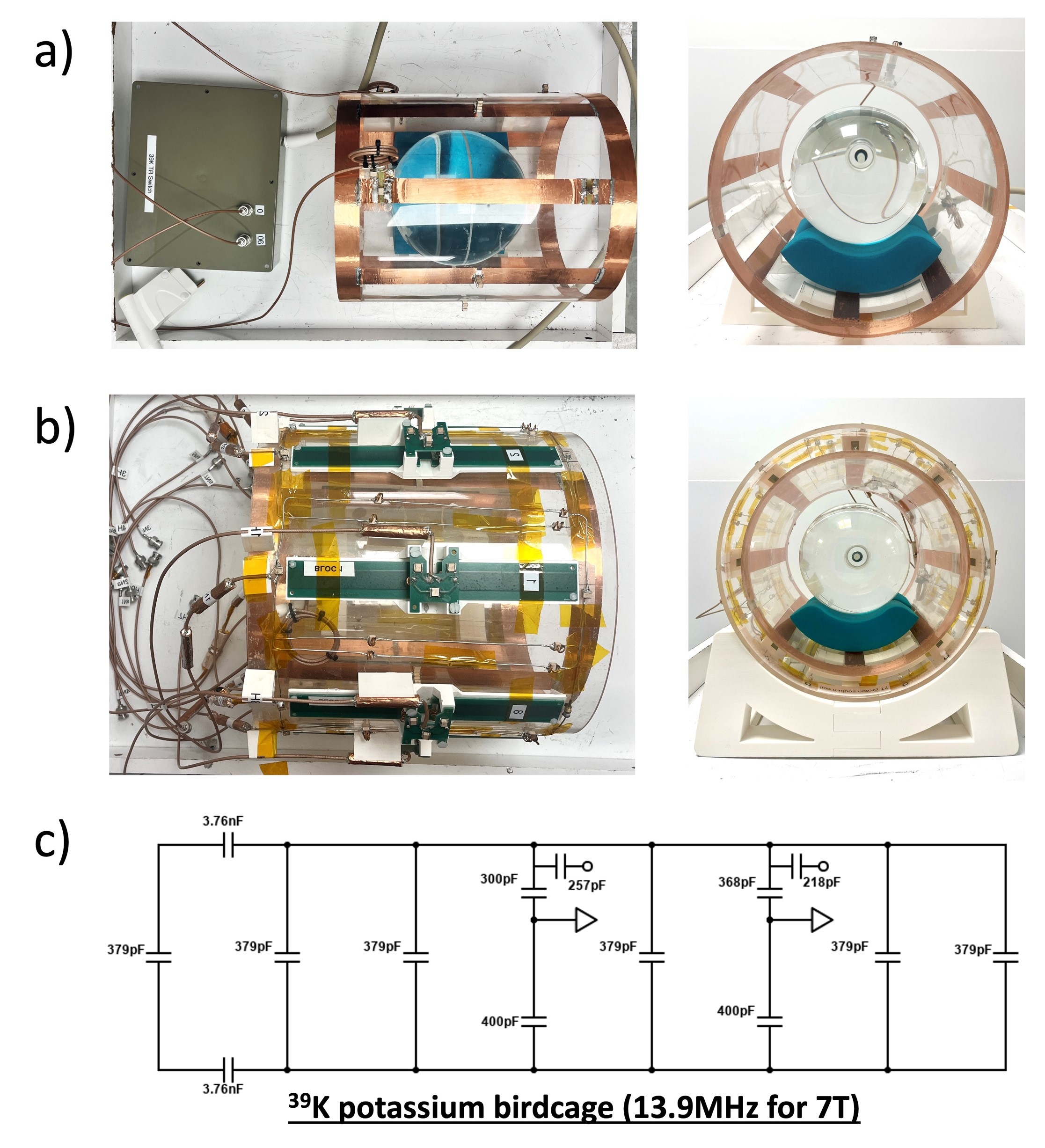
Birdcage construction A low-pass quadrature birdcage is designed and constructed with copper tape for potassium MRI at 7T. The resonator is composed of two end rings(d=25cm,w=2.5cm) and eight rungs(l=25cm,w=2.5cm) distributed equally around an acrylic cylinder that sat on custom-designed 3D printed holders(PLA,Ultimaker). The birdcage targets operation in two configurations, either performing on its own(Fig.3a) or inserting into an in-house developed 1H/23Na dipole/loop array(5) to allow image acquisition with three nuclei (Fig.3b). An analytical solution for the birdcage was derived with BirdcageBuilder6 and series capacitors of 379pF are inserted in the middle of the legs based on the calculation. A double probe was inserted in the middle transverse plane of the birdcage to ensure circularly polarised mode is tuned to the desired frequency. Two matching circuits made of a parallel and a series capacitor were then inserted into the sources that were 90 degrees apart on the legs to ensure a quadrature drive. Two capacitors are inserted symmetrically on both end rings to reduce eddy currents in the acquisition (Fig.3c). Cable traps tuned to the designated frequency are integrated along the coaxial cable feed (K_02252_D,Huber+Suhner, Switzerland) to inhibit common mode interference. A frequency shift of 0.27MHz was observed when the coil is placed at the centre of the bore, hence the birdcage was tuned to 13.61MHz on the bench to accommodate the shift and ensure maximum power efficiency in the acquisition.
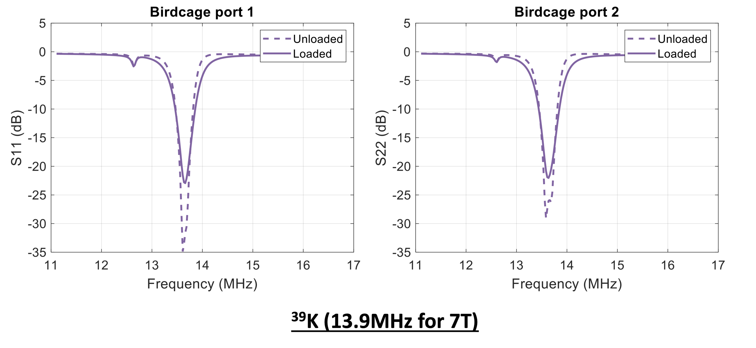
Bench measurement S-parameters when loaded by the phantom and unloaded were recorded from the VNA and optimised tuning and matching to 13.61MHz (shifted Larmor frequency within the bore) was achieved prior to MR acquisition (Fig.4).
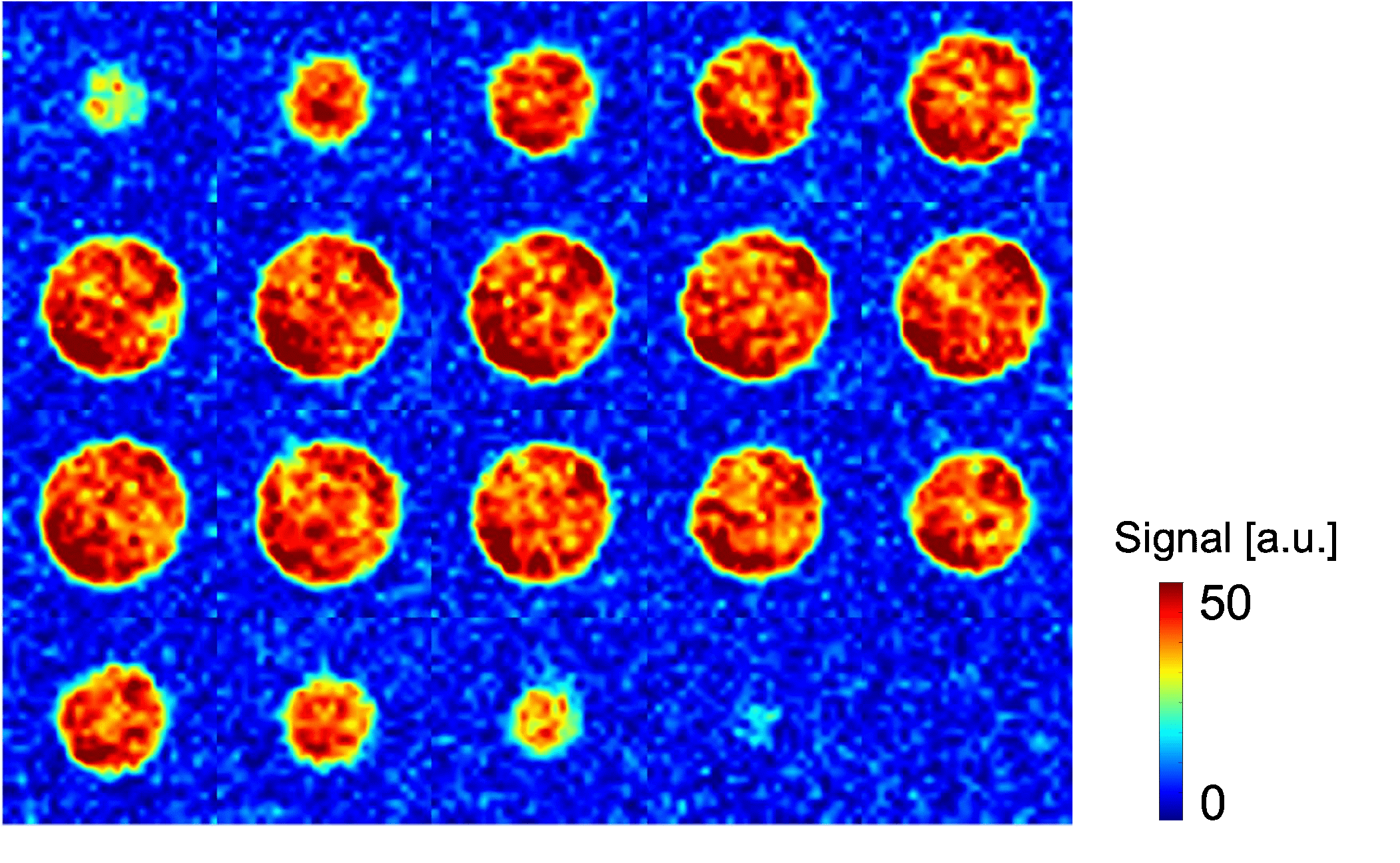
MR acquisitions MR images were acquired on a 7T MR scanner (MAGNETOM Terra, Siemens Healthineers AG, Germany) with the phantom. The coil was connected to a dedicated quadrature 39K TR switch (Fig.3a). A 3D ultrashort echo-time sequence (TR/TE=100ms/0.27ms, FA=90deg, 1 average, 8.6×8.6×8.6mm3, 58 slices, bandwidth=30Hz/pixel, 4000 radial spokes,TA=6min12s) was used to acquire the image. No post-processing steps were applied after the acquisition.(Fig.5)
Results and Discussion
The conductor comparison showed that copper strip and tube loops exhibited distinct Q ratios despite having identical geometry, which confirms the conductor's shape influences the Q factors, as noted in previous studies7.(Fig.2) Copper strip produced the highest Q factor with more than double of that for copper wires, therefore it was chosen to construct the birdcage resonator. Exceptional tuning and matching was demonstrated on the bench with less than -22dB in both ports.(Fig.4) Given the narrow 0.1MHz span of the peak at such low frequency, it is crucial to recognise the frequency shift when the resonator is transported from the bench to the scanner environment and make necessary adjustments during construction. Homogeneous. phantom image were acquired SNR of 11.7(mean signal/std noise=34.8/2.9), demonstrating great potential for in-vivo 39K quantification.(Fig.5) A phantom with double physiological concentration was used for this initial experiment, further assessment of the coil will be performed with phantom of sensible concentration.Conclusion
This study has demonstrated the feasibility of constructing a highly efficient birdcage resonator in-house for potassium MRI at 7T. Homogeneous images were acquired in a clinically feasible timeframe, underpinning the resonator's potential for in-vivo 39K quantification. The integrated configuration will be explored to enable simultaneous acquisition with 1H/23Na/39K in the future.Acknowledgements
This work was supported by King’s China Scholarship Council, by core funding from the Wellcome/EPSRC Centre for Medical Engineering [WT203148/Z/16/Z], Wellcome Trust Collaboration in Science grant [WT201526/Z/16/Z] and by the National Institute for Health and Care Research (NIHR) Clinical Research Facility based at Guy’s and St Thomas’ NHS Foundation Trust and King’s College London. JMW and GCE acknowledge funding from the NIHR Biomedical Research Centre at The Royal Marsden NHS Foundation Trust and The Institute of Cancer Research, London, and the Royal Marsden Cancer Charity. The views expressed are those of the author(s) and not necessarily those of the NHS, the NIHR or the Department of Health and Social Care.References
1. Benatar M. Neurological potassium channelopathies. QJM Mon J Assoc Physicians. 2000 Dec;93(12):787–97.
2. DiFranco M, Yu C, Quiñonez M, Vergara JL. Inward rectifier potassium currents in mammalian skeletal muscle fibres. J Physiol. 2015 Mar 1;593(Pt 5):1213–38.
3. Huang X, Jan LY. Targeting potassium channels in cancer. J Cell Biol. 2014 Jul 21;206(2):151–62.
4. Gast LV, Völker S, Utzschneider M, Linz P, Wilferth T, Müller M, et al. Combined imaging of potassium and sodium in human skeletal muscle tissue at 7 T. Magn Reson Med. 2021;85(1):239–53.
5. Wu M, Clément J, Vliem J. Proc. Intl. Soc. Mag. Reson. Med. 31. 2023. A 16-Channel Proton/Sodium Transmit/Receive Array Design for 7 Tesla Head Imaging.
6. Chin CL, Collins CM, Li S, Dardzinski BJ, Smith MB. BirdcageBuilder: Design of specified-geometry birdcage coils with desired current pattern and resonant frequency. Concepts Magn Reson. 2002;15(2):156–63.
7. Giovannetti G, Hartwig V, Landini L, Santarelli MF. Low-Field MR Coils: Comparison between Strip and Wire Conductors. Appl Magn Reson. 2010 Dec 1;39(4):391–9.
Figures