1580
Frequency Selective Flux Focusing Passive Lenz Resonators for Substantial MRI Signal-to-Noise Ratio Amplification1Physics, Lakehead University, Thunder Bay, ON, Canada, 2Chemistry, Lakehead University, Thunder Bay, ON, Canada, 3Thunder Bay Regional Health Research Institute, Thunder Bay, ON, Canada, 4Chemistry and Material Sciences, Lakehead University, Thunder Bay, ON, Canada, 5Northern Ontario School of Medicine, Thunder Bay, ON, Canada
Synopsis
Keywords: New Devices, New Devices, MRI, SNR enhancement, flux-focusing elements, Lenz lens, Lenz Resonator, Signal Amplification
Motivation: Despite numerous developments since MRI’s invention, low sensitivity remains the main limitation.
Goal(s): We aimed to improve upon the Lenz lens design for passive signal amplification in MRI, ultimately improving the signal-to-noise ratio.
Approach: We created a first-in-kind Lenz resonator, a passive frequency selective flux-focusing circuit, to isolate and enhance the signal from a desired nucleus.
Results: Performing RF testing with a vector network analyzer there was a 60 times signal amplification for the proton resonance frequency. At 3.0T, MRI demonstrated an experimental amplification of the signal-to-noise ratio by 3.9 times using an MRI insert of two coaxial Lenz resonators.
Impact: The substantial SNR boost produced by our Lenz resonators has a paramount importance for the field of MRI. The superior increase in SNR allows quicker scans, higher resolution scans, and precise disease detection.
Introduction
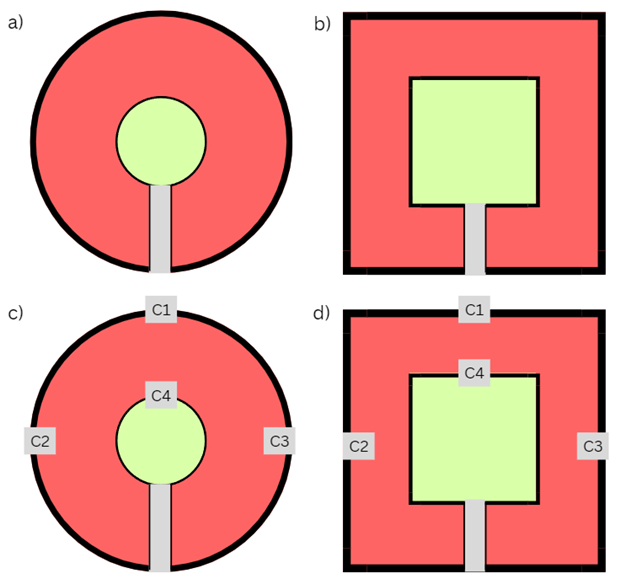
Since the original invention of MRI1, its main limitation remains the same: low sensitivity. Recently, the use of Lenz Lenses (LLs) – a passive flux focusing elements – have been explored for signal-to-noise (SNR) amplification for nuclear magnetic resonance (NMR) and magnetic resonance microscopy2-4. LLs are geometrically shaped loops of a conductor that focus the magnetic flux of the B1 field2-4, which is based on the reciprocity principle5 resulting in a subsequent MRI signal amplification. However, the non-selective nature of LLs makes the achieved SNR amplification quite limited4.To overcome this limitation, we designed a first-in-kind Lenz resonator (LR) by introducing a distributed capacitance (Fig. 1) tuned to the resonance frequency of protons (1H) at 3.0T. Using a system of two coaxial LRs, we obtained up to 3.9 times SNR amplification within a 150x150 mm2 field-of-view (FOV) in a clinical 3.0T MRI scanner. Moreover, we evaluated two different shapes of LRs – circular resonators and square resonators and compared their performance to LLs of the same size.
Method
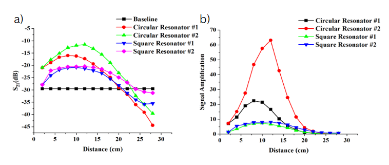
After establishing a preliminary design (Fig. 1), RF testing was performed to evaluate the performance of the designed LRs. Two custom-built rectangular RF coils tuned to 127.6 MHz (the resonance frequency of 1H at 3.0T) were connected to a two-port vector network analyzer (VNA) (Hewlett Packard 8751A) and were fixed at a separation of 28cm. The Lenz resonator or Lenz lens was inserted in between the coils and the transmission coefficient (S21) was measured as a function of the flux-focusing element position (Fig.2a). The obtained S21 dependences were recalculated into the signal amplification using the following equation:Amplification = 10(S21-S21baseline)/10 [1]
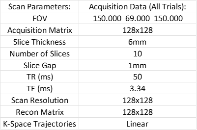
Following a preliminary RF evaluation, the performance of the designed LRs was assessed in a clinical Philips Achieva 3.0T MRI scanner. A pomegranate was chosen as a phantom for imaging. The pomegranate was placed in the middle of the quadrature body coil (QBC) and either two LRs or two LLs were placed 12 cm from the pomegranate, perpendicular to y-axis of the bore. A 2D multislice gradient echo imaging (GRE) was performed to assess the performance of the resonators. The imaging parameters are shown in Table 1. The images were reconstructed and analysed using custom scripts in Matlab 2021b (The Mathworks, Inc, Natick, MA).
Results
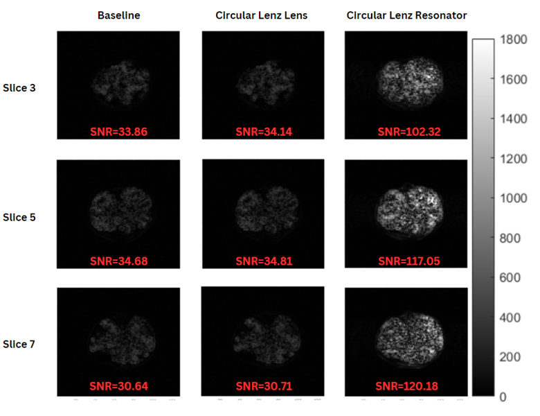
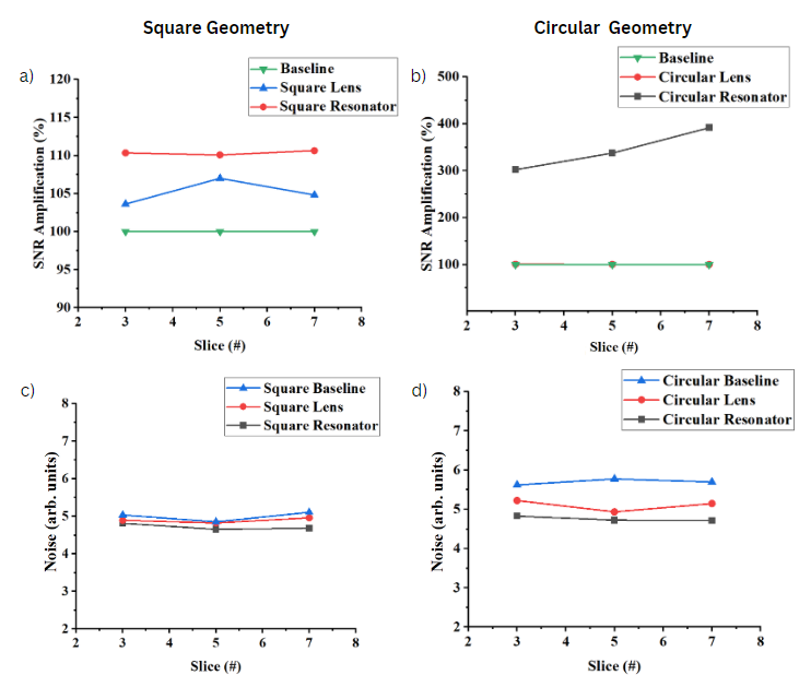
The measured transmission coefficient and its dependence on the LR position is shown in Fig. 2a. The circular LRs significantly outperformed the rectangular resonators in terms of signal amplification with a peak signal amplification at 8 cm and 12 cm from the receiver, respectively. It should be noted, however, that the obtained S21 dependence demonstrated a linear decrease after reaching its maximum amplification. On the contrary, the rectangular resonators demonstrated stable S21 enhancement for a wide range of distances (between 4 and 16 cm), albeit much lower enhancement compared to the circular resonators. The recalculated signal amplification dependence is shown in Fig. 2b. It can be clearly seen that rectangular geometry demonstrated much better reproducibility. The peak amplification achieved with the circular resonators was equal to approximately 64, whereas rectangular resonators demonstrated a peak amplification of 8. LRs substantially outperformed LLs (Fig. 2c,d).Fig. 3 shows representative slices of 2D GRE scans of the pomegranate. The first column shows the original image, the second column depicts images acquired with LLs, and the third column shows the respective slices acquired using two LRs. The image scale was selected to demonstrate the respective level of signal amplification from the LRs. The SNR of the original image was equal to 33.85, 34.68, and 30.64 for slices #3, #5, and #7 respectively. The implementation of LLs did not significantly improve the image SNR. Once the phantom was placed in between the two coaxial circular LRs, the image SNR increased by 302%, 337%, and 391% for slices #3, #5, and #7 respectively (Fig. 4 a,b), without a significant change in the noise level (Fig. 4c,d).
Discussion and Conclusion
The designed LRs completely outperformed LLs resulting in MRI SNR amplification up to 390%. While circular resonators demonstrated substantially higher SNR amplification, the rectangular geometry may have advantages for further preclinical and clinical usage due to its stable SNR performance over a wide range of distances from the signal source. The variability in the circular resonator’s performance can be attributed to small geometrical variations due to the difficulty of manufacturing a consistent circular shape. Overall, our pioneering work demonstrates the feasibility of novel LRs as a passive MRI insert for substantial SNR amplification (up to 3.9 times) in a clinically relevant FOV.Acknowledgements
This study was supported by a MITACS Accelerate grant (IT31144) and a MITACS Elevate grant (IT25574).
References
1. Lauterbur PC. Image formation by induced local interactions: examples employing nuclear magnetic resonance. Nature. 1973; 242:190-191.
2. J. Schoenmaker, K. R. Pirota, J. C. Teixeira. Magnetic flux amplification by Lenz lenses. Rev Sci Instrum. 2013; 84(8):085120.
3. J. Funk, P. While, N. Spengler, J. Korvink, RF Resonator with a Lenz Lens. US20170003360A1. May. 11, 2021
4. M. Jouda, Kamberger R., Leupold J., Spengler N., Hennig J., Gruschke O., Korvink JG. A comparison of Lenz lenses and LC resonators for NMR signal enhancement. Concepts Magn Reson Part B: Magn Reson Eng. 2017; 47B(3): e21357.
5. Hoult D., Richards R., The Signal-to-Noise ratio of the nuclear magnetic resonance experiment. J Magn Reson. 1976; 24:71-85.Figures