1576
Wireless MRI data transfer with an Integrated/Radio-frequency Wireless Spiral Coil Design for low-field portable MRI over cellular networks.1Duke University, Durham, NC, United States, 2Massachusetts General Hospital, Charlestown, MA, United States
Synopsis
Keywords: Low-Field MRI, RF Arrays & Systems, Wireless, Low-Field, Portable, Cellular
Motivation: Wireless transmission of MRI data acquired with low-field portable MRI scanners within EMT vehicles over cellular/satellite networks drastically decreases time between stoke onset and imaging for improved patient outcomes.
Goal(s): Our goal is to enable wireless communication with an iRFW coil design for simultaneously imaging and wireless cellular/satellite data transfer from within the scanner and an EMT vehicle.
Approach: iRFW-Cellular simulations within a portable 70 mT scanner are performed to evaluate its SNR and far-field gain patterns for wireless communication.
Results: The iRFW-Cellular simulations showed a uniform SNR in the head and gain patterns appropriate for the wireless transmission of MRI data.
Impact: The iRFW-Cellular spiral coil design potentially enables wireless MRI data transfer from a low-field portable MRI scanner inside, or out, of an EMT vehicle for better stroke onset to imaging times.
INTRODUCTION
Recent advancements in low-field portable point-of-care MRI scanners1 have enabled, for example, neuroimaging in hospital intensive care units or potentially of stroke patients in the back of Emergency Management Technician (EMT) vehicles to reduce the time to treatment for improved long-term outcomes. It has been estimated the probability of favorable outcomes for stroke patients is reduced by ~25% for every 30-minute delay between symptom onset and diagnoses by a radiologist after imaging3. Unfortunately, residents in rural populations are often far from a stroke center or hospital making them at higher risk for unfavorable stroke outcomes than those in urban areas4. Modern EMT vehicles in major urban areas are often equipped with expensive state-of-the-art satellite and cellular communication devices that allow first-responders to share patient data with physicians at the destination hospital to triage patients before arrival. Here, a low-field head-only MRI scanner located in the EMT vehicle and connected directly to on-board communication equipment could be used to image the brain of a stroke patient and wirelessly transmit the image data to a neuroradiologist for diagnosis. Unfortunately, rural EMT vehicles may lack on-board communication to transmit MRI data to radiologists, which increases the time between symptom onset and imaging. To address this, we propose a new multiband integrated radio-frequency/wireless (iRFW-Cellular) spiral coil design for simultaneous low-field MRI signal acquisition and wireless data transmission over 700 MHz Long-Term Evolution (LTE) and 2300 MHz satellite communication (S-band) networks for use in, and outside, an EMT vehicle. In this work, proof-of-concept iRFW-Cellular spiral coil design simulations in a portable head-only low-field 70 mT Halbach MRI 3.05 MHz scanner will be performed to evaluate its 1) signal-to-noise ratio (SNR) in a human head phantom and 2) ability to radiate wireless signals (i.e., far field Gain Patterns) from within the head-only scanner.METHODS
First, the portable MRI scanner main permanent magnet was modeled in an electromagnetic simulation program precisely placing more than 200 NdFeB rare-earth magnets within an RF-shielded enclosure that contours the human head to generate the field gradients for image encoding. The magnets and enclosure were assigned magnetic bias a perfect electric conductor (PEC) boundary conditions, respectively, to model the magnet’s internal magnetizations and the RF-shield. A spiral solenoid coil with 1-oz copper traces excited by 1 A 3 lumped port current source was modeled within the main magnet and close to a human water phantom. Next, the coil was tuned to resonate at 3 MHz, finite-element simulations of the model were performed to determine the electric and magnetic field in the head, which were exported onto a 2 mm isotropic Cartesian grid to calculate the average SNR7 and the SNR uniformity Coefficient-of-Variance (stdev/mean). The spiral solenoid was then modified into an iRFW-Cellular coil array by modifying the 18th turn to resonate at 700 MHz (i.e., LTE) by inserting along the trace a high-impedance filter resonate at this frequency, but short and open circuit at 3 and 2300 MHz. The 700 MHz feed structure was connected across the filter, RF-isolated at 3 MHz to maintain SNR and matched to 50 Ohms to maximize the gain pattern. Likewise, the 8th turn was modified to resonate at 2300 MHz (i.e., S-band) by inserting along the trace a high-impedance filter at this frequency, but short and open circuit at 3 and 700 MHz, with a similar feed structure as before but matched to 2300 MHz. Finally, the iRFW-Cellular SNR in the head and the far-field 700 and 2300 MHz gain and directivity were calculated to evaluate it wireless performance. Additionally, the gain patterns were plotted within the scanner to visualize the radiated power from the iRFW-Cellular design.RESULTS
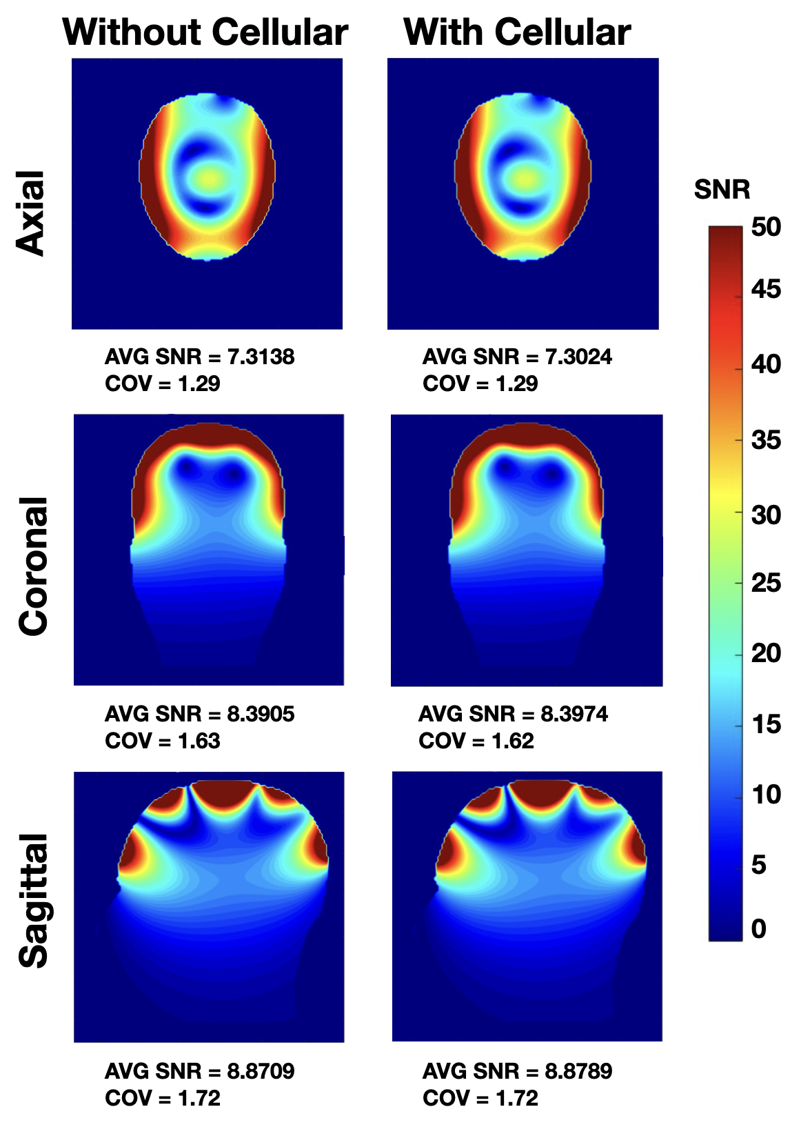
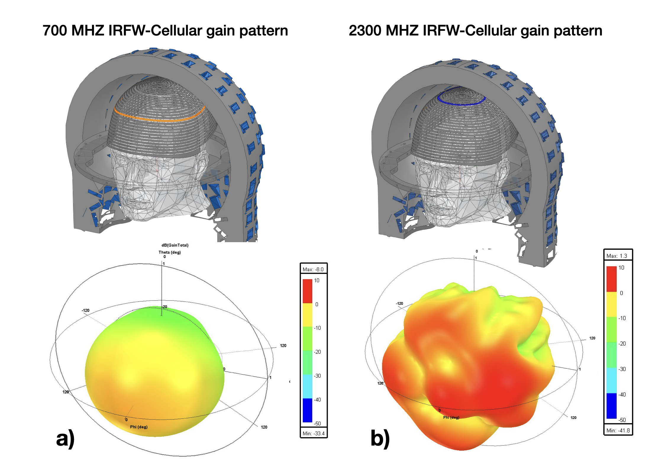
The MRI solenoid and iRFW-Cellular coil designs showed virtually no difference in SNR uniformity in the axial, coronal, or sagittal planes with COV values of 1.29, 1.63, 1.72 and 1.29, 1.62, 1.72, respectively (Fig. 1), which is a consequence of the high-impedance filter designs. Further, the 700 and 2300 MHz iRFW-Cellular far-field gain was -8.2 and 1.3 dB, respectively, and the corresponding patterns were directed outside the scanner bore (Fig. 2).DISCUSSION
The iRFW-Cellular solenoid coil design can be made to radiate in these, or other, cellular frequency bands by choosing turns with a circumference proportional to the operational wavelength that generate directed gain patterns outside the scanner. Here, solenoid turns of approximately 53 and 18 cm were chosen to resonate at 700 MHz and 2300 MHz because they generate gain patterns directed out of the scanner and toward the windows of an EMT vehicle for wireless transmission of MRI data.Acknowledgements
No acknowledgement found.References
1. Cooley CZ, McDaniel PC, Stockmann JP, Srinivas SA, Cauley SF, Śliwiak M, Sappo CR, Vaughn CF, Guerin B, Rosen MS, Lev MH, Wald LL. A portable scanner for magnetic resonance imaging of the brain. Nat Biomed Eng. 2021 Mar;5(3):229-239. doi: 10.1038/s41551-020-00641-5. Epub 2020 Nov 23. PMID: 33230306; PMCID: PMC8597947.
2. Ribo M, Molina CA, Cobo E, Cerdà N, Tomasello A, Quesada H, De Miquel MA, Millan M, Castaño C, Urra X, Sanroman L, Dàvalos A, Jovin T; REVASCAT Trial Investigators. Association Between Time to Reperfusion and Outcome Is Primarily Driven by the Time From Imaging to Reperfusion. Stroke. 2016 Apr;47(4):999-1004. doi: 10.1161/STROKEAHA.115.011721. Epub 2016 Mar 8. PMID: 26956258.
3. Georgakakos PK, Swanson MB, Ahmed A, Mohr NM. Rural Stroke Patients Have Higher Mortality: An Improvement Opportunity for Rural Emergency Medical Services Systems. J Rural Health. 2022 Jan;38(1):217-227. doi: 10.1111/jrh.12502. Epub 2020 Aug 5. PMID: 32757239; PMCID: PMC7862426.
4. Roemer PB, Edelstein WA, Hayes CE, Souza SP, Mueller OM. The NMR phased array. Magn Reson Med. 1990 Nov;16(2):192-225. doi: 10.1002/mrm.1910160203. PMID: 2266841.
Figures