1564
Making RF coils MR-invisible by additive manufacturing using magnetically filled polymer1ETH Zurich and University of Zurich, Zurich, Switzerland
Synopsis
Keywords: Non-Array RF Coils, Antennas & Waveguides, Challenges, Materials
Motivation: In MRI of tissues with short T2, materials of RF coils may be detected and can cause background artefacts. Current solutions to this problem either compromise sequence performance or impose restrictions on coil design.
Goal(s): Making RF coils MR-invisible.
Approach: The material used for constructing the coil housing is filled with ferrimagnetic material, leading to effective signal spoiling. Coil formers are created by additive manufacturing using custom filaments made from magnetite-filled polymer.
Results: Unwanted signals from the RF coil are eliminated by using coil formers made from magnetically filled polymer. Hence, background-free short-T2 imaging is enabled.
Impact: RF coils made MR-invisible by using magnetically-filled materials simplify coil design and manufacturing, and improve the performance of MRI of tissues with short T2, such as bone, tendon, lung, or myelin.
Introduction
In MRI, in addition to signals from the targeted object, unwanted signals from parts of the MR scanner – in particular RF coils – may also be detected, and cause artefacts. Usually, for such parts, materials that exhibit relatively fast signal decay are used, preventing detection with common echo times (TE). However, there is continuously growing interest in sequences with ultra-short or zero TE1, which are employed to image tissues with short T2. Therefore, background artefacts from RF coils have become a significant issue1-4.Approaches to avoid this problem at the sequence level include using a large field of view (FOV)1,5,6, subtraction of background signal7, T2-selective magnetisation preparation8, or outer volume suppression9. However, these methods require sequence modifications, compromise sequence performance, or offer only limited efficacy. Alternatively, materials with minimal 1H content such as glass and PTFE are used4,10-14, but this imposes restrictions for coil design, manufacturing, and handling.
In the present work, a new concept for eliminating background signal from RF coils is proposed, utilising local B0 distortion by magnetic materials. Such signal spoiling has previously been achieved with metallised textiles or by wrapping with magnetic wire12. Here, this idea is exploited more rigorously in that material used for constructing the coil housing is filled with ferrimagnetic microparticles15.
We demonstrate this concept by filling a polymer with magnetite and using it for additive manufacturing of coil formers. Formers with different base materials and filler load were assessed with respect to background signal, coil performance, and field homogeneity. The resulting utility is demonstrated by imaging in a phantom and in vivo.
Methods
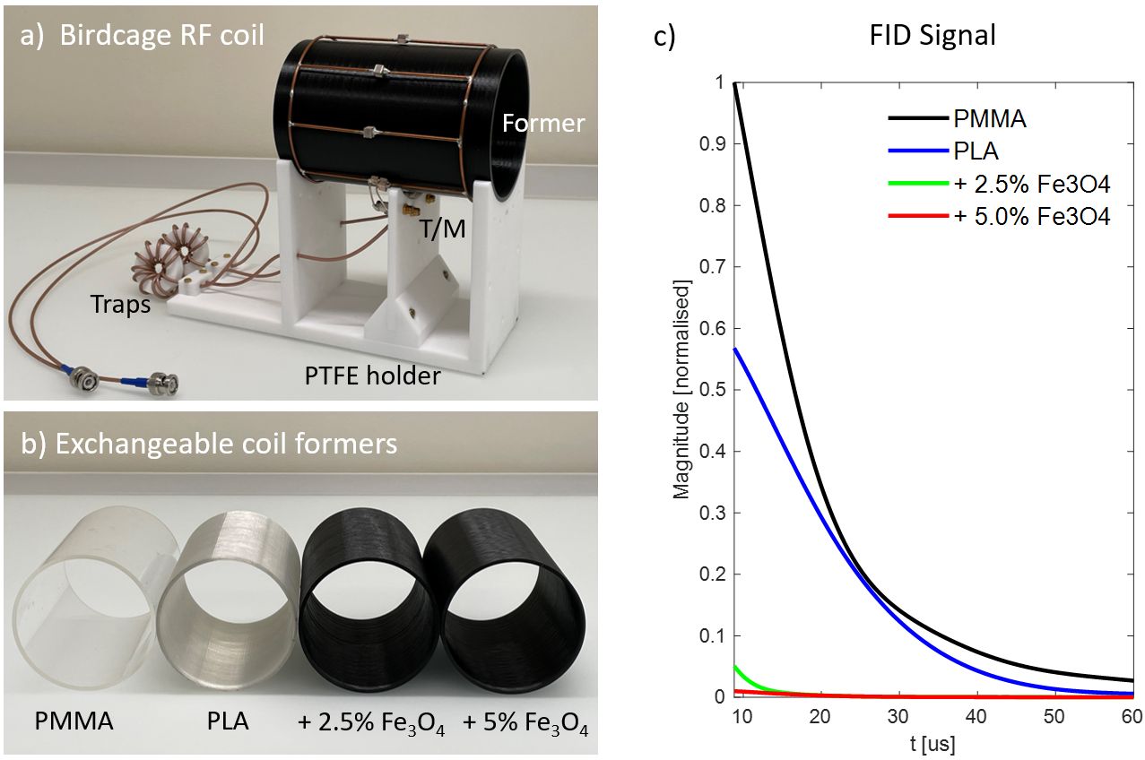
To compare formers made from different materials, a 1H-free birdcage coil was designed to be mechanically self-supporting. This allowed for exchanging a hollow cylinder which would normally act as the former (Figure 1a, b). The quadrature, low-pass birdcage was equipped with a matching network allowing for a large loading range.For the custom-made materials, polylactic acid (PLA, (C3H4O2)n) pellets were heated and extruded to produce filaments for additive manufacturing (Precision 350, 3devo, Utrecht, The Netherlands). For magnetic filling, either 2.5vol% or 5.0vol% of magnetite (Fe3O4) powder (particle size 1-20 µm, Kremer Pigmente, Germany) were added to the PLA pellets before heating.
Experiments were performed using a 3T Philips Achieva system equipped with a high-performance gradient16, a custom RF chain5, and fast transmit-receive switches17. For short-T2 imaging, a PETRA18 sequence with 3D isotropic geometry and a dead time of 10 µs was used.
Results
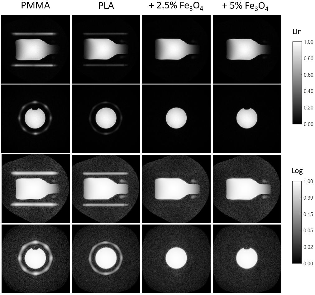
With all formers, comparable coil performance concerning quality factor, power efficiency and image SNR was found. Magnetic forces on the magnetite-filled formers were weak relative to their own weight.Figure 1c shows free induction decay signals for four different formers. PMMA, which serves as a reference, exhibits higher signal than PLA. With magnetite filling, the PLA signal amplitude at 10 µs is reduced by 91% and 98%, respectively. Accordingly, in the large-FOV images in Figure 2, the pure PMMA and PLA formers are clearly visible. With magnetite filling, the signal drops to noise level or below.
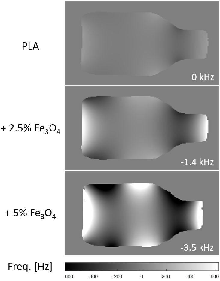
Figure 3 shows effects of the magnetic material on the static field in the imaging volume after second-order shimming. Field distortion is observed, which is larger than usually aimed at for standard MRI. However, in short-T2 imaging considerably higher gradient strengths are applied, making local resonance offsets less critical.
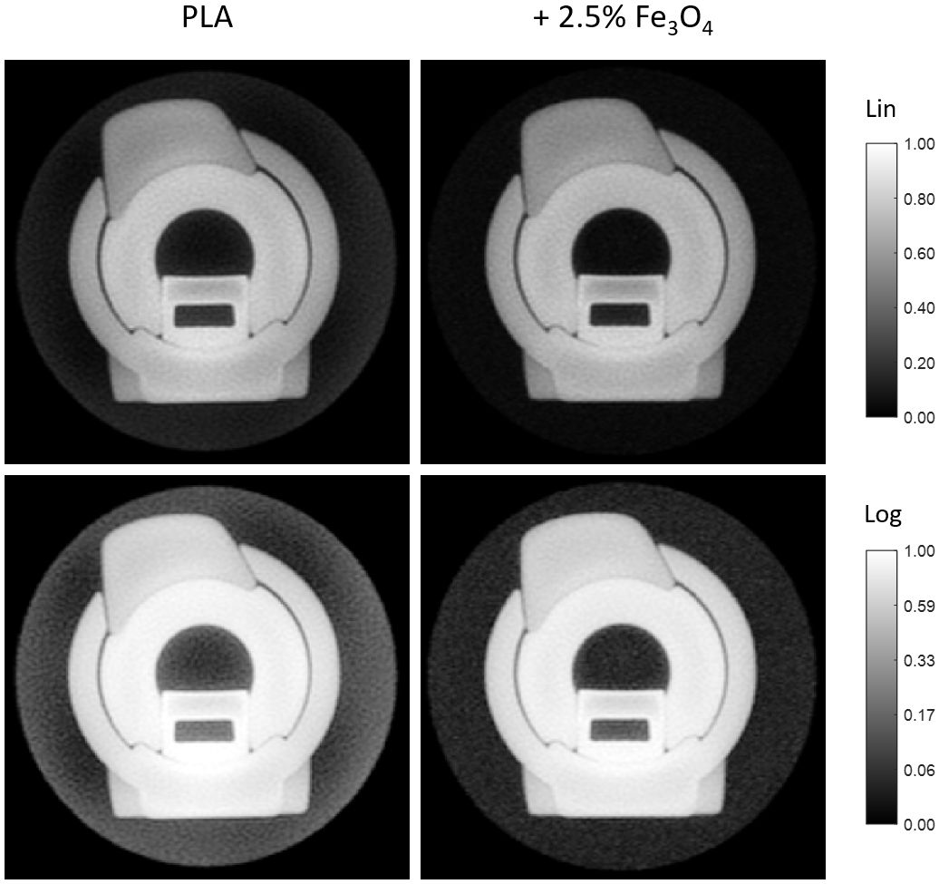
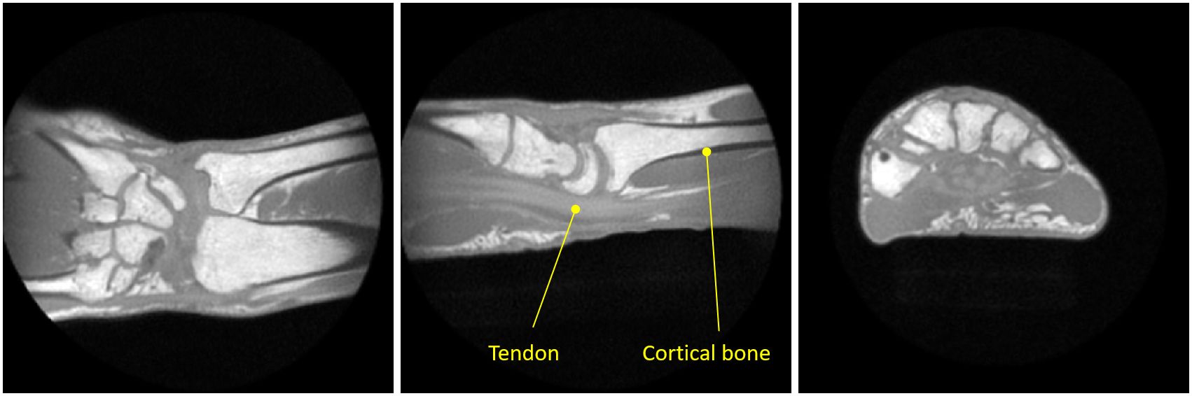
Given the results of Figures 2 and 3, the material with lower filling load is considered a good compromise between signal reduction and field distortion and was therefore used for successive experiments. Figure 4 demonstrates that in short-T2 imaging with object-matched FOV, aliased background artefacts are successfully eliminated with the proposed approach. Similarly, in-vivo experiments yielded clean images unaffected by background signal (Figure 5).
Discussion
In this work, a new concept for making RF coils MR-invisible was proposed and successfully demonstrated in short-T2 imaging with short dead time and high bandwidth. The particular implementation by filling polymers for additive manufacturing with magnetic material offers high flexibility for coil design and production.The main side effect of adding magnetic materials is reduced main field homogeneity in the imaging volume. Hence, the choice of the filling load should balance signal suppression and field distortion. However, the effect on homogeneity could be greatly reduced by increasing the former length and profiling its thickness. In addition, the amount of magnetic material could be reduced by choosing base materials with lower proton density or by printing in lower-density mode, including small-scale voids.
Acknowledgements
References
1. Weiger M, Pruessmann KP. Short-T2 MRI: Principles and recent advances. Prog Nucl Mag Res Sp 2019;114-115:237-270.
2. Robson MD, Bydder GM. Clinical ultrashort echo time imaging of bone and other connective tissues. NMR Biomed 2006;19:765-780.
3. Larson PEZ, Han M, Krug R, Jakary A, Nelson SJ, Vigneron DB, Henry RG, McKinnon G, Kelley DAC. Ultrashort echo time and zero echo time MRI at 7T. Magn Reson Mater Phy 2016;29:359-370.
4. Wu YT, Hrovat MI, Ackerman JL, Reese TG, Cao HH, Ecklund K, Glimcher MJ. Bone matrix imaged in vivo by water- and fat-suppressed proton projection MRI (WASPI) of animal and human subjects. J Magn Reson Imag 2010;31:954-963.
5. Weiger M, Brunner DO, Dietrich BE, Muller CF, Pruessmann KP. ZTE imaging in humans. Magn Reson Med 2013;70:328-332.
6. Ljungberg E, Damestani NL, Wood TC, Lythgoe DJ, Zelaya F, Williams SCR, Solana AB, Barker GJ, Wiesinger F. Silent zero TE MR neuroimaging: Current state-of-the-art and future directions. Prog Nucl Mag Res Sp 2021;123:73-93.
7. Weiger M, Wu M, Wurnig MC, Kenkel D, Jungraithmayr W, Boss A, Pruessmann KP. Rapid and robust pulmonary proton ZTE imaging in the mouse. NMR Biomed 2014;27:1129-1134.
8. Weiger M, Pruessmann KP. MRI with zero echo time. eMagRes. Volume 1. Chichester, United Kingdom: John Wiley & Sons, Ltd; 2012. p 311-322.
9. Dreher W, Bardenhagen I, Huang L, Bäumer M. On the suppression of background signals originating from NMR hardware components. Application to zero echo time imaging and relaxation time analysis. Magn Reson Imag 2016;34:264-270.
10. Eichhorn T, Ludwig U, Fischer E, Grobner J, Gopper M, Eisenbeiss AK, Flugge T, Hennig J, von Elverfeldt D, Hovener JB. Modular coils with low hydrogen content especially for MRI of dry solids. PloS one 2015;10.
11. Rösler MB, Weiger M, Schmid T, Brunner DO, Froidevaux R, Pruessmann KP. Ultrasonic soldering on glass for the construction of MRI coils with minimized background signal in short-T2 images. In Proceedings of the 33rd Annual Scientific Meeting of ESMRMB, Vienna, Austria, 2016. p.87.
12. Weiger M, Brunner DO, Schmid T, Froidevaux R, Rösler MB, Gross S, Pruessmann KP. A virtually 1H-free birdcage coil for zero echo time MRI without background signal. Magn Reson Med 2017;78:399-407.
13. Horch RA, Wilkens K, Gochberg DF, Does MD. RF coil considerations for short-T2 MRI. Magn Reson Med 2010;64:1652-1657.
14. Ozen AC, Ilbey S, Jia F, Idiyatullin D, Garwood M, Nixdorf DR, Bock M. An improved intraoral transverse loop coil design for high-resolution dental MRI. Magn Reson Med 2023;90:1728-1737.
15. Weidenfeller B, Höfer M, Schilling F. Thermal and electrical properties of magnetite filled polymers. Composites Part A: Applied Science and Manufacturing 2002;33:1041-1053.
16. Weiger M, Overweg J, Rösler MB, Froidevaux R, Hennel F, Wilm BJ, Penn A, Sturzenegger U, Schuth W, Mathlener M, Borgo M, Börnert P, Leussler C, Luechinger R, Dietrich BE, Reber J, Brunner DO, Schmid T, Vionnet L, Pruessmann KP. A high-performance gradient insert for rapid and short-T2 imaging at full duty cycle. Magn Reson Med 2018;79:3256-3266.
17. Brunner DO, Furrer L, Weiger M, Baumberger W, Schmid T, Reber J, Dietrich BE, Wilm BJ, Froidevaux R, Pruessmann KP. Symmetrically biased T/R switches for NMR and MRI with microsecond dead time. J Magn Reson 2016;263:147-155.
18. Grodzki DM, Jakob PM, Heismann B. Ultrashort echo time imaging using pointwise encoding time reduction with radial acquisition (PETRA). Magn Reson Med 2012;67:510-518.
Figures